ENGLISH-musculoskeletal disorder part-2
1)Definition of Septic Arthritis

- Septic arthritis is an infection and inflammation of the joint and is mainly caused by bacteria, fungi, mycobacterial, viral and other pathogens.
- This infection mainly travels from another part of the body through the blood stream and creates an infection in the joint.
- Septic arthritis It occurs when germs travel into a joint through a penetrating injury, creating an infection.
- Septic arthritis primarily affects the knee, but septic arthritis can also involve the hip, shoulder, and other joints.
- Septic arthritis is primarily monoarticular, meaning it primarily involves one large joint, such as the hip or knee.
Explain Etiology
- Staphylococcus Aureus bacteria,
- If there has been any type of infection or injury to the joint before,
- Kingella kingae gram negative bacteria
- Kingella kingae gram negative bacteria
- Arthroscopic surgery,
- Neisseria gonorrhoea,
- Arthrocentesis,
- Streptococcus pneumoniae,
- Joint replacement surgery,
- Intravenous drugs abuse,
- Some types of medication,
- Any skin condition including eczema, psoriasis.
- Any chronic medical illness,
- Hemodialysis,
- Hemophilia,
- Immunosuppressive therapy,
- corticosteroids drug tacking,
- H. IV infection,
- Joint replacement surgery.
explain pathophysiology (Explain pathophysiology.)
Due to any etiological factor and risk factor
 |
\/
Infection and inflammation occur and due to this synovitis (= infection and inflammation of the synovial members) and joint effusion (joint effusion) occur.
 |
\/
The infecting organism multiplies in the synovial fluid and synovial lining.
 |
\/
The infecting organism produces virulence factors (adhesion).
 |
\/
Because of this, bacteria penetrate the joint and infect the joint.
 |
\/
This leads to the formation of abscesses in the synovial tissue and bones, leading to the destruction of the affected joint.
 |
\/
Septic arthritis.
explain clinical manifestation/ sign and symptoms (लक्षणो और चिन्हो वर्णारण)
- Reduced range of motion,
- Joint redness,
- Joint pain Pain from side to side,
- A lot of pain in the affected joint, especially when you are about to move.
- The affected joint cannot be moved.
- Fatigue.
- Weakness.
- Fever and chills.
- Pain.
- Joint swelling.
explain diagnostic evaluation (write diagnostic evolution)
- history tacking and physical examination,
- evaluation of affected joint,
- complete blood count,
- white blood count,
- erythrocyte sedimentation rate,
- ultrasonography,
- synovial fluid analysis,
- blood culture,
- culture of joint fluid,
- X Ray,
- ct scan,
- MRI,
Explain the management of septic arthritis
- provide antimicrobial therapy,
- administration intravenously antibiotics,
- fluid aspiration from affected joints,
- if needed surgery should be done,
- arthrotomy,
- arthroscopy,
- Provide NSAID ( NON STEROIDAL ANTI INFLAMMATORY DRUG),
Nursing management
- Encourage the patient to exercise.
- Provide education to the patient to maintain aseptic technique when changing dressings.
- Instruct the patient to wash hands frequently.
- Instruct the patient to use ambulatory aids safely.
- Instruct the patient to do range of motion exercises Tell.
- Assess the patient’s pain level and provide proper treatment.
- Ask the patient to use a pillow.
- Ask the affected joint to be elevated.
- Use a splint to immobilize the joint.
- Provide the patient with anti-inflammatory medicine To do.
- Provide analgesic medicine to the patient.
- Provide various types of comfort measures to the patient.
- Apply hot or cold to the patient.
- Massage the affected area and change its position.
- Provide a pillow for support to the patient.
- Provide the patient with a splint.
- Provide the patient with relaxation techniques and diversional activities.
- Instruct the patient to take complete bed rest.
- Instruct the patient to rest in a chair if necessary.
- Instruct the patient to change positions frequently.
- Ask the patient to move on the bed.
- Provide support above and below the affected joint to avoid jerky moments.
1) Define gonococcal artheritis.
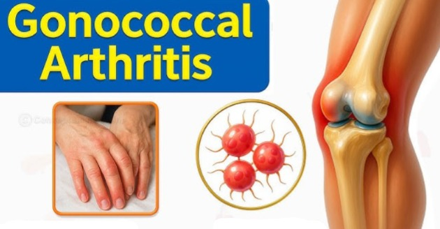
- Gonococcal arthritis is inflammation of the joints and is mainly caused by gonorrhea infection. It is caused by.
- Gonococcal arthritis is a complication of sexually transmitted infections.
- It mainly causes painful inflammation of the joints and tissues.
- Gonorrhea is spread through sexual contact.
- Gonococcal arthritis is a disease caused by the gonorrhea bacteria, which spread through the bloodstream to the joints.
- Gonococcal arthritis is a form of septic arthritis.
Explain the Etiology of gonococcal arthritis.
- gonorrhea infection,
- Nigeria gonorrhea,
- It is spread from an infected mother to her child.
- Women and teenage girls are more likely to be infected.
- multiple sex partners.
- unprotected sexual activity.
explain the Clinical manifestation/ sign and symptoms. (Explain the symptoms.)
- Joint pain,
- Fever,
- Feeling cold,
- Fatigue,
- Pain in hands and feet,
- gonococcal bacteria migraine to tendon,
- Red and swollen joints,
- tender and painful joints,
- decreased range of motion,
- fever and chills,
- Skin Leasion
Explain the diagnostic evaluation. Write a diagnostic evaluation.
- History taking and physical examination,
- throat culture,
- cervical gram stain,
- urin and blood tests,
- complete blood count tests,
- white blood cell count,
- blood cultural test,
- imagine test.
explain management of gonococcal artheritis. (Write the management of gonococcal artheritis.)
- In this, fluid is drained from the joint.
- Immobilize the joint with a splint.
- Give the patient antibiotic medicine.
- Give the patient analgesic medicine.
- Provide Azithromycine to the patient.
1) Explain osteomyelitis.
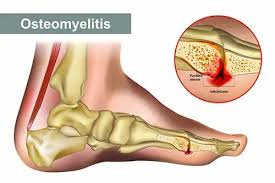
- Osteomyelitis of the bone and is a pyogenic infection of the surrounding tissues.
- Osteomyelitis is an infection of the bone and mainly involves the cortex and medullary cavity.
- Osteomyelitis is an acute infection of the bone.
- It mainly occurs in acute, subacute and chronic processes.
explain Etiology
- staphylococcus Aureus ( Staphylococcus Aureus ),
- E -coli ( E coli ),
- pseudomonas ( Pseudomonas ),
- proteus ( Proteus ),
- salmonella ( Salmonella),
- rheumatoid arthritis,
- people over 50 years of age.
- sickle cell disease.
- obese or malnourished patients.
- patients receiving haemodialysis.
- whose immune system Impaired.
- Due to a post-operative wound.
- Chronic disease (diabetes, rheumatoid arthritis).
- Alcoholism,
- intravenous drug use or drug abusers.
explain clinical manifestation/ sign and symptoms (Describe the clinical manifestation.)
- Bone pain,
- Fever,
- General discomfort.
- Fatigue.
- Swelling and warmth in the affected area.
- Swelling in the local area.
- Redness and warmth.
- Loss of range of motion.
- Feeling cold.
- Profuse sweating.
- Low back pain.
- Swelling of ankle, feet and legs,
- Drainage of pus from the skin.
- General discomfort.
- Nausea.
- Profuse sweating.
- Swelling in the ankles, feet and legs.
- Changes in gait.
- Constant, pulsating pain present.
explain Diagnostic evaluation (Write a diagnostic evaluation.)
- History taking and physical examination,
- bone X Ray,
- ct scan,
- MRI,
- blood test,
- blood culture,
- needle aspiration,
- Biopsy,
- bone scan,
- Bone Biopsy,
- bone X Ray,
- complete blood count,
- c reactive proteins,
- erythrocyte sedimentation rate ( ESR ),
- MRI of bone,
- needle aspiration,
Explain the medical management of osteomyelitis. (Write the medical management of osteomyelitis.)
- administration antibiotic,
- cefriaxone,
- Ciprofloxacine,
- clindamycin,
- vancomycin,
- Lenezolid
- administration intravenously antibiotics,
- implantation of antibiotic beads or pumps.
- Provide hyperbaric oxygen.
surgical management
1)sequestrectomy
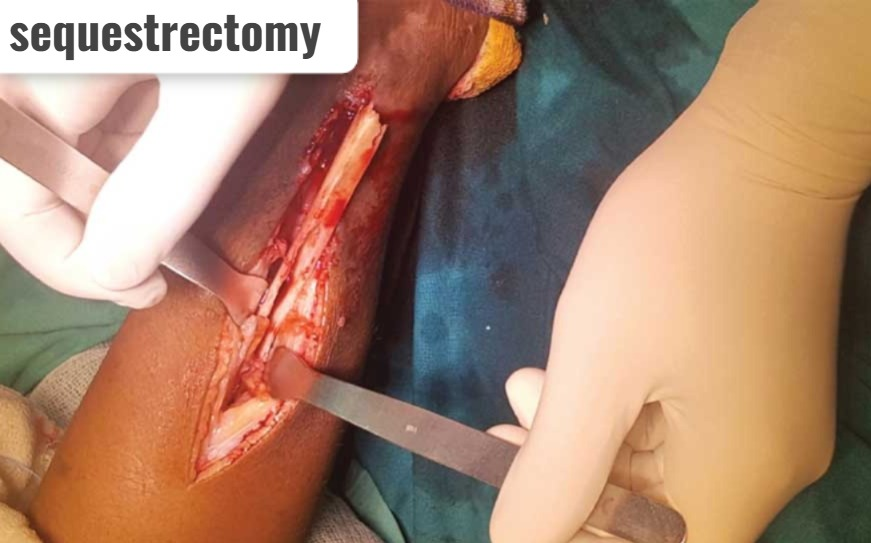
- In this, dead bones are removed.
2) debridement

- In this, as much of the dead bone as possible is removed.
3) drainage (drainage)
- In this Open wounds and accesses are drained by needle aspiration.
4) provide internal fixation and external supportive devices.
Nursing management
- Provide opioids if the patient is in pain.
- Body area ne examination To check for any tenderness, warmth, and swelling.
- Ask the patient to explain his/her feelings.
- Ask the patient to do self-care.
- Maintain strict aseptic technique when changing dressings and irrigating the wound.
- Provide the patient with full information about the disease condition.
- Ask the patient to rest.
- Ask the patient to adopt relaxation techniques.
- Ask the patient to adopt non-pharmacological techniques, including relaxation techniques, guided imagery, and deep breathing.
- Provide support to the affected limb with pillows.
- Elevate the affected area to reduce swelling.
- Check the patient for any pressure ulcers.
- Assess the vascular status of the affected area.
- Instruct the patient to take complete bed rest.
- Check the patient’s vital signs.
- Instruct the patient to take complete bed rest.
- Provide the patient with a high protein, vitamin C rich diet, and a well-balanced diet to promote proper healing.
- Provide a splint to the affected area to reduce pain and muscle spasm.
- Provide the patient with prescribed antibiotics.
- Instruct the patient to do range of motion exercises every four hours.
- Provide support to the affected extremity.
1)Explain the “potts’s spin”. (Describe Potts spine.)
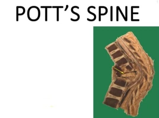
Potts spine Also called tuberculosis in spinal cord.
Potts’s disease is named after Percival Potts’s (who was a surgeon in London).
percival potts’s( who was a surgeon in London) .
Potts’s spine is also called tuberculosis in the spinal (intervertebral joints) cord.
This is a disease of the spinal cord that occurs when Mycobacterium tuberculosis reaches the spinal cord and affects the spinal cord.
Another name for Potts’s spine
- Spine tuberculosis ( spine tuberculosis) ,
- tuberculosis spondylitis ( tuberculosis spondylitis ) ,
- potts’s caries ( potts’s caries) ,
- David disease ( David disease ( David disease) Dizziness),
- potts’s curvature ( Potts’s curvature) . It is called.
Bone is the main site of involvement in Pott’s disease.
The hip ( hip := waist) and the knee ( knee:= knee) are also affected.
It mainly involves the lower thoracic and upper lumbar vertebrae.
2) Etiology of potts’s spine. Causes.)
- The most common cause is Mycobacterium tuberculosis.
- People with poor socioeconomic conditions.
- Hiv infection,
- drug addiction,
- alcoholism,
- spread through lymphatic and hematogenous (by blood),
- endemic tuberculosis,
Explain the signs and symptoms. (Symptoms and signs.)
- The symptoms start gradually.
- Back pain.
- Fever.
- Night sweats.
- Loss of appetite.
- Weight loss.
- Paravertabral swelling.
- Stiff position.
- Abscess formation.
- Spinal moment restricted.
- Weakness.
- Spinal deformity causes muscles wasting.
- muscles weakness of legs.
- compressive myelopathy.
- bone necrosis.
- osteomyelitis.
- Kyphosis (abnormal curvature of spine).
- numbness,
- paresthesia (feeling of tinglingandnumbness),
explain the diagnostic evaluation.(Write a diagnostic evaluation.)
- History taking and physical examination,
- previous exposure to tuberculosis.
- spin X Ray.
- CSF test := to find out bacteria to.
- tuberculin test.
- ct scan.
- MRI.
- radionuclide scanning.
- gallium and tuberculosis bone scan.
- Complete blood count.
- Elevated erythrocyte sedimentation rate.
- strong positive montoux skin test.
Explain the management of potts’s spin.
Medical management of potts’ spin.
- provide antituberculosis chemotherapy.
- ( isoniazid and rifampicine and additional drug at least for two months)
- To take complete bed rest to the patient Tell.
- Immobilize the affected joint.
- Tell the patient to take high protein rich food.
- Drainage of abscess if present.
- Do physiotherapy to the patient.
surgical management
- 1) laminectomy,
- 2) surgical decompression,
- 3) Anterior radical focal debridement also done.
nursing management
1.Disturbed body image.
- Ask the patient to describe his feelings.
- Explain the treatment and its complications to the patient.
- Allow the patient’s family members to stay with him.
2) self care deficit.
- Provide sponge bath to patient.
- Provide back care to patient.
- Provide mouth care to patient.
- Provide eye care to patient.
- Change patient’s position every two hours.
3)Acute pain related to inflammatory process.
- Assess the patient’s pain level.
- Provide the patient with a comfort device.
- Ask the patient to lie down on a hard bed.
- Give the patient the prescribed analgesic medicine.
- Ask the patient to change position frequently.
- Provide moist compression to the patient’s left side.
4) Impaired physical mobility.
- Advice the patient to use assisting device ex:= walker, cans, wheel chair, according to level of mobility of patient.
- Ask the patient to do early ambulations.
1) define sprain.

- A sprain is a condition in which the supporting ligaments of a joint are injured.
- A sprain is a stretching (stretching := pulling) or tearing (tearing := breaking) of one or more ligaments and supporting muscle fibers surrounding a joint.
- A sprain is mainly caused by any sudden injury, and hyperextension or twisting motion.
- In this, the ligament is partially or completely torn. torn (torn).
- Sprain mainly occurs in the ankle or knee joint.
- In a sprain, the ligaments surrounding the joint are injured.
- Usually, the ligaments maintain the stability of the joint, but when a ligament is torn, the stability disappears.
- Edema is seen in the joint. Due to sprain, tenderness is present in the joint and the joint is very painful, along with swelling and Bleeding is also seen.
degree of sprain:=
- 1) first degree sprain,
- 2)second degree sprain,
- 3) Third degree sprain.
1) First degree sprain:=
- In this, the ligament is stretched to a mild amount and there is no instability of the joint.
2) Second degree sprain:=
- In this, the ligament ruptures laterally but there is no instability of the joint.
3) Third degree tear:=
- In this, there is complete rupture of the ligament and instability of the joint is also present.
2) provide Etiology
- Due to twisting of the joint.
- Due to a fall.
- Due to a sports injury.
- Due to over stretching.
- Due to a forceful blow.
- age.
- height.
- weight.
- ankle:= due to walking and exercising on uneven surfaces.
- knee:= during athletic activity.
- wrist:= due to weight on the hand due to falling.
- thumb := Due to playing sports.
explain clinical manifestation/ sign and symptoms.
- Joint pain.
- Swelling.
- In the joint Stiffness.
- Skin discoloration.
- Loss of muscle strength.
- Muscle cramps or spasms.
- Loss of ability to move or use the joints.
- Tenderness.
- Pain during movement.
- Swelling.
- Brushing.
- Discolouration.
- Reduced ability to move the affected joint.
- Swelling.
- Pain.
explain diagnostic evaluation .(Write diagnostic evaluation.)
- History taking and physical examination.
- X Ray truly out contusion, sprain, and strain.
- MRI.
Explain the management of sprain.(Write the management of sprain.)
medical management:=
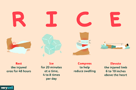
RICE
- R := REST ,
- I:= ICE ,
- C:=COMPRESSION ,
- E:=ELEVATION .
1)REST:=
- Resting can prevent further injury and promote healing.
2)ICE:=
- Applying ice can stop the erection as well as relieve the discomfort in the ED.
- To reduce swelling, ice is applied wrapped in a clean cloth.
- Do not apply ice directly to the skin.
- Apply ice for 10 to 15 minutes.
3) COMPRESSION:=
- Provide support to the injured tissues with a bandage.
- Wrap the injured area with a bandage.
- Elastic compression bandages control bleeding and reduce edema and provide support to the injured tissues.
4) ELEVATION:=
- Elevation reduces swelling.
- Elevation reduces pain levels and improves muscle tone.
- Provide support to the affected area with a belt.
- Provide physiotherapy to the patient.
- Surgical repair.
- Immobilize the affected area with a cast.
- Provide hot compression.
- Apply an electric heating pad.
- Retreat with active or passive exercise after two to five days.
- A sprain or strain may take a week or even a month to heal.
- Assess the neurovascular status of the injured extremity.
- If the sprain is severe, immobilize for one to three weeks.
- Provide the patient with analgesic medication.
- Provide the patient with analgesic medicine, such as NSAID (NON STEROIDAL ANTI INFLAMMATORY DRUG) or COX- 2 (CYCLOOXYGENASE).
- If the sprain is severe, repair it surgically or with a cast. Immobilization.
- Application of splint to prevent further injury.
Nursing management
- Provide education to the patient on the correct use of crutches, walkers, canes, and sleighs.
- Provide a comfortable position to the patient.
- Palpate the skin for warmth.
- Check the neurovascular status of the affected extremity every four hours.
- Check the patient’s capillary refill time.
- Check the patient’s sensation level.
- Check that the swelling does not increase on the affected extremity and that it is properly seen.
- Check that the patient can move the body part properly or not.
- Ask the patient to do range of motion exercises.
- Identify the body part that is under pressure and take care not to put pressure on it.
- Provide prescribed medicine to relieve pain.
- Elevate the affected body part.
- Apply a splint to mobilize.
- Provide cold applications for 20 to 30 minutes in the first 24 hours.
- Instruct the patient to take the prescribed analgesic medicine.
- Provide education to the patient on the proper use of crutches.
- Initially provide rest to the affected body part but gradually and slowly increase activity.
- Provide diversional therapy to the patient.
- Provide emotional support to the patient. To do.
- Provide a comfortable position to the patient.
- Check the patient’s proper vital signs.
- Ask the patient to move the affected body parts.
- Teach patient to use ( RICE :=REST, ICE, COMPRESSION, ELEVATION) therapy to care for injury.
- Proper follow-up of the patient to say.
1)define dislocation
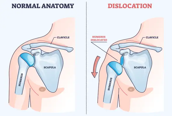
- Dislocation is a condition in which the articular surface separates from the joint.
- Dislocation of a joint occurs when the bone forming the joint dislocates and separates from its anatomical contact.
- This is an emergency situation in which the blood and nerve supply is disrupted ( disruption := disruption ( ulter)) is.
- In subluxation, partial and complete displacement of the joint surface occurs.
- The displaced bone ruptures its blood supply, ruptures blood vessels, damages nerves and muscles.
type of dislocation
- 1)CONGINATAL DISLOCATION (Congenital Dislocation).
- 2)TRAUMATIC DISLOCATION (Traumatic Dislocation).
- 3)PATHOLOGICAL DISLOCATION (Pathological Dislocation).
- 4)PARALYTIC DISLOCATION (Paralytic Dislocation)
1) CONGINATAL DISLOCATION ( Congenital Dislocation):
- This dislocation is mainly congenital and is mainly found in the hip bone and knee bone.
- This can be mainly due to any genetic factor or due to improper development of the fetus.
2) TRAUMATIC DISLOCATION ( Traumatic Dislocation):
- Traumatic dislocation can be caused by a fall, any kind of sharp injury, or due to injury or severe violence.
3)PATHOLOGICAL DISLOCATION ( Pathological Dislocation).
- Pathological dislocation can be mainly due to any infection, rheumatoid arthritis and neuromuscular disease and other diseases.
4)PARALYTIC DISLOCATION (Paralytic dislocation)
- This is mainly due to the muscles It may be due to power imbalance.
- Ex:= poliomyelitis.
explain Etiology (explain the causes)
- due to falling,
- due to severe beating,
- Due to the application of force,
- Due to an accident.
- age,
- sports participation,
- hereditary ,
- Sports injury,
- downhill skiing,
- gymnastics,
- Volleyball,
- due to some disease,
- trauma.
Clinical manifestations:
- Pain,
- Deformity,
- Change in length of extremities,
- Loss of normal movement,
- Length of extremities altered.
- Severe pain.
- Swelling of the affected extremity.
- Empty joint.
- Change in contour of joint.
- Loss of normal mobility.
- Visible deformity is present.
- Deformity.
- Changes in the length of extremities
- Alteration in normal moments.
explain Diagnostic evaluation (Write a diagnostic evaluation).
- History tacking and physical examination,
- X Ray,
- MRI,
Management:
- The doctor immobilizes the part of the joint with a splint for a few weeks.
- How long the splint is left in place depends on the involvement of the joint, nerves, blood vessels, and supporting tissue.
P R I C E treatment

P:= PROTECT
- Properly protect the joint that is dislocated to prevent further injury.
R := RICE
- Proper rest to promote healing.
I := ICE
- To remove discitis.
C:=COMPRESSION
- To reduce pain.
E := ELEVATION
- To reduce swelling. And to relieve discomfort.
- Instead of attempting to relocate a dislocated bone, seek immediate referral to a physician so that proper treatment can be taken.
REDUCTION:=
- In this process, the dislocated bone is brought back into position and this is mainly done by the doctor.
- In the reduction position, the joint is kept in its stable position using bandages, splints, casts and traction.
- Providing analgesic medicine to the patient.
- Providing relaxants medicine to the patient.
- Ask the patient to do range of motion exercises.
- Assess the patient’s neuromuscular status.
Nursing management.
- Assess muscle weakness and severity of weakness.
- Provide the patient with assistive devices such as splints, wheel chairs that increase mobilization.
- Instruct the patient to do range of motion exercises.
- Instruct the patient to do exercises that improve muscle strength.
- Ask the patient to do as much activity as possible that will help the patient become independent.
- Assess the patient’s respiratory rate every four hours.
- Take measures to prevent skin breakdown.
- Assess the patient’s neurovascular status.
- 5 “p”s
- P:= pain
- P:= pallor ,
- P:= pulse ,
- P:= paralysis
- P:= parestheaia
- Provide patient with immobilization device.
- Proper physiotherapy to patient Provide.
- Provide the patient with prescribed medicine.
- Provide the patient with comfort devices to reduce pain levels.
- Protect the injured joint.
- Refer the patient for physical and occupational therapy.
Complication:
- Traumatic arthrotomy,
- fracture within the joints,
- Avascular necrosis,
- arthritis,
- nerve and blood vessels damage.
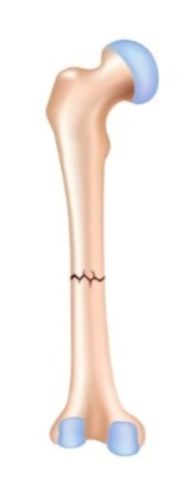
1) Define fracture. (Define fracture).
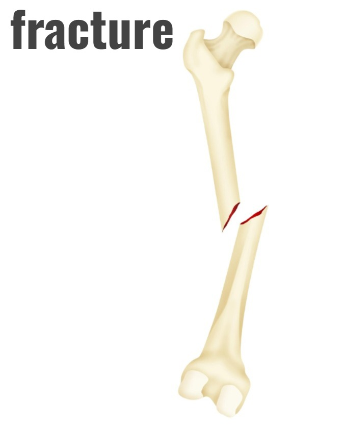
- Fracture is a breakdown in the continuity of a bone.
- A fracture is a break in the structure of a bone.
- A fracture involves the bone, its tissues, bone marrow, and periosteum.
- A fracture of a bone can be partial or complete.
explain Etiology .(Give reasons.)
- Due to trauma,
- Due to road traffic accident,
- Due to fall,
- Due to injury,
- Due to any disease condition,
- Osteoporosis,
- osteomalacia,
- Cancer, other bone infections due to long-term use of corticosteroids.
- Direct trauma.
- crushing force.
- torsion (twisting).
- Excessive muscle contraction Due to.
- bending force.
- Compression force.
- Accident.
- Bondage.
- Old age.
- Occupation.
Explain the Classification of fracture. (Explain the classification of fracture.)
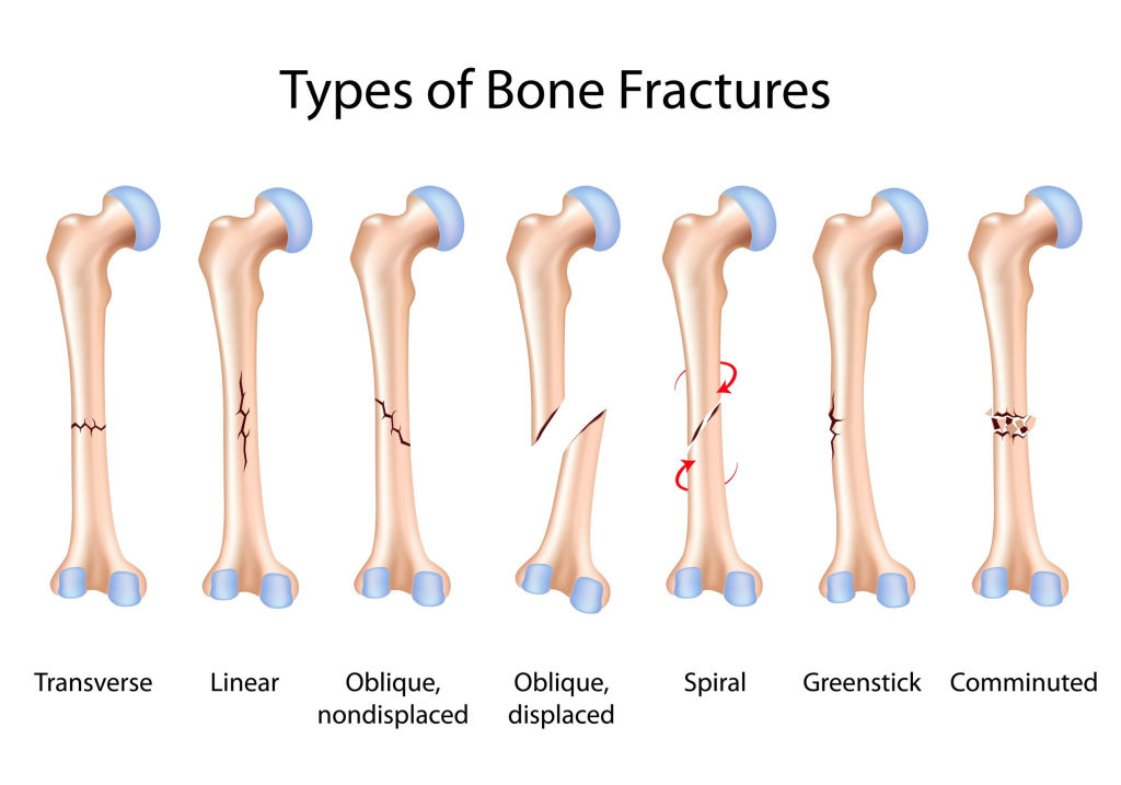
1) complete fracture:=
- In this, the bone breaks down in cross section.
- In a complete fracture, the bone divides into two parts.
2)Incomplete fracture:=

- In this, the bone does not break down completely.
- In an incomplete fracture, the bone cracks but does not break down completely.
3)closed fracture:=
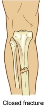
- Close fracture is also called simple fracture.
- In this, the bone is the background.
- But it It remains inside the skin, meaning the skin is intact so the wound is not open or visible and the skin on the fracture side is intact.
4)open fracture:=
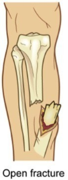
- An open fracture is also called a compound fracture.
- In this, the bone breaks down through the skin and is visible outside.
- The fracture site is the interrupted skin.
- In an open fracture, bacteria can enter through that open site. and creates infection.
According to grade:=
- grade 1 := In this case, the wound is clear and smaller than one centimeter.
- grade 2:= In this case, the wound is in a moderate amount and is larger than one centimeter.
- Grade 3:= The wound is highly contaminated with extensive soft tissue, nerve, and tendon involvement and the wound is larger than 6-8cm.
5) Displaced fracture:=
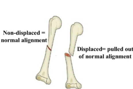
- This fracture is a type of fracture in which the ends of the broken bone separate from each other and this is mainly seen due to fall down.
6)comminuted fracture:=
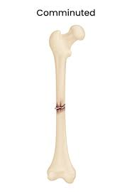
- In this, the bone fragments are crushed and broken down in many parts.
- This is mainly seen in elderly people due to falls.
Classification by fracture pattern:=

1)linear fracture:=
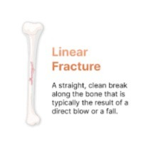
- In this, the fracture is parallel to the long axis of the bone.
- And this is mainly seen due to direct force applied to the bone.
2) Transverse fracture:=

- In this Fractures are seen at 90 degrees.
- Ex:= paget’s disease,
Osteomalacia.
3)Oblique fracture:=
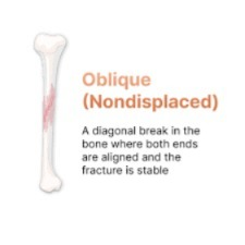
- In this, the fracture is seen at an angle of 45° degrees.
- The fracture is mainly seen due to the application of twisting force.
4) spiral fracture:=
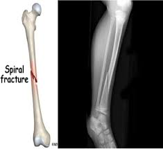
- Spiral fracture is called torsion fracture.
- This is called bony fracture.
- This is mainly seen due to the application of twisting force.
5)depressed fracture:=

- This fracture is mainly seen due to depression in the skull bone and facial is.
6)longitudinal fracture:=
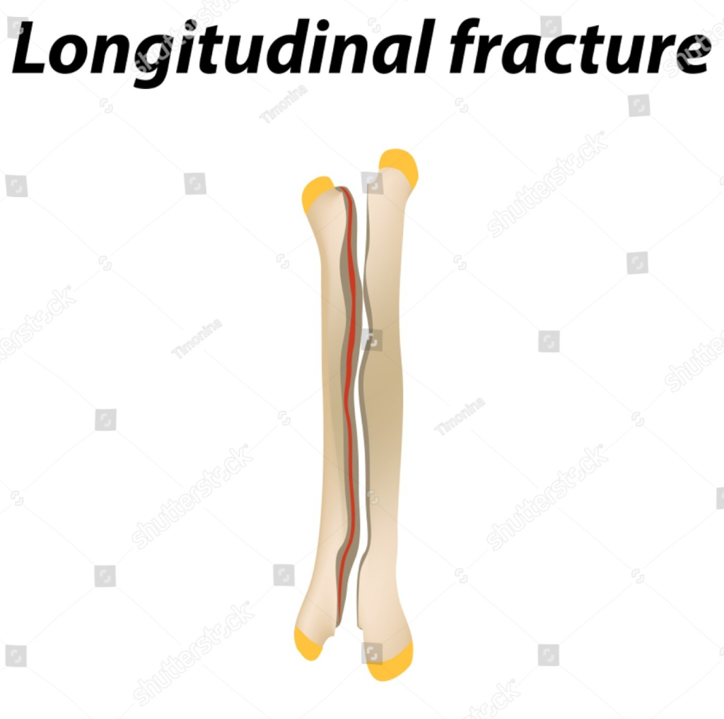
- It is mainly a fracture that occurs in the long axis of the bone.
- In this, the fracture line is longitudinal.
Classification by type of fracture:=

1)Avulsion fracture:=

- This is a fracture in which a segment of bone breaks down through a ligament and tendon.
2)Compression fracture:=
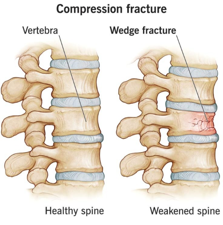
- Compression fractures are also called crush fractures and are mainly caused by compression of the bone.
3)Green stick fracture:=
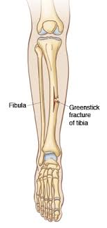
- In this, the bone breaks down on one side and the bone bends on the other side.
4) Impacted fracture:=
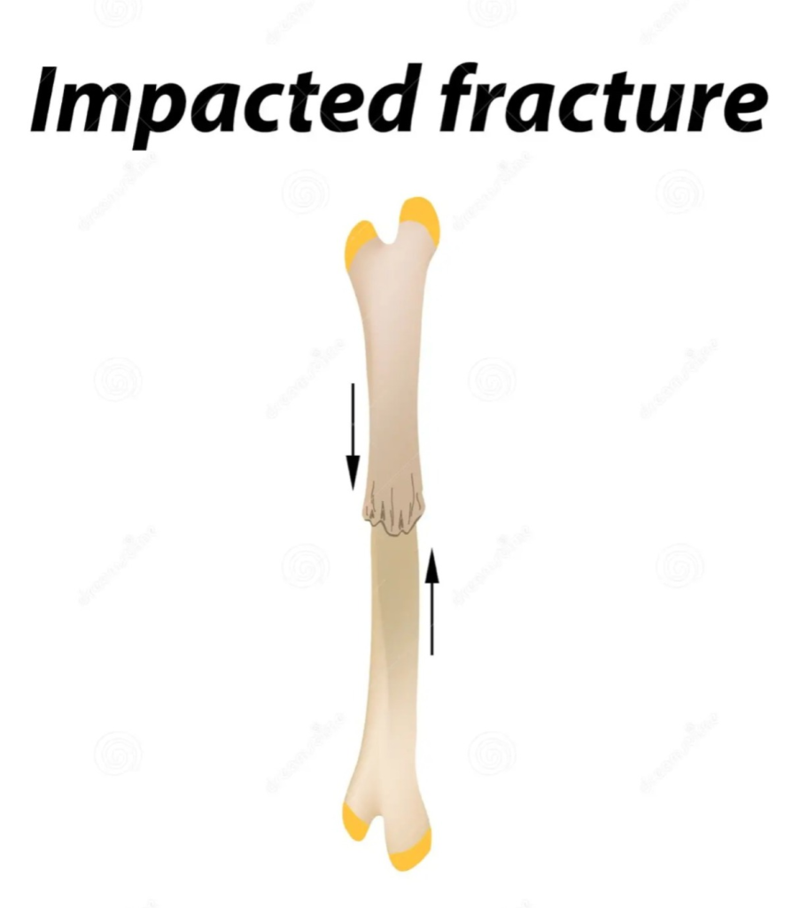
- In an impacted fracture, the continuity of the bone is lost.
5)Pathological fracture:=
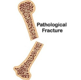
- This fracture is mainly caused when there is a breakdown and fracture where there is a weak bone.
6) Stress fracture:=
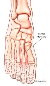
- Stress fractures occur due to repeated loading on the bone.
Classification by eponym.
1)colles’s fracture:=

- Colles’ fracture is also called a broken wrist.
- The radius bone is fractured about one centimeter from its wrist to the articular surface.
2)pott’s fracture:=
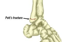
- Potts fracture is mainly found in the medial melleiolus of the tibia and fibula is.
Classification by anatomical location:=
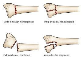
1)articular fracture:=
- This fracture involves the articular surface of the joint.
- This fracture mainly damages the articular cartilage but also damages the subchondral bone.
2) extracapsullar fracture:=
- This fracture is mainly near the capsule of the joint but does not involve the joint capsule and this type of fracture is mainly in the hip.
3) intracellular fracture:=
- This fracture is mainly found within the joint capsule and is mainly found at the neck level and on the head of the femur bone.
4)Epiphysial fracture:=
- This fracture mainly involves the epiphyseal plate of the long bone is.
- This fracture is also called a Selter fracture.
explain Clinical manifestation /sign and symptoms . (Explain the signs and symptoms of the fracture.)
- Pain,
- Tenderness at site of fracture.
- Swelling.
- Increase in body temperature.
- Loss of function.
- Deformity.
- Blood loss.
- Deformity.
- Swelling.
- Pain.
- Impairment in function.
- Empty breath.
- Crepitus.
- Hypovolemic shock.
- shortening of extremities.
- discolouration.
- Impaired sensation.
- abnormal mobility.
- shock.
- diminished capillary refill.
- pallor.
Diagnostic Evolution (Diagnostic Evaluation):
- History taking and physical examination.
- Clinical examination.
- radiographic examination.
- ct scan .
- MRI.
Management :
emergency care of fracture:=
- Immobilize the fractured part immediately when the fracture is detected.
- Provide proper support to the fractured part.
- If there is severe trauma, take measures to reduce it.
- If there is an open fracture, provide it with a sterile dressing immediately.
- In this, apply a dressing to the fracture to control bleeding.
- If bleeding occurs, apply pressure.
- Cover the patient to preserve body heat.
- Check the movement, warmth, circulation, and color of the fracture site and extremity.
- Apply a sufficient amount of splint to the affected joint.
- Immobilize the affected joint.
- Immobilize the affected limb.
- Do a complete physical assessment to rule out any further injuries.
medical management
1)REDUCTION:=
- Reduction restores the fractured part to its anatomical alignment.
1) closed reduction: =
- In closed reduction, the fracture part is given proper position on its anatomical site and a splint is applied to it.
2)open reduction:=
- In this, internal fixation is used to fix the bone fragment.
- In which metal, pin, wire, screen, rod, etc. are used.
2) Immobilization:=
- Properly immobilize the bone fragment after fracture.
- This Immunization is done as an external fixator and an internal fixator.
External fixator included:=
- In this external fixator, bandages, casts, splints are used.
Internal fixation:=
- Internal fixators use metal pins, wire, screen rods, etc.
3)maintaining and restoring function:=
- Elevate the affected extremity to reduce swelling.
- Provide the patient with ice application.
- Assess the patient’s neurovascular status.
- Ask the patient to express his/her feelings.
- Change positions frequently to reduce the patient’s pain level.
- Administer tetanus injection to the patient as a prophylactic.
- Provide the patient with antibiotic medicine.
- Provide the patient with analgesic medicine.
- Provide the patient with calcium and iron supplementation.
- Provide cold application to the patient’s affected extremity.
- Provide the patient with education on alternative treatments for pain management like relaxation and guided imagery.
- Tell the patient to do exercises to reduce muscle wasting.
4) Pharmacological management:=
- Provide narcotic medicine to the patient.
- Provide analgesic medicine to the patient.
- Provide the patient with non-steroidal anti-inflammatory medicine (NSAID).
- Administer antibiotic medicine to the patient.
- Administer anticoagulant medicine to the patient.
- Give the patient a school softener.
Nursing management
- Elevate the affected extremity to reduce swelling.
- Provide the patient with a comfortable position.
- Maintain SMT technique when handling the patient.
- Assess the patient’s neurovascular status.
- Check the patient’s vital signs.
- Provide prescribed antibiotics, analgesics, calcium supplements.
- Maintain the patient’s intake output chart.
- Provide the patient with a protein and calcium rich diet.
- Provide reassurance to the client.
- Repeatedly assess the patient for any infection.
- Ask the patient to do some daily routine activities and exercise.
- Check the patient’s vital signs.
- Check the patient’s pain level using a pain scale.
- Slowly elevate the affected limb.
- Have the client breathe deeply. Tell.
- Ask the patient to adopt relaxation techniques.
- Check the patient’s capillary refill time frequently.
- Examine the limb to see if there is any swelling in the heel.
- Keep checking the tightness of the cast.
- Keep the affected limb above heart level.
- Maintain aseptic technique when dressing the patient.
- Instruct the patient to perform range of motion exercises for the affected extremity.
- Instruct the patient to perform early ambulation.
- Provide education about the patient’s assistive devices such as crutches, walkers, canes, slings, etc. To do.
- Keep changing the patient’s position every two hours.
complications
- shock,
- fat embolism,
- compartment syndrome,
- volkmans contracture,
- deep vein thrombosis,
- infection,
- Delayed Union,
- avascular necrosis of bone,
- reflux sympathetic dystrophy.
1) define spinal fracture. (Define spinal fracture.)

- A spinal fracture occurs when the bones of the spine, called vertebrae, break or collapse.
- This can mainly be caused by any trauma, injury or fall, and can also be caused by a car accident.
2) Explain Etiology (Explain the cause.)
- Due to abnormal curvature of the spine (Kyphosis, Scoliosis, Lordosis).
- Due to a tumor in the spinal cord.
- Due to any injury.
- Due to an accident.
- Due to a fall.
- Due to an assault.
- Due to a sports injury.
- Due to any injury to the spinal cord.
explain clinical manifestation/sign and symptoms (Describe the symptoms and signs.)
- The symptoms and signs of spinal fracture depend on its severity and location.
- Severe pain.
- Empty back.
- Tingling and numbness sensation.
- Muscle spasm to occur.
- Weakness.
- Changes in the bowel and bladder.
- Decreased energy.
- Paralysis.
explain diagnostic evaluation Write the diagnostic evaluation.
- History taking and physical examination.
- X Ray.
- ct scan.
- MRI.
explain management of fracture of spine.
- There are three things to keep in mind in the management of spinal cord.
- Maintain the alignment of the spinal cord.
- Immobilization of the spinal cord should be maintained during healing.
- Pain should be controlled by restricting movement.
- Instrumentation and fusion are surgical procedures used to correct unstable fractures.
- Plates,
- Rods,
Hooks,
Pedicles,
Screws,
And cages are used to join two fractured bones.
These applications can take several months for the bones to fuse. - Vertebroplasty and kyphoplasty is an invasive procedure. It is performed when there is a fracture of the spine.
in vertebroplasty :=
- In vertebroplasty, bone cement is inserted into the fractured vertebrae through a hollow needle.
in kyphoplasty:=
- In kyphoplasty, a balloon is first inserted and then inflated so that the compressed vertebrae can be moved to a normal position.
Explain the degenerative conditions of joint and spine.

1) Explain Osteoarthritis. (Define osteoarthritis)
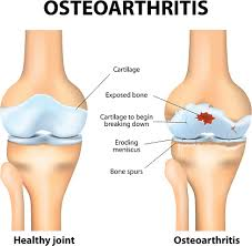
- Osteoarthritis is a chronic, noninflammatory, and slowly progressive disorder that causes deterioration of articular cartilage.
- Osteoarthritis mainly affects the hip bone and knee bone.
- Osteoarthritis is also called degenerative joint disease.
- It is a slow, progressive, non-inflammatory disorder that occurs primarily in mobile joints, particularly articulation joints. is.
- Osteoarthritis is mainly caused by the breakdown of cartilage around the joint.
type of Osteoarthritis:=
1)primary Osteoarthritis:=
- Primary Osteoarthritis is mainly seen in elderly people and mainly in women.
- This is mainly due to any trauma, hereditary, and due to obesity, age etc. Osteoarthritis is seen.
2)secondary Osteoarthritis:=
- Secondary osteoarthritis is seen at any age.
- This is mainly seen due to any previous injury, repeated strain or sprain, joint dislocation, fracture, inflammation, congenital dislocation of hip, Disorder of nervous system, Use of corticosteroids etc.
provide Etiology (Give reasons)
- Due to older age,
- more commone in women,
- Due to genetic factors,
- Due to excess weight,
- Due to obesity,
- Due to septic arthritis,
- Trauma Due to,
- due to strenuous and repetitive exercise,
- due to joint injury,
- due to decreased estrogen levels.
- due to increased parathyroid hormone.
- due to metabolic diseases such as diabetes, gout, and other hormonal disorders.
explain clinical manifestation/ sign and symptoms
- Joint pain,
- Joint stiffness,
- Pain increases with activity and decreases with rest.
- Stiffness in the joints in the morning.
- parestheia ( tingling and numbness sensation),
- swelling, weakness in the muscles, bony deformity,
- swelling in the joints (warmth, effusion, synovial thickening).
- tenderness and soreness in the joints.
- break
- Reduced flexibility of the joint.
- Reduced range of motion in the affected joint.
- Crapitus (a sound made by friction between two bones).
- Swelling in the bone.
- Grating sensation.
explain diagnostic evaluation (write diagnostic evaluation)
- History taking and physical examination
- X Ray,
- ct scan,
- MRI,
- Blood test,
- analysis of synovial fluid,
- erythrocyte sedimentation rate (ESR Test),
- Radionuclide imagine.
Explain the management of Osteoarthritis Write the management of Osteoarthritis.
- If the patient is in pain, provide him with analgesic medicine.
- Ex:= acetaminophen
- Provide medicine to relieve inflammation and pain.
- Ex:=NSAID( Non steroidal anti inflammatory drug),
- Ibruprofen, Naproxen.
- Tramadol.
- Cox 2 inhibitor drug.etc.
- Reduce weight properly to avoid stress on joints.
- Exercise properly Exercise increases joint movement and muscle strength in the muscles surrounding the joints.
- Simple exercises like swimming and walking i.e. doing them on a flat surface are less stressful on the joints.
- Tell the patient to take proper nutrition, get proper sleep and reduce stress, due to which the belching improves.
- If the patient is obese, tell him to lose weight.
- Due to weight loss, the stress on the knee joint, hip joint and spine is reduced and due to this Pain is also relieved.
- When the patient’s pain level increases excessively, it is also necessary to manage the patient surgically.
- Using supportive devices such as splints, shoes, due to which the pain level is reduced.
- Using supportive assistive devices which reduce pressure on the joint and are mainly used to stabilize the ligament and reduce pain levels.
- glucosamine and chondroitin:= These are mainly used to reduce pain in people with osteoarthritis.
- Providing hot and cold applications to the patient.
- Providing hot therapy reduces joint stiffness and is mainly provided two to three times throughout the day. should be done.
- Cold therapy is mainly used to reduce swelling and cold application should not be applied for more than 20 minutes.
explain the surgical management of Osteoarthritis (सुर्जिकल मेंडियाने नि विश्वादने के लिए.)
1)Osteotomy:=
- In osteotomies, the bone is cut from above and below and the weight is reduced, which reduces the pain level.
2) joint fusion:=
- In joint fusion, the damaged joint is removed and the two bones are fused and this is mainly done where joint replacement is not effective. is.
3)Arthroscopy:=
- In arthroscopy, the damaged cartilage is mainly cleaned and the tissues are repaired.
4)joint replacement:=
- In joint replacement surgery, the damaged joint surface is removed and replaced with a plastic or metal device called a prosthesis.
Nursing Management:
- Assess the patient’s pain level.
- Measure the location and intensity of the patient’s pain using a pain scale.
- Provide the patient with hot or cold applications.
- Ask the patient to change positions frequently and to assume a comfortable position.
- Ask the patient to take complete rest.
- Ask the patient to take the prescribed analgesic medicine.
- Provide the patient with comfortable mattresses and pillows and to take proper rest.
- Ask the patient to practice good body mechanics while walking, sitting, moving, or lifting anything.
- Ask the patient to use devices such as splints, braces, traction, etc. properly.
- Ask the patient to take complete rest.
- Ask the patient to take the prescribed analgesic medicine.
- Ask the patient to adopt stress management techniques.
- Provide education to the patient to reduce activities that increase the pain level.
- Provide hot and cold applications to the patient.
- Provide education to the patient on correct position and body mechanism.
- Ask the patient to roll a towel and then place it at the neck level and rest.
- Ask the patient to reduce weight.
- Ask the patient to do as much activity as possible.
- To exercise the patient’s proper range of motion.
- Provide education to the patient to take adequate rest, sleep and nutrients.
- Check the patient for inflammation at the joint site.
- Assess the patient’s range of motion of the affected joint.
- Ask the patient to do range of motion exercises.
- Provide the patient with a safe environment, for example, raise the chair, use high grip and tub and toilet, the use of mobility aids/wheelchair rescue.
- Provide the patient with the opportunity to do active and passive exercises.
- Ask the patient to do early ambulation by use of assisting devices like crutches, walker and canes.
- Provide education to the patient to do exercises.
- Ask the patient to use assistive devices.
- Ask the patient to maintain a comfortable position.
- Ask the patient to maintain proper posture.
- If the patient is obese due to tuition, ask him to lose weight.
- Ask the patient to take the prescribed medicine.
- Ask the patient to make modifications in his lifestyle.
Define Rheumatoid Arthritis.
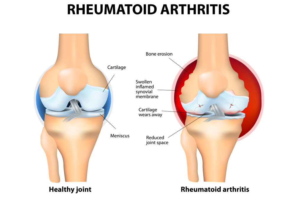
- Rheumatoid Arthritis is a chronic, systemic, autoimmune connective tissue disorder that causes inflammation of the tissues surrounding the joints, including the synovial membrane, and destruction and proliferation of synovial members, resulting in joint destruction, ankylosis (stiffness of the joint) and deformity (physical deformity).
- In autoimmune disease, the body’s immune system produces antibodies against normal cells and damages normal cells. This is called autoimmune disease. Joint pain, stiffness, and immobility occur.
- Rheumatoid arthritis also affects other organs of the body, including the skin, eyes, lungs, and blood vessels.
Explain Etiology of Rheumatoid Arthritis (Causes of Rheumatoid Arthritis):
- Rheumatoid The exact cause of arthritis is unknown.
- Due to genetic factors (if a parent has this disease, there is a possibility of it in their child),
- Due to stress.
- Sex: women are more likely to develop Rheumatoid Arthritis.
- Due to any infectious agent.
- Age mainly between 30 and 60 years In age.
- Due to environmental factors.
- Due to family history.
- Due to hormonal effects.
- Due to long-term smoking.
- Due to metabolic and biochemical abnormalities.
- Any bacteria, Due to fungal, viral infection.
- Due to immunological response.
Explain the Stages of Rheumatoid Arthritis (Explain the Stages of Rheumatoid Arthritis).
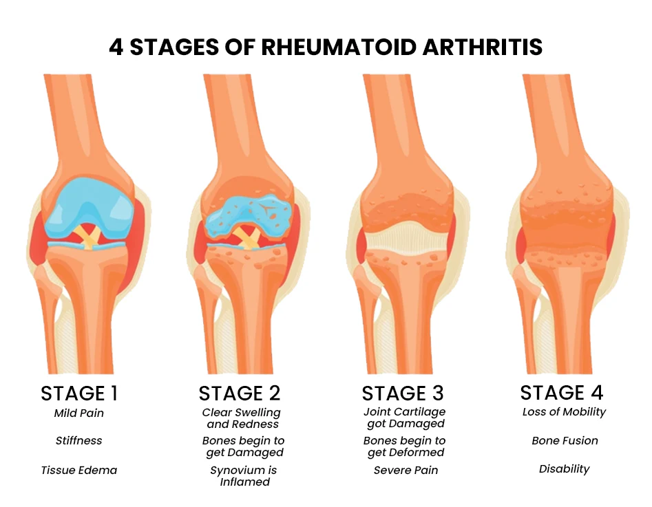
1) Synovitis,
2) Pannus formation (Panus formation),
3) Fibrous tissues Ankylosis,
4) Bony Ankylosis.
1) Synovitis:
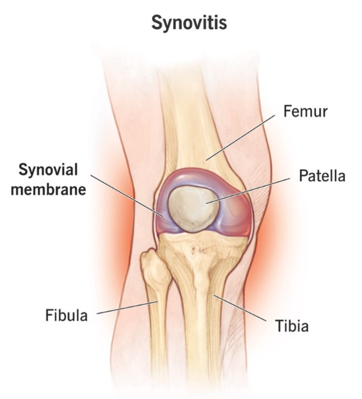
- In the synovitis stage, when there is an infection in the body, that infection affects the synovial membrane of the joint and due to this, the synovial members become infected and inflamed and due to this, synovitis occurs and synovial fluid increases.
2) Pannus formation:
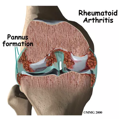
- In this, the synovial fluid gets invaded and becomes very thick and this fluid increases around the capsule of the joint.
3) Fibrous tissues Ankylosis:
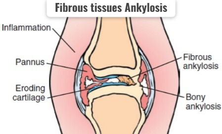
- In this, the synovial fluid increases a lot and it becomes stiff and it gets stuck around the joint and forms a hard structure.
4)Bony Ankylosis (Bony Ankylosis):
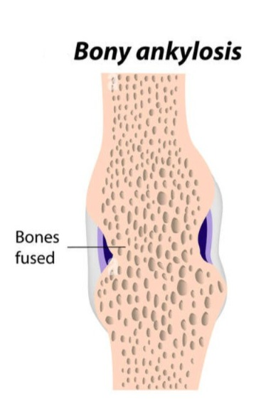
- In this, the fibrous tissue forms a very hard structure and forms a bone-like formation and due to this the joint becomes immobilized And stiffness occurs in it.
Due to any etiological factor.
|
\/
Infection of synovial membrane occurs.
|
\/
Inflammation of synovial members occurs.
|
\/
Synovial fluid is secreted from synovial members.
|
\/
This fluid progresses and accumulates in the bone.
|
\/
Then the bone becomes very hard and stiff Which is an immobilized bone.
|
\/
Rheumatoid arthritis .
Explain Clinical Manifestation/ sign and symptoms (सल्कश्नो तथा सिन्हो परिष्टो):
- The affected joint becomes red, warm.
- The joints become swollen and stiff and tender.
- Joint pain
- Morning stiffness in the joints.
- Arthritis occurs in more than three bones.
- The joint becomes swollen (sponge like).
- Arthritis occurs in the joints of the hands.
- firm bumps of tissues under the skin on Arms.
- Rheumatoid nodules form.
- Rh factor positive.
- Fluid accumulation occurs in the ankles.
- The joint loses its range of motion and becomes deformed.
- Muscular atrophy around the affected joint.
Ulnar Deviation:
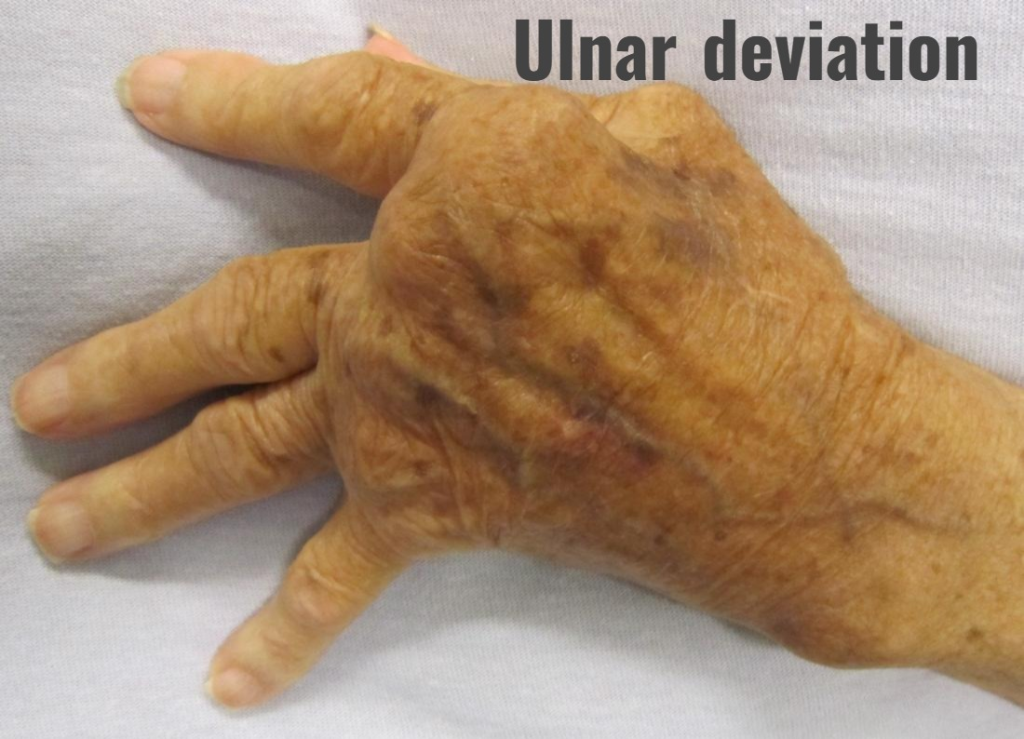
In this, the finger deviates towards the ulnar surface.
Swan Neck Deformity:
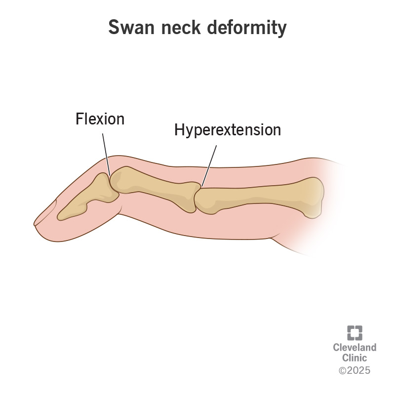
In this, the finger becomes a swan’s head.
Bouterine Deformity:
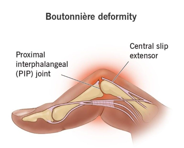
Ama, finger is bent.
- knock knee.
- Difficulty in Sleep .
- Numbness and tingling sensation.
- Burning sensation in hand and foot.
- Module formation within the skin.
- Burning sensation in the eyes.
- Itching and discharge.
- Dry mouth and eyes.
- Chest pain.
- Weakness.
- Difficulty breathing.
- Fatigue.
- Loss of appetite.
- Weight loss.
- Low grade fever.
- Malaise.
- Depression.
- Lymphadenopathy.
- Inflammation of blood vessels.
- Multiple organ involvement (pericarditis, Osteoporosis, Anemia, subcutaneous nodules, vasculities, neuropathy, fibrotic lungs disease).
Explain Diagnostic Evaluation (Write diagnostic evaluation):
- history tacking and physical examination (History Taking and Physical Examination).
- Rheumatoid factor test: RA positive (Rheumatoid Factor Test).
- Antinuclear Antibody test (Antinuclear Antibody Test).
- Erythrocyte sedimentation rate (ESR – Erythrocyte Sedimentation Rate ).
- C-reactive protein.
- complete Blood count test.
- comprehensive metabolic panel ( to monitor kidney and liver function).
- synovial fluid analysis (synovial fluid changes from transparent to milky ,cloudy, and dark yellow fluid).
- Arthroscopic examination (Examination ) .
- X Ray ( X Ray ) .
- joint Ultrasound ( Joint Ultrasound ) .
- MRI ( M.R.I.) .
Explain the management of rheumatoid arthritis (Write the management of rheumatoid arthritis):
Medical Management (Medical Management):
1) NSAID (NON STEROIDAL ANTI INFLAMMATORY DRUG):
This medicine is used to relieve pain and inflammation.
Ex: ibuprofen,
Naproxen sodium.
2) DMARDs ( Disease modifying Antirheumatic Drugs):
These are mainly given in conditions such as moderate to severe Rheumatoid Arthritis.
Ex: imuran,
Anti malarial medication,
Panicillamine and mithotrexate.
3) Antimalarial medication :
This medicine mainly uses hydroxychloroquine ( hydroxyquloroquine ) along with methotrexate.
4) Corticosteroid ( Corticosteroid ) :
Corticosteroid is used to relieve inflammation.
5) Biological agent ( Biological agent) :
Tnf-a antagonist targets B cells, T cells.
- Actemra in biological agent,
- Rituxan,
- Remicade,
- Enbrel,
- Includes Kindred.
6) Immunosuppressants (Immunosuppressants):
The immune system is out of control in rheumatoid arthritis. This medication weakens the immune system.
Ex: Azathioprine (imuran, azasan),
Cyclosporine.
7) Tumor necrosis factor a:
This medicine inhibits the inflammatory chemical that is tumor necrosis factor.
This medicine inhibits the inflammatory chemical that is tumor necrosis factor.
Explain Surgical management (Explain Surgical Management):
1) Joint fusion:
In this, the joint is surgically fused to stabilize the joint.
2) Synovectomy:
In this, the joint lining is removed is removed.
This is mainly used to remove the inflamed tissues that are creating pain.
Synovectomy is mainly used to reduce swelling and slow down joint damage.
3) Tendon repairs:
The surgeon repairs the tendons around the joint that are inflamed and damaged, causing loss of tendons. and keeps it stable.
4) Total joint replacement:
In joint replacement, the surgeon removes the damaged joint part and inserts a prosthesis made of metal or plastic in its place.
Explain nursing management:
- Encourage the patient to join self-help groups and support groups.
- Assess the patient’s pain level.
- Assess whether the patient has morning stiffness.
- Provide the patient with a comfortable position.
- Anchor the patient for non-pharmacological management Do.
Such as yoga, relaxation techniques, guided imaginary, and rhythmic breathing. - Advise the patient for hot and cold applications.
- Provide the patient with prescribed medicine.
- Instruct the patient to rest between activities.
- Instruct the patient to rest frequently.
- Instruct the patient to rest frequently.
- Instruct the patient to Ask the patient to do physical activities such as walking, swimming, etc.
- Ask the patient to use an assistive device.
- Ask the patient to verbalize his/her feelings.
- Provide the patient with complete education about the disease and its treatment.
- Ask the patient to maintain strict aseptic technique.
- Provide education to the patient to participate in self care activities.
1)Define bone tumor. Define bone tumor.
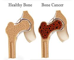
INTRODUCTION ( Introduction ) :
Bone cancer can occur in any part of the body. But bone cancer mainly affects the long bones, such as the bones of the arms and legs. If the cancer originates in the bone, it is called primary bone cancer, and if the bone cancer spreads from another part of the body and spreads to the bone, it is called secondary bone cancer.
type of bone cancer:
1)benign bone tumor ( Benign bone tumor):
Benign bone tumor include:
- osteomas ( osteomas),
- osteoblastomas ( osteoblastomas),
- osteoidosteoma ( osteoid osteoma),
- osteochondromas ( Osteochondromas),
- enchondroma ( in enchondros)
2)malignant bone tumor( malignant bone tumor ):
The most common bone tumor
- osteosarcoma ( osteosarcoma),
- chondrosarcoma (chondrosarcoma),
- fibrosarcoma (fibrosarcoma),
- chordoma (chordoma)
3)Metastasis bone cancer :
Almost all types of cancer spread to the bone but mainly
Bone(bone),
Breast,
Lungs,
Kidney, Thyroid And Prostate
These are the main organs from which cancer spreads to the bone.
Etiology:
occurs at 10-25year age,
exposure to radiation,
inherited genetic disorder,
some drugs,
clinical manifestation:
pain,
mass or lump felt in the bone,
weak bone,
fever,
chills,
night sweat,
anemia,
anorexia,
fatigue,
tenderness,
weight loss,
neurological symptoms may present with nerve root compression (neurological symptoms may also be seen).
Swelling,
limited motion,
increase skin temperature over mass.
Diagnostic evaluation:
- History taking and physical examination,
- bone scan,
- X Ray,
- ct scan,
- MRI,
- myelography,
- arteriography,
- Biopsy,
- Elevated serum alkaline phosphate.
Management:
- Radiation therapy,
- Chemotherapy,
- Biotherapy
- Bone marrow transplantation
- Immunotherapy,
- Gene therapy.
Surgical management:
- limb sparing surgery,
- amputation,
- lymph node dissection,
- Reconstructive surgery,
- tumor curettage,
- bone grafting,
- limb salving procedures.
Nursing management:
- Checking the patient’s vital signs.
- To see how much blood loss the patient has had.
- To see if the patient has any complications. like deep vein thrombosis, Pulmonary embolism, Infection, Etc.
- Give the patient analgesic medication.
- Provide the patient with intravenous fluids.
- Provide support to the affected extremity with pillows.
- Provide the patient with a splint for additional protection.
- Ask the patient and his family members to ask all his fears and questions.
- Provide psychological support to the patient.
- Prepare the patient in advance for the patient’s body image to change.
- Encourage the patient to do their daily routine activities
- Provide complete information about the disease and its treatment to the patient and his family members.
- Maintain the patient’s nutritional and hydration status.
- Provide work and a comfortable environment to improve the patient’s sleep pattern.
- Ask the patient to move the extremities that are not part of the body.
- Provide support to the patient’s joints.
- Be careful that the patient does not fall.
- Create a hazard-free environment for the patient.
- Provide a supportive environment for the patient.
- Provide psychological support to the patient.
- Answer all the patient’s questions.
- Provide psychological support to the patient to improve coping abilities.
1) Explain the spinal cord tumor.
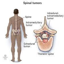
- Spinal cord tumors are abnormal growths of tissues.
- And they are mainly found near the spinal cord because the spinal cord is a rigid, bony structure and abnormal growth causes a sensation of pressure and impairs the function of the spinal cord.
- Spinal cord tumors are mainly found inside the spinal cord and adjacent to it.
- A spinal cord tumor is a cancerous growth found within the spinal cord and can be either benign or malignant.
- If the tumor is causing compression of the spinal cord or nerve roots, immediate medical attention is required because compression of the nerves can lead to paralysis. Even if the tumor is benign, it can still affect the nerves.
- A spinal cord tumor usually originates from another part of the body and metastasizes and transfers to the spinal cord.
type of spinal cord tumor.
1)primary spinal cord tumor:=
- A tumor that arises from the spinal cord is called a primary spinal tumor.
- Only 10% of primary spinal cord tumors arise from the spinal cord.
2)secondary spinal cord tumor:=
- Secondary spinal cord tumor is a tumor in another part of the body that becomes metastatic to the spinal cord.
- Tumors in the breast, lungs, prostate, thyroid gland spread to the vertebrae, i.e., metastasis occurs.
- Metastatic tumors mainly compress the spinal cord and nerve roots.
- Lymphomas also spread to the spinal cord and compress the spinal cord
type:=
- 1) Intramedullary tumor.
- 2) intradural extramedullary tumor. ( Intradural Extra Medullary Tumor)
- 3) Extradural Spinal Tumor
- 4) Secondary Bone Cancer
1) Intramedullary Tumor:=
- This tumor arises mainly in the spinal cord.
- And it is mainly located in the nave of the spinal cord.
- There are many types of cancer in the spinal cord, but the main common ones are two.
- 1)astrocytomas (astrocytomas).
- 2)ependymomas ( Ependymomas)
2) intradural extramedullary tumor.( Intradural Extramedullary Tumor) :=
- This tumor mainly arises from the dura but is located outside the spinal cord, so it is called an intradural extramedullary tumor.
- This is mainly inside the covering of the spinal cord but is located outside the spinal cord itself.
- Ex:=
- 1)meningiomas (meningiomas) 2)schwannomas (schwannomas)
3) extradural spinal tumor (extradural spinal tumor)
:= Extradural spinal tumors are mainly metastases but they arise from the vertebrae.
These mainly start from the bones of the spine.
Ex:=
benign tumor includes:=
- 1) Chordomas (Chordomas)
- 2) osteomas ( osteomas)
Malignant tumor includes:=
- 1)chordosarcomas
- 2)fibrosarcomas
- 4)secondary bone cancer
This cancer mainly spreads to the spinal cord from other parts of the body.
This is called secondary bone cancer.
This cancer mainly affects the vertebrae in lungs, breast, lymphoma, prostate cancer, and myeloma.
Myeloma is a cancer of plasma cells that mainly affects the vertebrae.
2) explain Etiology (Write the reasons:=
- Due to a genetic defect,
- Due to exposure to certain types of chemicals.
- Due to hereditary disorders.
- Due to tumor metastases from other parts of the body.
- Due to a history of previous cancer.
explain clinical manifestation/sign and symptoms
- Symptoms and signs depend on the location of the tumor and its type.
- Pain occurs in the back and neck.
- Pain also occurs in other parts of the body.
- Numbness, tingling sensation.
- Muscle weakness and weakness, especially in the muscles of the arms and legs.
- Decreased sensation.
- Difficulty walking.
- Progressive weakness.
- Sensitivity to pain, cold, and heat decreases.
- Loss of bowel and bladder function.
- Difficulty walking.
- Paralysis.
- Muscle contractures and spasms.
- Nerve damage.
- Pain
- Swelling.
- Weakness.
explain diagnostic evaluation (Write diagnostic evaluation.)
- History taking and physical examination.
- X Ray.
- ct scan.
- MRI.
- emissions tomography.
- spinal tap.
- Biopsy.
- myelogram.
3) Explain the management of spinal tumor. (Write the management of spinal tumor.)
medical management
- strictly monitoring of patient.
- Radiation therapy,
- Chemo therapy,
- Steroids Therapy,
- surgery:= Decompressive laminectomy.
Nursing Management:
- Continuously assess the patient’s neurological status.
- Continuously check the patient’s vital signs.
- Check the patient’s intracranial pressure.
- Maintain strict aseptic technique when handling the patient.
- Check the patient’s level of consciousness.
- Check the patient’s visual acuity.
- Perform a sensory assessment of the patient.
- Check whether the patient has a condition such as headache, vomiting, or nausea.
- Check the patient’s continuous intake output.
- Check the patient’s electrolyte level.
- Maintain the patient’s hydration status.
- Keep the patient’s head up 15 to 30 degrees to reduce cerebral venous congestion.
- If the patient has photophobia, keep him in a dark room and provide him with sunglasses.
- Provide the patient with a quiet environment.
- Ask the patient to take adequate rest.
- Keep the patient in an upright position to allow proper expansion of the lungs.
- Provide chest physiotherapy to remove mucus.
- Ask the patient to do pursed lip breathing.
- Ask the agent to take small and frequent meals.
- Provide the patient with a comfortable and workable environment.
1) Explain Amputation. Explain Amputation
introduction:=
- Amputation is a surgical reconstructive procedure in which a total or partial extremity is removed.
- Amputation is an acquired condition in which a limb is lost and can be caused primarily by injury, disease, or surgery.
- The word amputation is derived from the Latin word
” amputare “ - Which means
- ” to cut off”
comes from. - Amputation means the partial or complete removal of a deformed body part.
- Amputation is any acute condition
(traumatic event), - or chronic condition such as
(peripheral vascular disease, diabetes mellitus, or surgery)
explain Etiology(Explain Etiology:=
- Injury.
- Due to malignant tumor.
- Due to congenital deformity.
- Inadequate tissues percussion.
- Crush injury.
- Due to burn.
- frost bite.
- due to bone tumor.
- electric burn.
- due to gangrene.
- chronic osteomyelitis.
- due to feral vascular disease.
- diabetes.
- gas gangrene.
- due to electrical injury.
- to remove any tumor.
explain the purpose of amputation (Write the purpose of amputation.)
- To remove tissues that do not receive the proper amount of blood supply.
- To remove a malignant tumor if it is present.
- To remove a severe trauma to a body part.
explain the level of amputation (write the level of amputation)
- 1)circulation in the part.
- 2)It’s usefulness.
- 3) Level of maximum available tissues for wound healing.
- 4) develops a functional, non tender, pressure tolerant residual limb.
1) Partial foot amputation (Partial Foot Amputation)
- The lower part of the ankle joint is separated.
2) Transversal Amputation (Trans Tarsal Amputation):=
- In this, the tarsal bone of the foot is amputated mainly
3) syms ‘s Amputation or modified ankle disarticulation Amputation (Syms ‘s Amputation or Modified Ankle Disarticulation Amputation) .
- In this, mainly if there is very severe trauma to the foot, then it is cut and it is mainly the lower limb that is cut.
4)Ankle disarticulation
- In this, the lower limb is mainly cut from the ankle joint.
5) Below knee Amputation
- In this, the lower limb is cut between the knee joint and the ankle joint.
6)Knee disarticulation (disarticulation of the knee) :=
- In this, the lower limb is mainly cut from the knee joint.
7) ubow knee amputation (above knee amputation) :=
- In this, the amputation is mainly done between the hip joint and knee joint.
8) hip disarticulation hip dish articulation:=
- In this, the lower limb is amputated from the hip joint but the pelvis is intact.
9) trans pelvic amputated (trans pelvic amputation)
- In this, mainly the full lower limb is cut and part of the hemi pelvis is also cut.
10) Partial hand Amputation:=
- In this, the amputation of the upper limb is done and it is done distally from the wrist joint.
11) Wrist disarticulation
In this, the upper limb is mainly cut from the wrist joint.
12)Below Elbow Amputation:=
- In this, the upper limb is mainly cut from between the wrist joint and the elbow joint.
13)Elbow disarticulation (Elbow disarticulation):=
- In this, disarticulation is mainly done from the elbow joint.
14) Above elbow Amputation (Above elbow amputation):=
- In this, disarticulation is mainly done from the elbow joint and The cut is made from the middle of the shoulder joint.
15)Shoulder disarticulation (Solder Disarticulation) :=
- In this, the cut is mainly made from the solder joint.
16)Forequarter Amputation (Forequarter Amputation) Imputation)
- In this, the scapulothoracic and sternoclavicular joints are mainly cut.
17)staged Amputation: =
- In this, the body part is mainly amputated if there is any gangrene or infection. is.
type on the basis of amputated part
- amputed finger ( amputated finger),
- amputed thumb ( amputated thumb),
- amputed arm ( amputated hand),
- amputed toe ( amputated toe),
- amputed leg ( amputated foot),
- amputed lower leg ( amputated lower leg) leg),
on the basis of type of surgery
1)open Amputation:=
- Open amputation is also called guillotine amputation.
- This type of amputation is mainly done when any infection is present or there is a chance of developing.
- In this, the bone is cut at the same level and the cavity is not closed but left open to drain any secretions.
- In this, a lot of dressing is done on the end of the stump and when the infection is not present for a long time, this wound is closed and then the drainage of fluid stops.
2) Closed Amputation: =
- Closed or flap amputation is the preferred method because it heals quickly and the patient gets the opportunity to fit the prosthetic device quickly.
- In this, when any tissue or bone is cut, that part is covered by the skin and it is sutured.
- Therefore, it is called closed amputation. is.
Explain the diagnostic evaluation Explain the diagnostic evaluation
History taking and physical examination.
- routine blood test.
- biochemistry.
- blood cross marching.
- Coagulation study .
- blood pressure.
- arteriography.
- venogrsphy.
- Complete blood count.
- White blood cell count.
- vascular doppler ultrasonography.
explain complication Explain complication
- hemorrhage,
- infection,
- into hematoma,
- Skin breakdown,
- Delayed healing.
- Contracture.
- Skin flap necrosis.
- Joint deformity.
- Chronic pain.
- Phantom Pen.
explain rehabilitation of patient. Explain rehabilitation of patient
- Rehabilitation is the process of taking measures to restore the patient to the highest level of mobility and function.
- A person who is young and healthy and has had an amputation may need immediate rehabilitation.
- Elevate the foot for 24 to 48 hours to reduce edema.
- Advise the patient to exercise.
- Exercising can prevent contractures and strengthen muscles.
- And the amputated part can be prepared for prosthesis.
- The stump should be bandaged properly.
- Train the patient to walk properly.
- Psychological counseling of the patient.
Nursing Management:
To check patient’s vital sign.
1) Monitoring fluid balance:=
- Check the patient’s blood pressure and any abnormality in any vital sign.
- Check the amount of drainage the patient has.
- Check the patient’s intake output.
2) Reliving pain
- Assess the patient’s pain level.
- Check the patient’s pain level, location, and intensity.
- Provide the patient with prescribed medicine.
- Elevate the affected part or provide support with a pillow.
- Provide comfort measures to the patient.
- Ask the patient to adopt stress management techniques (deep breathing exercise, visualization and guided imaginary).
- Ask the patient to take analgesic medicine.
- Ask the patient to administer medication.
- Check the patient’s vital signs.
- Palpate peripheral pulse, noting strength and equality.
- Periodically assess the patient’s neurovascular status.
- Assess the patient’s pulse, skin color, and temperature.
- Elevate the affected limb.
- Ask the patient to adapt to the changes.
3)maintaining adequate peripheral tissues perfusion.
- Instruct the patient to use proper dressing.
- Soft, soft with pressure wrap, semi rigid or rigid.
- Have the patient inspect the dressing site.
- Provide adequate fluid to the patient.
- If there is hemorrhage, apply direct pressure to the bleeding site.
- Provide adequate fluid to the patient.
- If there is hemorrhage, apply direct pressure to the bleeding site.
- Apply direct pressure to the bleeding site with a bandage.
- Provide intravenous fluid to the patient.
4)Preventing wound infections:=
- Use sterile dressings.
- Maintain aseptic technique.
- Instruct the patient to eat a protein-rich diet.
- Handle the affected limb gently.
- Maintain aseptic technique when changing the dressing.
- Inspect the wound.
- Note the characteristics of the drainage.
- Check the patency of the drainage device and empty it.
- Check the patient’s vital signs.
- Provide antibiotic medicine to the patient.
5)Promoting physical mobility
- Provide stump care on a routine basis to the patient.
- Change the patient’s position frequently in bed.
- Ask the patient to do range of motion exercises.
- Instruct the patient to do small amounts of daily routine activity Say.
6)Promote independent self care.
- A mutation changes the body image, so it is necessary to maintain proper coordination with the patient. It is necessary to counsel the patient in a proper manner.
- Proper care of the patient Provide counseling.
- Instruct the patient to stay active.
- Tell the patient to try to do dressing and bathing on their own as they become independent.
- Tell the patient to use a wheelchair and do self-care activities.
- Tell the patient to be careful not to injure the remaining extremity.
explain health education Explain health education.
- Inspect the stump daily for any redness, blisters, or abrasions.
- Use a mirror to examine the stump.
- Maintain the patient’s daily hygiene Said.
- Clean the stump properly using mild soap.
- Do not apply alcohol, cream, lotion, powder on the stump after bathing.
- For the cleanliness and comfort of the patient, woolen stump shocks should be worn on the stump.
- Ask the patient to exercise continuously.
- Is prosthetics Keep the socket properly clean and dry.
- Provide education to the patient or wrap the stump properly.
- Instruct the patient to do exercise and physical activity.
- Instruct the patient to use assistive devices.
- Instruct the patient to make some modifications to the home environment.
1) explain prosthesis

- This is a rehabilitation device in which an artificial device is placed in a whole or partially device.
- This device is manufactured.
- A prosthesis is a device that replaces a missing body part and improves body work.
- Prosthesis is made of metal, titanium, stainless steel, chromium, plastic.
- This prosthesis is placed in place using acrylic cement and helps the bone grow.
- Once the joint is placed in its place, its physical activity can be restored.
- Bone substitutes are called biologics.
parts of prosthesis :=
- 1) interface (interface) ,
- 2) components ( component) ,
- 3)cover( cover) .
1)interface:=
The prosthesis is attached to the body through an interface.
The interface consists of a socket (shocket) and a rigid frame.
The shocket :=
This is mainly made of plastic or laminated material and is attached to the working parts of the prosthesis.
the frame:=
- The frame is mainly made of graphite or a similar material that provides structural support to the socket
- The linear is worn between the remaining part of the limb.
- The socket acts as a cushion.
- The linear is made of polyurethane and silicone.
- The linear clings to the skin and also provides friction. No.
- The interface is a device that holds the prosthesis securely in place.
A)suction valve:=
- When the stump is placed into the socket.
- Air is forcibly entered through the opening at the bottom of the stump.
- The one-way suction valve keeps the socket closed and open and holds the process in place.
B)Linear with a locking pin:=
- Most of the liners are locked at the bottom of the socket and are locked primarily by a notched pin.
- Because the pin is packed into the stub of the title.
- In this case, the body part is stumped in the body part.
2) components:=
A component is the working part of the prosthesis.
The compound consists of a terminal device, a joint, and a metal shaft.
3)cover :=
Some people who wear a prosthesis have a cover.
Classification :=
1)Endo prosthesis :=
- These are mainly implants that are also used to replace joints.
- Ex:= Austin Moore prosthesis.
2)exo prosthesis :=
- In this, a part of the limb that has been lost is used to make an external place.
type of prospective
- 1)trans tibial prosthesis.
- 2)trans femoral prosthesis.
- 3) trans humeral prosthesis .
- 4)transdial prosthesis.
- 5)finger prosthesis.
- 6)Partial hand prosthesis.
- 7)Partial feet prosthesis.
- 8) hip prosthesis.
New innovation
1)Myoectric Prosthesis (Myo Electric Prosthesis)
- Myoelectric prostheses use electromyographic signals to contract muscles and control the movement of the prosthesis.
- Ex:=elbow flexion ,
- Wrist supination ,
- Opening or closing of finger.
advantage:=
- Provides natural response and requires less effort than traditional mechanical systems.
- Myoelectric system Increases mobility and helps in maximum function.
Ex:= transhumeral,
forequarter amputees and shoulder disarticulation.
2)cable operated prosthesis:=
- This prosthesis is held in the socket by a slung
- And this type of prosthesis is Opened and closed by a cable.
- A cable prosthesis is mainly an upper extremity prosthesis.
3)Ossiointegration: =
- This prosthesis is mainly implanted in the bone.
4)Robotic limbs:=
- In this, the robot arm is mainly used by pressing a button.
- In this, the robot arm has been designed to work more effectively and is operated by the person’s mind.
5)neuroprosthesis:=
- Neural prosthesis is a series of devices is.
- These are mainly motor, sensory or cognitive modality.
- And these can be damaged mainly if any disease or injury occurs.
Ex:= cochlear implants.
explain the care of prosthesis. (Describe the care of prosthesis.)
- Prosthesis is a very expensive and precise device.
- If the prosthesis is cared for a little, its life can be increased.
- Prosthesis should be checked by a prosthetist every six months.
- Wash everything that comes in contact with the skin properly.
- Wash everything properly with soapy water and then dry it properly.
- Do not use anything that contains alcohol.
- Keep checking the prosthesis properly.
- On the day the prosthesis is removed, clean the socket properly with warm water and a cloth and dry it properly.
- Make sure that the shoe size is The prosthesis should be correct.
- Keep the things that are needed in an emergency handy (ex:= stumps, socks, pull socks , bandages, antibiotics, antihistamine, ointment).
- If you have a prosthesis and there is any trouble, do not try to make changes to it on your own and go to the prosthetist immediately.
- Check the prosthesis once a week and check its attachment properly.
- Remove shoes and socks and do not leave anything on the prosthesis. But check whether there is any abnormality or not.
explain skin care during prosthesis
- When wearing a prosthesis, wash the limb properly with soap and water once a day.
- Dry the washed limb properly.
- If the washed limb is not dried properly, there are chances of fungal infection.
- Keep checking the site of the prosthesis for any redness or soreness.
- Keep checking the site where the prosthesis is attached for any screen breakdown, especially if the patient is diabetic.
- If the skin is very dry, apply a cream to soften the skin.
- Do not use Telcum Powder on the area where the prosthesis is attached, as this can cause abrasion.
- Do not use alcohol or any other chemicals on the affected limb.
- Keep the limb covered with the prosthesis at all times. Keep it and apply sunscreen if exposed to the sun.
- If there is any problem with the prosthesis, inform the prosthetist immediately.
- If there is a red spot on the prosthesis, there is a chance of soreness on that area, so avoid the prosthesis and inform the prosthetist immediately.
- If there is no sensation on the limb where the prosthesis is, assess it frequently and do not expose it to hot water and sun exposure as it can cause burns and blisters.
- Proper care should be taken of the skin with the prosthesis.