ENGLISH-msn-2-Musculoskeletal (Part-1)-definition
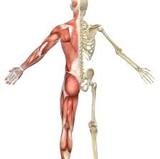
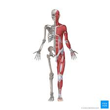
Chapter := 9 Musculoskeletal system
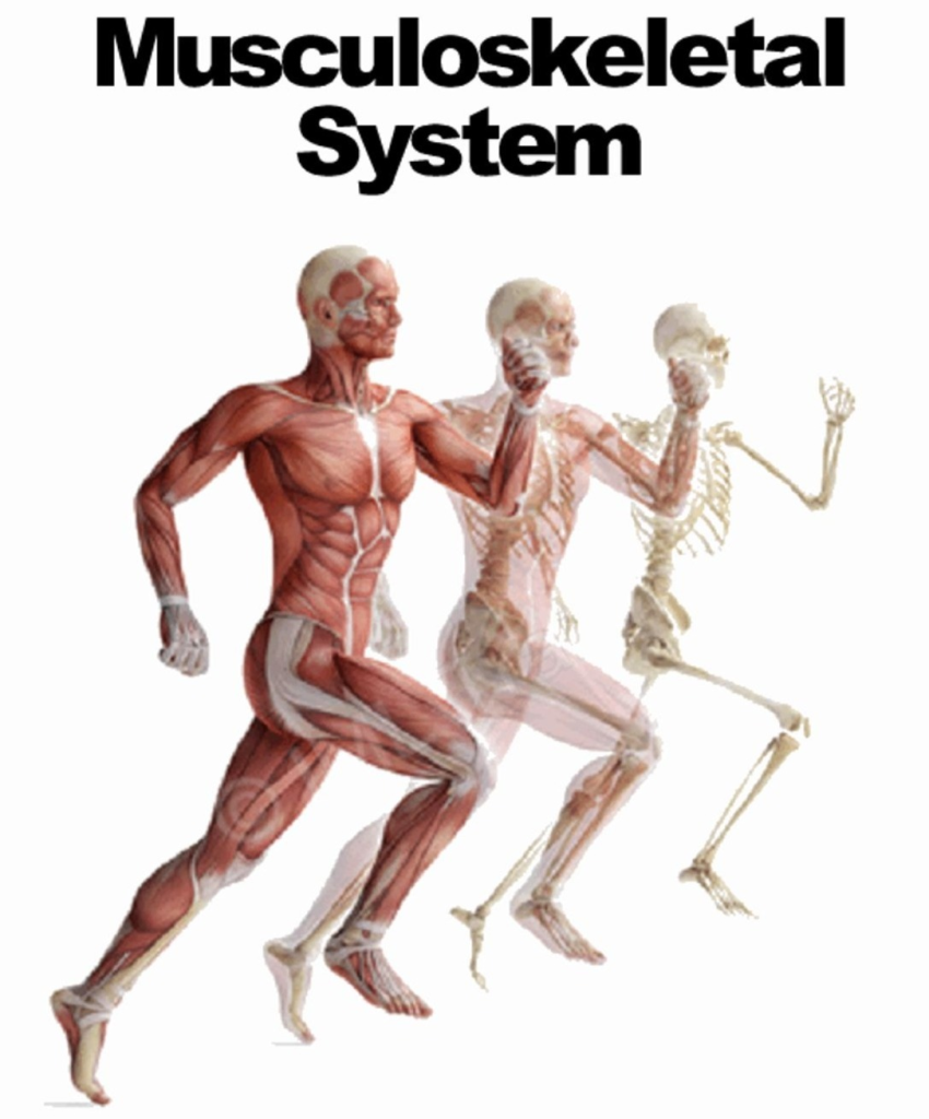
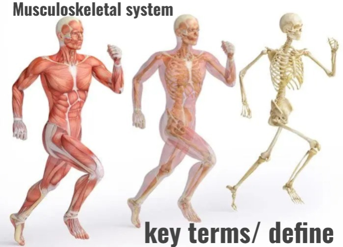
key terms/ define.
1) Define tendinitis:=

- Inflammation of muscle tendons Tendinitis is called.
2) Define traction
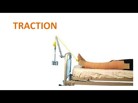
- The application of force on a part of the body is called traction.
3) Define strain

- Musculo stenotic injuries is called strain (musculo stendinous injury its called strain).
4)define sprain (Define sprain)

- Sprain is an injury to the ligaments and soft tissues of a joint.
5) Define Kyphosis (Define Kyphosis)
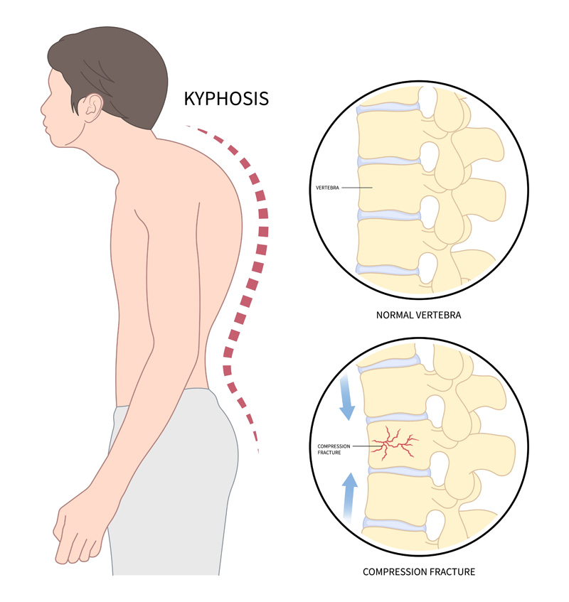
- Kyphosis is a deformity of the spine, in which there is a posterior (backward) curvature of the spine, especially in the thoracic region, which causes the upper back to appear rounded or hunchbacked.
6)Define Scoliosis
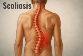
- Scoliosis is a deformity in which the spine curves laterally and its structure It becomes ‘S’ or ‘C’ shaped. It may manifest as unbalanced shoulder and hip height, pelvic tilt and rib hump. This condition is especially common in adolescent patients and is known as idiopathic scoliosis.
7) Define Lordosis. (Define lordosis)
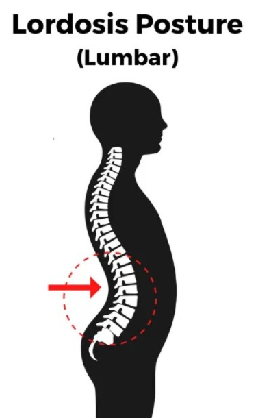
- In lordosis, the inward curvature of the lumbar spine increases. Therefore, swayback (swayback: = swayback means the pelvis tilts forward and the abdomen protrudes) is seen in lordosis.
8) define Abduction. (Define abduction)
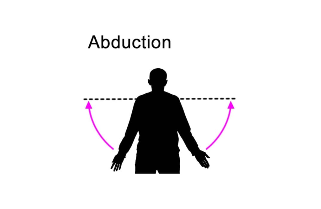
- Away moment is given to the body from the center line or median line of the body.
9) define Adduction. (Define adduction).
- In this, the body parts are momentarily moved towards the center part or median part of the body.
10) define splint. (Define splint).
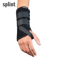
- A type of device that is used to maintain the alignment of affected body parts and to immobilize them. Therefore, it is designed to provide support.
11)define allograft. (Define allograft).
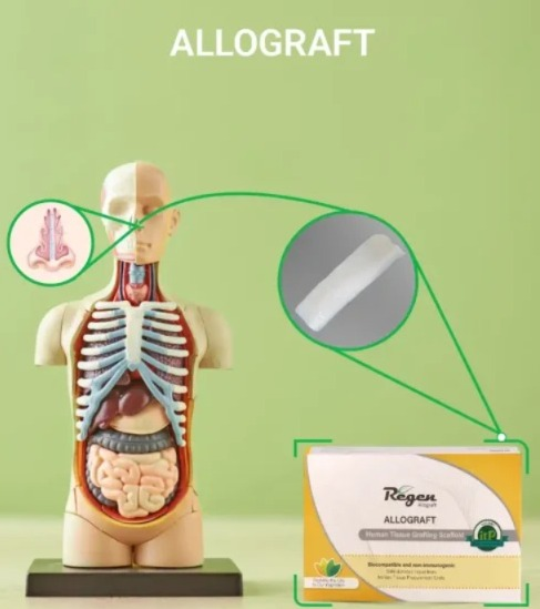
- In this, tissue is obtained from a donor to provide a graft in any other donor.
12) define arthroscope. (Define arthoscope).
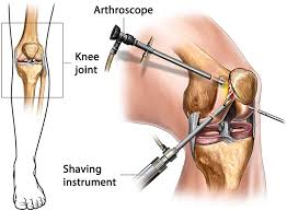
- A surgical instrument used to examine the internal structure of a joint.
13)define Amputation. (Define amputation).
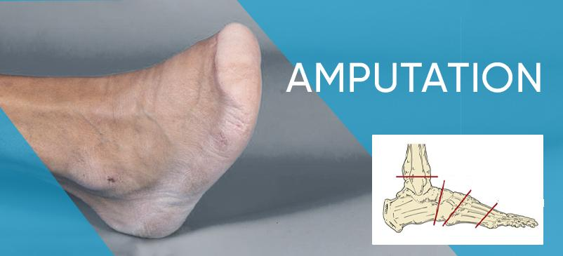
- In this, parts of the body are removed (removal of body part ( limb)).
14) define RICE.
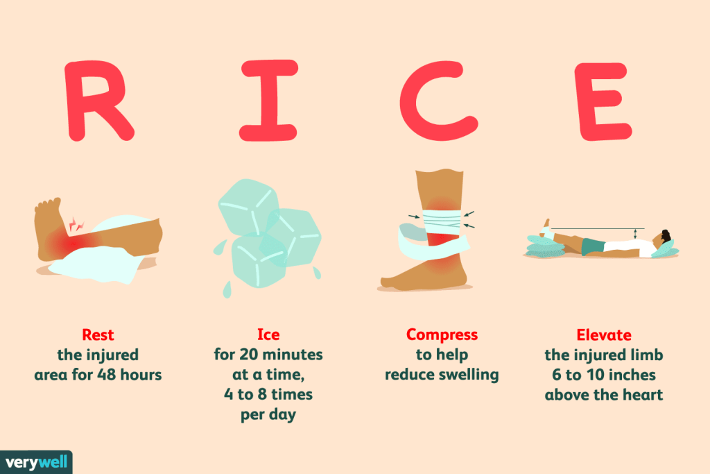
- R:=REST,
- I:=ICE ,
- C:=COMPRESSION,
- E:=ELEVATION .
15)Define arthrodesis. (Define arthrodesis).
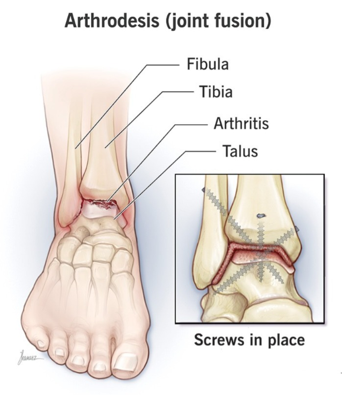
- In this, the joint is surgically fused.
16) Define arthroplasty:=
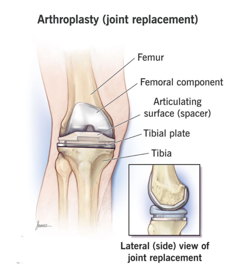
- This is a joint replacement in which an artificial joint i.e. prosthesis is used.
17) define Resorption
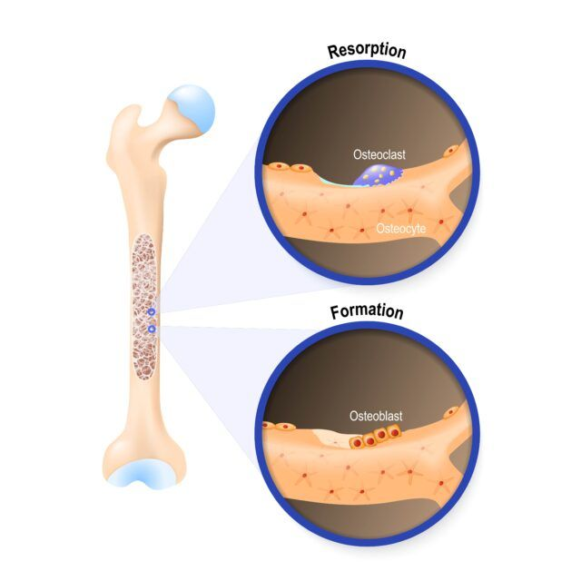
- This involves bone destruction.
18)define phantom limb pain. (Define Phantom Limb Pain)
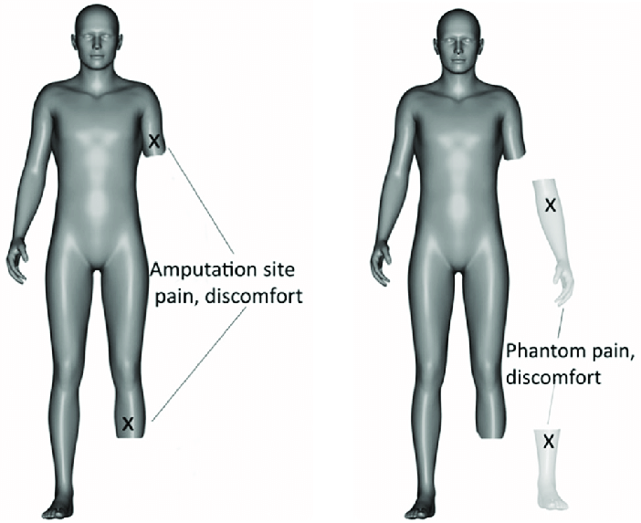
- In this case, pain is received from the amputated limb.
19) define atonic. (Define atonic.)

- There is no tone in the muscles.
20) define periosteum. (Define periosteum).
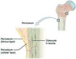
- Fibrous connective tissue that covers bones.
21) Define Atrophy. (Define atrophy).
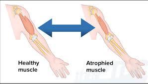
- In this, the muscles shrink and their size decreases.
22) Define paresthesia. (Define paresthesia).
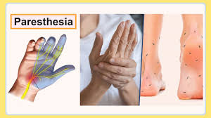
- This involves feeling abnormal sensations. Such as burning, tingling, numbness sensation.
23) Define Avascular necrosis. (Define avascular necrosis.)
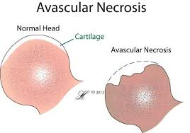
- In this, tissues die due to insufficient blood supply.
24) Define osteomyelitis. (Define osteomyelitis.)
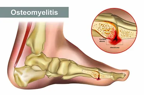
- This is an infection of the bone.
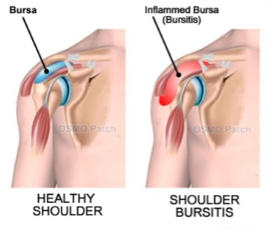
25) define bursa. (Define Burza)
- It consists of a fluid-filled sac that is mainly composed of connective tissues of the joint.
26)Define osteoporosis. (Define osteoporosis).
- In this, there is loss of bone tissue.
27)Define bursitis. (Define Bursitis.)
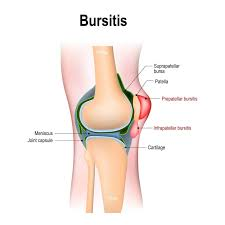
- Inflammation of the bursa is called bursitis.
28) Define osteogenesis. (Define Osteogenesis).
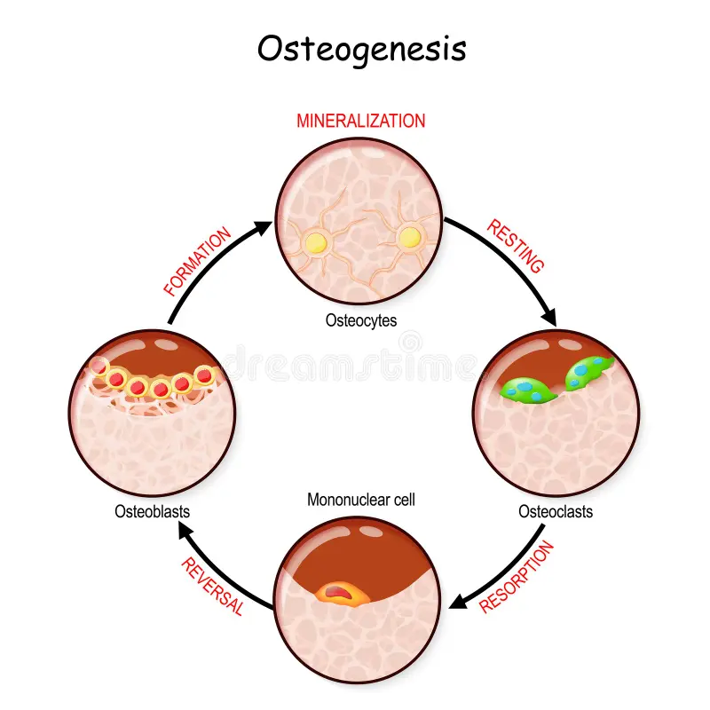
- The formation of bone is called osteogenesis.
29)define brace. (define braces)
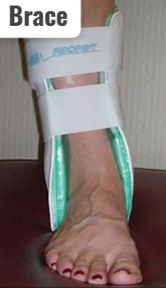
- A brace is a device that is applied externally to the body to prevent injury and to support the back.
30) define osteocytes. (Define osteocytes)
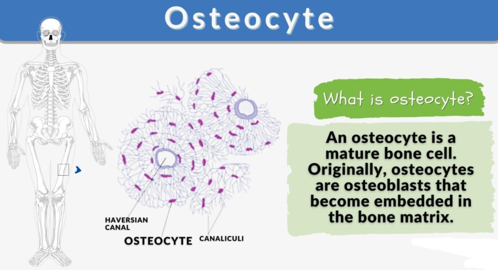
- There are mature bone cells.
31)define cancellous bone. (Define cancellous bone).
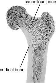
- This is a lattice bone structure.
32) Define osteoclast. (Define osteoclast.)
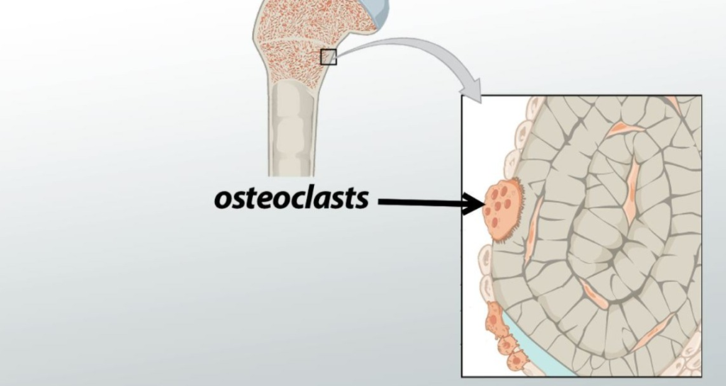
- This is bone reabsorption cell.
33)define callus.
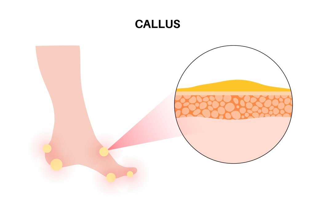
- This fibrous tissue is located on the fracture side.
34) define osteoblast. (Define osteoblast.)
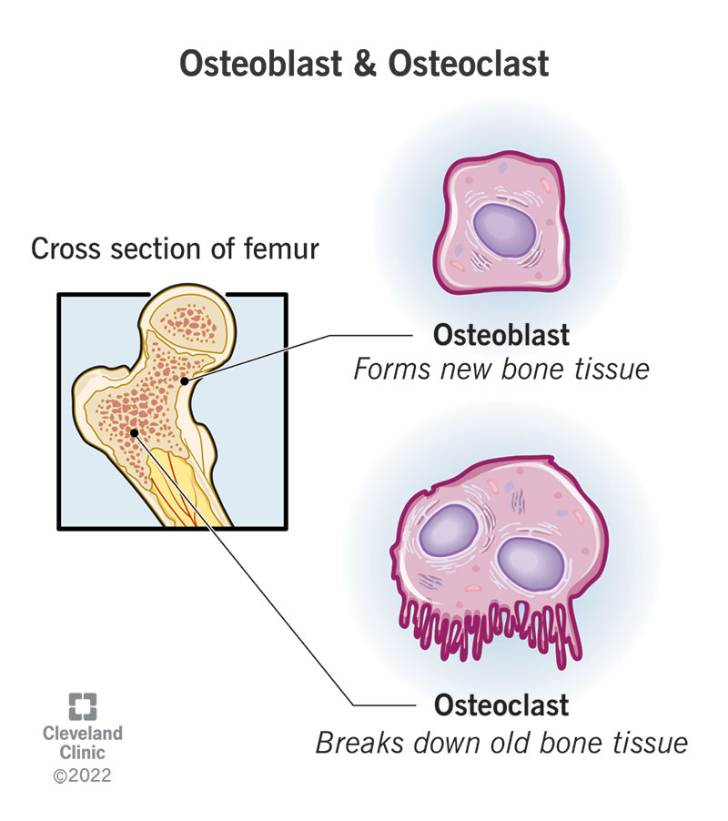
- This is a bone forming cell.
35) define cast.
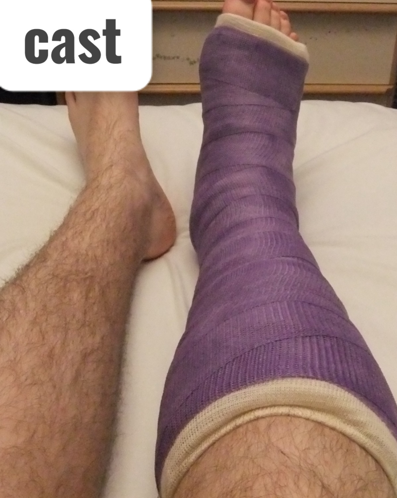
- Cast is a rigid immobilizing device that the body Keeps the part countour.
36) Define ossification. (Define ossification).

- Ossification is the physiological process of bone formation, in which calcium salts and organic matrix are deposited, converting cartilage or fibrous tissue into hard bone.
37) define contracture .
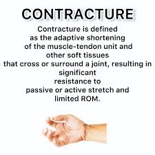
- In this, there is abnormal shortening of the muscles or the joint or both.
38) define meniscus. Do)
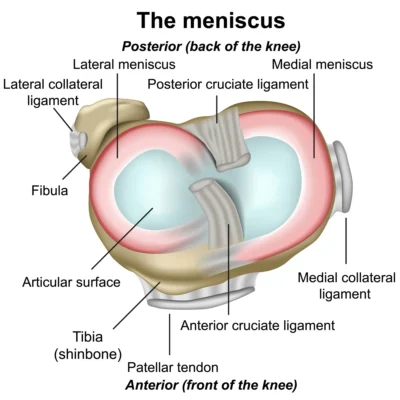
- The meniscus is a crescent-shaped fibromuscular cartilage that is located in the knee joint. is.
39) define contusion. (Define contusion)
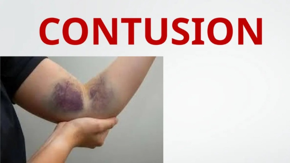
- This is a blunt force injury that occurs to the soft tissue.
40) define joints. (Define joints).
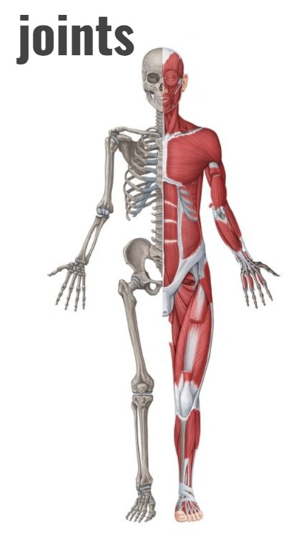
- Joints are places where two or more bones, or bones and cartilage, attach to each other, providing stability to the body and allowing necessary movement.
41)define crepitus
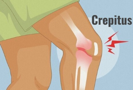
- Crepitus is a grating sound that occurs when bony fragments rub against each other. It occurs due to rubbing.
42) Define fracture reduction. (Define Fracture Reduction).
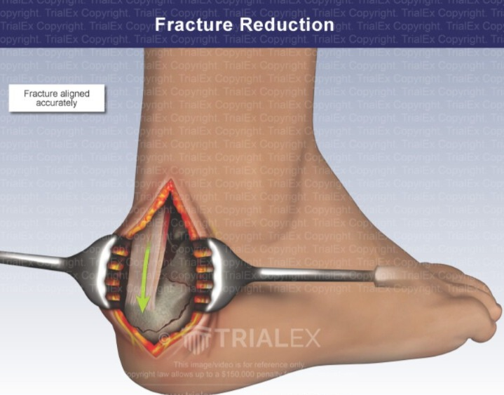
- In this, the restoration of the fracture fragment is done on its anatomical position.
43) define debridement
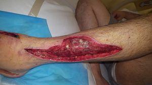
- In this, contaminated and devitrified tissue and foreign material are surgically removed.
44) define fracture. (Define the facture)
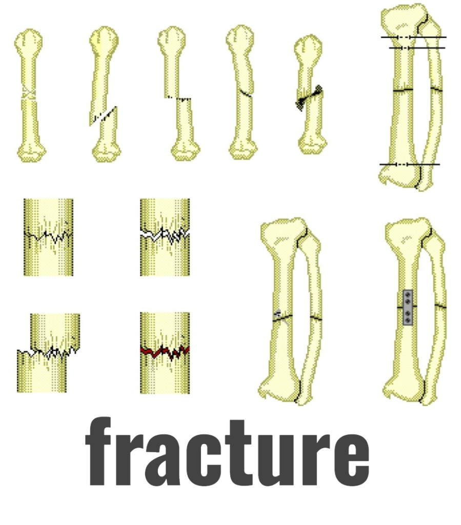
- A break in the continuity of a bone is called a fracture.
45) Define Diaphysis
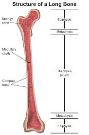
- The shaft of a long bone is called the diaphysis.
46) Define fasciotomy. (Define fasciotomy).
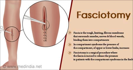
- Fasciotomy is a surgical procedure in which the muscle fascia is constricted, which reduces the pressure on the muscles tissues.
47) Define disarticulation. (Define Disarticulation)
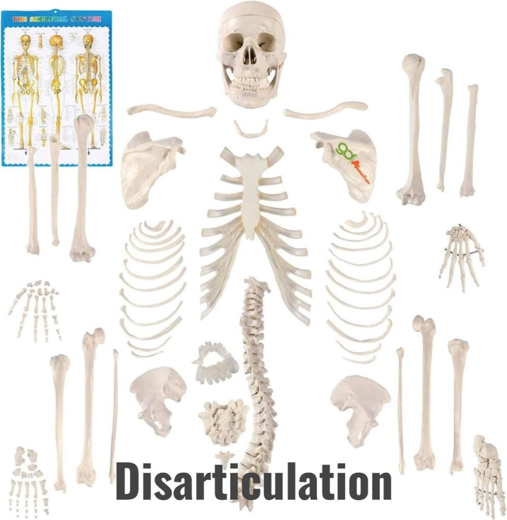
- Amputation from a joint is called disarticulation.
48) define facia
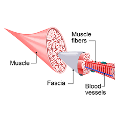
- Fascia is a fibrous tissue that covers, supports, and separates muscles.
49) Define dislocation. (Define Dislocation)
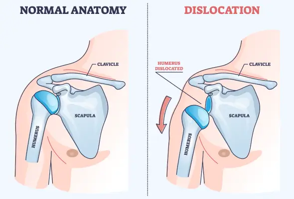
- In this, the joint surface is separated.
50) define external fixator. (Define external fixator)
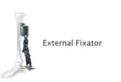
- This is an external metal frame that is attached to a bone fragment to maintain the stability of the bone.
51) define Endosteum. (Define endosteum)
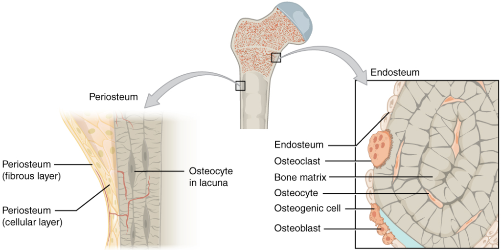
- This is a thin membrane that covers the cavity of the bone marrow that is in the space of the cancellous bone.
Nursing management of patients with musculoskeletal disorder and disease.
- assessment.
- history ,
- physical examination,
- diagnostic test. subjective data
explain musculoskeletal assessment
1. Assess the patient’s pain level:
- location( location of pain),
- characteristics( characteristics of pain),
- duration( duration of pain),
2) Assess the range of motion ( range of motion )
- Limited mobility of the patient or whether it affects his daily routine activities.
- Mobility of the joint.
- Moment is altered.
- Stiffness.
- Swelling of muscles.
3) Associated symptoms
- Any bony nodules or deformities.
- Weight loss.
- Fever and malaise.
- Any Assess for any type of sensory or motor deficit such as weakness in climbing.
explain how to take health history
medical history
- Ask the patient if there are any chronic diseases that could cause immobility of the body, such as casting, traction, surgery.
- Ask the patient if he/she has any type of prostate, breast, or lung cancer.
- If female, ask if he/she has used any type of estrogen replacement therapy.
- Ask if he/she has used any type of corticosteroids.
- Ask the patient about their cultural beliefs.
- Ask whether any family members have osteoporosis or arthritis.
- Ask whether the patient is exposed to the sun.
- Ask whether the patient has any smoking habits.
- Ask whether the patient participates in any sports activities.
- Ask about the patient’s lifestyle.
- Ask about the patient’s occupation.
objective data (focused on musculoskeletal system)
- Assess the patient’s posture, gait, and cerebral function.
- Normal structure of the patient,
- Bony deformity,
Length discrepancies, - Alignment,
Amputation. - Check for abnormal motion of the patient. Limb measurement
Assess joints and test joint movements.
- Assess the patient’s range of motion exercises.
- Assess the patient’s joint stability.
- Note deformity associated with contracture or dislocation.
Assess muscle strength and range of motion.
- Assess the patient’s coordination of movements.
- Inspect the size and contour of the muscles.
- Palpate muscle tone.
- Identify range of motion movement.
neuromuscular aspect includes
- Assess the patient’s circulatory status, including assessing the skin color, temperature, peripheral pulse, capillary refill response and pain level of the extremities.
- Assess the reflexes of the patient’s extremities.
- Compared to all uninjured/unaffected extremities.
Skin component
- Assess whether the patient has any traumatic injury (cuts/bruises).
- Assess whether the patient has any chronic conditions (dermatitis/stasis/ulcer).
- Assess the patient’s hair distribution and nail condition.
- Assess the warmth and coolness of the skin.
- Assess the patient’s range of motion exercises.
explain how to talk history of patient with musculoskeletal system disorder
- Collect information about the patient’s biographical data, his chief complaint, past and present family and whether he has a current disorder or not in the patient’s health history.
1) Biographical data
- Biographical data of the patient
- Ask the patient about Age, Sex, Occupation, Dietary pattern, Habit, etc.
2) Chief complaint
- Ask the patient whether there is any pain, weakness, muscle weakness, joint stiffness, loss of sensation, and sensory changes.
- If the patient has any of these problems, take a complete description of it.
2) Pain level
- Take a description from the patient, pain intensity, location, duration and what steps can be taken to relieve the pain.
3) Sensory changes
- To ensure that the patient does not have any abnormal sensations, obtain complete information about numbness, tingling, or any other such sensations.
4) Swelling
- Ask the patient about any swelling in the bones and joints. Ask about factors that reduce swelling, including elevating the swollen part.
5) Deformity and immobility
- Ask about how the deformity occurs and how it affects mobility.
- Do you need any type of supportive equipment such as crutches, Ask about whether the patient uses a walker, or wheel chair.
6) Past health history
- In the past health history, ask the patient whether he has had any trauma, accident, or surgery of the musculoskeletal system.
- In the past health history, ask whether the patient uses any type of medication, including Ask about the use of corticosteroids, NSAID drugs, and relaxants.
7)Family health history
- In this, the patient should be asked whether there is any disorder such as arthritis,
- Osteoporosis,
- Gout,
- Ankylosis and spondylitis, muscular dystrophy and Scoliosis.
8) Psychosocial history
- Ask the patient about his/her daily activity, exercise, nutrition, habits, and safety.
Physical examination.
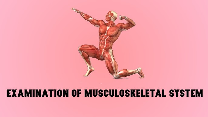
- Three areas of musculoskeletal assessment are:
- Inspection,
Palpation,
And range of motion are used to assess the patient’s
posture, gait, bone integrity, joint function and muscles strength and size.
inspection.
- This includes inspecting the patient’s posture and gait.
- Assessing whether the patient uses mobility aids such as a cane or walker.
- When inspecting the spine, examine the back, buttocks, and legs.
- Curvature of the spine Assess for lordosis, scoliosis, and kyphosis.
- Check for any deformity and temperature increase in any joint or muscle in the hands, feet, or feet.
- If there is severe pain in the joint, check for any effusion, swelling, and inflammation.
- Joint deformity is mainly contracture, dislocation, subluxation, and disruption of structure.
- Assess the patient’s general nutritional status.
- Assess the strength of the muscles, their movement, and whether there is contraction or deformity.
- Assess the patient’s neurological function, including reflexes, sensation and motor ability.
palpation.
- After inspection, palpation of warmth, Swelling, Tenderness,
Redness, Muscles mass of the patient. - The tissue surrounding joints is nodule formation.
- Rheumatoid arthritis, Gout, Osteoarthritis may produce characteristic nodules.
range of motion.
- Assess the mobility of the patient’s joint.
- Assess the range of motion exercises.
- See if there is any abnormality in the patient’s range of motion.
- Assess the patient’s daily routine activity.
- Assess the patient’s daily routine activity.
- Assess the patient’s range of motion
Exercises in the patient’s daily routine activity Observe. - Assess the patient’s temporomandibular joint, cervical spine, lumbar spine, finger, wrist,
elbows, shoulder, toes, ankles, toes, knees, hip joint.
muscles strength and size.
- In this, the muscular system is to assess the strength and coordination of the muscles.
- Weakness of a group of muscles might such as polyneuropathy,
Electrolyte imbalance,
Myasthenia gravity,
Polio,
Muscular dystrophy is to be assessed.
explain the diagnostic evaluation of musculoskeletal system( write musculoskeletal system diagnostic evaluation).
explain Arthrography (Arthrography)
- Arthrography is mainly performed to assess joint pain.
- It is mainly performed to identify whether there is an acute or chronic tear in the joint capsule and its supporting ligaments such as the knee, shoulder, ankle, hip, or wrist.
- In this, a contrast iodine solution is injected into the joint area to highlight the joint area. For.
- An x-ray of the joint is taken using a fluoroscope.
- And in this, an image of the joint is shown.
explain Bone densitometry.
- This test is mainly done to assess bone mineral density. Comes.
- And it is done to assess the amount of osteoporosis.
- DEXA:= DUAL ENERGY X RAY absorptiometry
- (Dual Energy X-ray Absorptiometry) This is mainly done to assess the density of the wrist, hip, and spine.
explain X Ray ( X-ray Explains Do)
- A bone x-ray is done to assess the density of the bone, its texture, erosion, integrity and whether there are any changes in the bone.
explain ct scan
- CT scan is mainly done to assess whether there is any tumor or injury to ligaments and tendons.
- CT scan is mainly done using contrast agent.
- CT scan is mainly done to assess whether there is any tumor or injury to ligaments and tendons.
explain MRI (Explain MRI.)
- MRI. It is mainly done to diagnose any avascular necrosis, osteomyelitis, tumor, disk abnormality, ligament or cartilage tear.
explain Bone scan
- A bone scan is mainly done to identify if there is unusually active bone formation.
- This is mainly done to identify if there is any arthritis, osteomyelitis, metastatic or primary bone tumor and necrosis.
- This is mainly done to identify if there is metabolic or primary bone tumor and necrosis, bone tumor, osteomyelitis, some fractures, and necrosis.
explain endoscopy studies
- This is mainly done to assess the internal surface of the joint.
- This is mainly done to assess whether there is any disease of the patella, meniscus, and synovial membrane.
explain ultrasonography
- It is mainly done to identify any inflammation around the joint.
explain Biopsy
- A biopsy is a It is mainly used to identify the structure of the bone marrow, its composition, bone, muscles and synovium, if any disease is present.
- After the biopsy, ice is applied to the biopsy site.
gulline thallium scans (Explain gulline thallium scan.)
- This test is mainly similar to a bone scan but It is a more specific and sensitive diagnostic test.
- Glulium is mainly found in the bone but also in the brain and breast tissue and can be easily diagnosed due to this.
electromyography (Describe electromyography)
- Electromyography is primarily used to assess the electrical impulses of muscles.
explain myelogram
- In a myelogram, contrast media is injected into the subarachnoid space, which allows the spine and spinal cord to be easily visualized.
- This involves telling the patient to keep their head in a down position so that the contractile medium can reach the level of the neck.
explain arthrocentesis. (Explain arthrocentesis).
- In arthroscopy, synovial fluid is removed from the joint for examination and to remove extra fluid in the joint.
- Synovial fluid is usually clear, pale, straw colored, and scanty in volume.
explain arthroscopy.
- In arthroscopy, the surgeon directly visualizes the joint.
- In this, the internal surface of the joint is directly examined using an endoscope.
explain laboratory test of musculoskeletal system (Write laboratory test of musculoskeletal system.)
explain RA latex test.
- RA latex blood test is mainly done to assess whether RA factor antibodies are present in a person who has rheumatoid arthritis.
- If the rheumatoid factor level is very high, then the person has rheumatoid arthritis and any other autoimmune disease.
explain serum muscles enzyme
- When muscle tissue is damaged, large amounts of enzymes are released into the blood.
- In which skeletal muscles creatinine kinase ( ck-mm),
Aldosterone ( Ald),
Aspartate aminotransferase ( AST),
Lactate dehydrogenase ( LDH),
mainly increase in muscle diseases such as muscular dystrophy, polymyositis, and dermatomyositis.
explain myoglobin.
- Myoglobin is a protein that is present in skeletal and cardiac muscles.
- And this mainly gives the red color to the muscles.
- When skeletal and cardiac muscles are damaged, the myoglobin level in the blood increases.
explain alkaline Phosphate.
- Alkaline phosphatase is an enzyme that increases when bone and liver tissue are damaged.
explain serum calcium and phosphorus.
- Bone disorders are mainly performed to assess whether there are any changes in calcium and phosphorus levels.
- Calcium and phosphorus levels are regulated by calcitonin, which is released from the thyroid and parathyroid glands.
- When these glands are not functioning properly, the levels of calcium and phosphorus are altered.
explain ccp (cyclic citrullinated protein antibody)
- The Ccp test helps diagnose rheumatoid arthritis.
explain sedimentation rate.
- This test is primarily used to evaluate the nonspecific activity of an infection.
- It is primarily used to assess inflammatory conditions, autoimmune disorders, and plasma cell dyscrasias.
explain antinuclear antibody test.
- This is mainly done to check the immune system.
- It measures whether the body’s immune system has made antibodies that are attacking the body’s own cells.
explain complete blood count (Complete blood count).
- Complete blood count is mainly done to check if the hemoglobin level has decreased after any trauma or surgery.
- Wbc level increase when any inflammatory disease, acute infections, trauma, hemorrhage, and tissue necrosis is assessed.
explain uric acid test.
- The uric acid test is mainly done to assess the amount of uric acid present in the blood.
- Uric acid is mainly a waste product that occurs due to the breakdown of cells and is mainly present in the blood and urine.
- If the amount of uric acid is elevated, it is diagnosed as gout, which is a form of arthritis.
- If the patient has osteomalacia, ulcerated parathyroid gland then serum calcium level increases
- When there is any tumor in the bone and when the fracture is healing, phosphorus level increases.
- Rheumatoid factor is done to assess rheumatoid arthritis.
- CRP is done to rule out any inflammation and arthritis.
- ESR is done when there is any inflammation Increases.
- Uric acid helps in the diagnosis of gout.
- Creatinine kinase (CK) is used to diagnose muscle trauma or disease.