ENGLISH-MSN : 2 (UNIT NO. 10 ) EMERGENCY MANAGEMENT
EMERGENCY MANAGEMENT.
Define triage

- Triage is derived from the French word ‘trier’ which means ‘to short out’ or ‘to sift out’.
- Triage is a process used in emergency and medical settings in which patients are given priority based on the severity of their condition.
- Triage involves separating patients who need early treatment from those who need it most, using a color code, so that early treatment can be provided.
Write types of triage
1) Simple triage:
- Simple triage is used when there is a mass casualty incidence, the patient needs to be quickly assessed and categorized based on their needs, and resources are limited. Simple triage uses methods such as the START (Simple Triage and Rapid Treatment) method as well as a color code system (red, yellow, green and black).
2) Advanced triage:
- Advanced triage is mainly used in hospital settings where more resources are available, in which more detailed examinations are performed. The Emergency Severity Index (ESI) method is primarily used in advanced triage. In which patients are categorized from level 1 (most urgent) to level 5 (least urgent).
Write about color code of triage
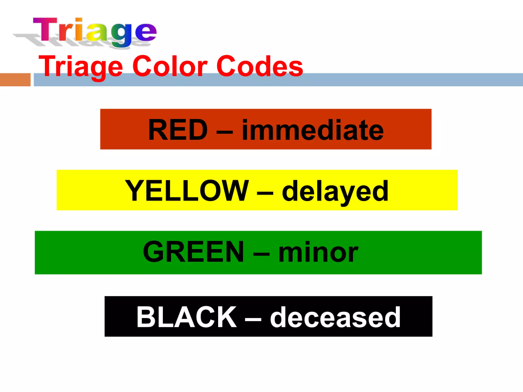
- red color :
Red Mins Emergency. Red color indicates ‘immediate word’ . That is, red color is given to victims with life-threatening conditions. That is, such people need immediate intervention. For example, severe respiratory depression, brain hemorrhage
- Yellow color :
Yellow means urgent. Such people have serious conditions but are not life-threatening, meaning that immediate intervention is not required.
- Green color :
Green means minor. Green color is given for minor injuries such as wounds.
- Black color :
Black means deceased or expectant. People who are dead or who are seriously injured and cannot survive are given black color.
Write advantages of triage
- Patients are given priority based on their severity and urgency so that they can be treated early.
- Useful in making difficult decisions easier is.
- Rapid initial assessment is provided.
- Ensures that resources are used effectively and their waste is prevented.
- Minimizes waiting time for critical services.
Define obstruction of airway (Define Obstruction of airway)
- This is a life-threatening emergency. In which the respiratory passage is blocked due to airway obstruction, which prevents free flow of the airway.
Write causes of airway obstruction
- Foreign Object: Inhaling or ingesting a foreign object that blocks the airway, such as a piece of food, toy, or small item
- Infection: Epiglottitis and infections of the larynx and pharynx cause edema and spasm, which can lead to airway blockage.
- Trauma: Upper airway obstruction can occur due to injuries to the face and neck. Such as fractures and dislocations
- Allergic reaction: Severe allergic reaction causes swelling in the airways, leading to obstruction.
- Tumor: Abnormal growth and tumor in the upper airways can cause blockage of the air passages.
Write clinical manifestations of airway obstruction Obstruction)
Choking is seen as the main sign due to upper airway obstruction.
- Choking: The universal sign for choking is ‘hand clutches to the throat’, meaning the person holds their throat while choking.
- Difficulty in breathing
- Cuffing or gagging
- Cyanosis
- Distress
- Restlessness
- Difficulty in speaking
- Aphonia
- Respiratory distress and death
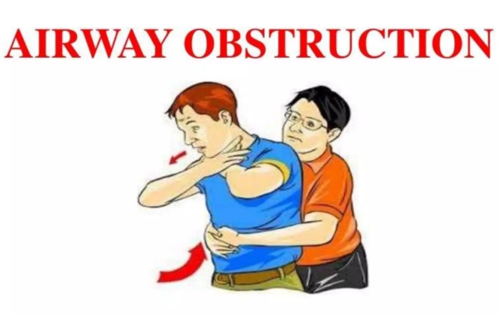
Write management of airway obstruction
- Assessment: Assess the severity of the obstruction. Check for signs of respiratory distress.
- Clear the airway: Perform the Heilmitch maneuver to clear the airway.
- Stand behind the adult patient. To create balance, keep one foot slightly ahead of the other. If it is a child, sit behind him with your legs bent at the knees.
- Hold the arm next to the patient’s waist.
Then make a fist with one hand and place the thumb side of that fist against the person’s abdomen, above the navel and below the ribs. - With the help of the other hand, hold the fist and provide rapid upward thrusts into the abdomen.
- If the object has not yet come out, provide repeated thrusts.
- Apply less pressure to the child.
- Provide chest thrusts to the patient if the patient is pregnant and the arm is not around the stomach. Chest thrusts involve placing the hand on the breastbone and providing thrusts.
- Providing back blows to newborns and infants.
- In which the baby is placed in the forearm and if grasped, five black blows are given with the hand.
- Open the airway: If the patient is conscious and has difficulty breathing, ask the patient to forcefully expel the cuff so that the obstruction is removed.
- Call for Help: If the obstruction does not clear and the patient’s condition worsens, call emergency services.
- Oxygen therapy: If oxygen is available, provide supplemental oxygen to improve oxygen levels.
Define Hemorrhage
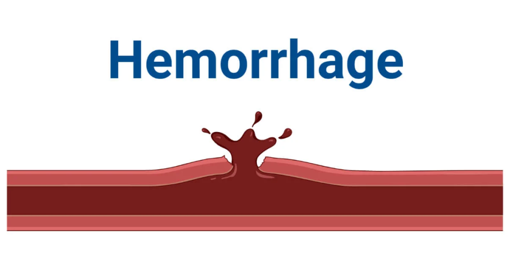
- Hemorrhage means bleeding. In which blood loss occurs from damaged blood vessels. Which is found inside or outside the body.
Write types of hemorrhage
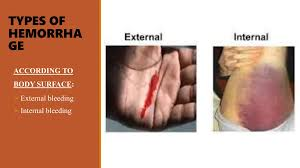
Hemorrhage is mainly divided into two types:
1) External hemorrhage
2) Internal hemorrhage
1) External hemorrhage:
- Bleeding in external hemorrhage is The wound, abrasion or injury site is visible from the outside of the body.
2) Internal Hemorrhage:
- In internal hemorrhage, bleeding is seen inside the body. It cannot be immediately visible like external hemorrhage. Which includes intracranial hemorrhage, gastrointestinal hemorrhage, pulmonary hemorrhage.
✓ Arterial hemorrhage: Bleeding seen in an artery is known as arterial hemorrhage. Which is seen in bright red color. Arterial hemorrhage is severe and life threatening. Which is difficult to control. (Because the blood pressure in the artery is high.
✓ Venous hemorrhage: Bleeding seen in veins is known as ‘venous hemorrhage’. Which is seen in dark red color. Venous hemorrhage is less severe than arterial hemorrhage.
✓ Capillary hemorrhage: Bleeding seen in the smallest blood vessels capillaries is known as capillary hemorrhage. In capillary hemorrhage, small amounts of bleeding are seen from minor cuts and abrasions.
Write causes of hemorrhage
- Trauma or injury (accident, fall down, violence)
- Medical condition (hemophilia, blood clotting disorder, liver disease)
- Medication (blood thinners – warfarin, aspirin)
- Surgery
- Aneurysm
- High Blood Pressure
- Cancer
- Ulcer
- Childbirth
Write sign and symptoms of hemorrhage
✓ External bleeding
- Bleeding from open wound
- Visible bleeding
- Bruising
- Discoloration of skin
- Swelling around affected area
✓ internal hemorrhage :
Intracranial hemorrhage :
- Severe headache
- Confusion or altered mental status
- Vision problems
- Seizures
- Weakness or numbness on one side of body
Gastro Intestinal hemorrhage:
- Blood in vomiting
- Blood in stool
- Abdominal pain and swelling
Pulmonary hemorrhage:
- Blood on Cough
- Chest Pain
- Breathing Difficulty
✓ general symptoms :
- Rapid and Shallow Breathing
- Rapid Pulse
- Hypotension
- Pale or scaly skin
- Weakness
- Fatigue
- Disorder
- Lightheadedness
Write diagnostic evaluation of hemorrhage
- History collection
- Physical examination
- Complete blood count
- Coagulation study
- Ultrasound
- CT scan
- MRI
- Angiography
- Endoscopy
Write management of hemorrhage
✓ Immediate First Aid Care:
- Apply Pressure: Apply pressure to the bleeding site using a clean cloth or bandage to control bleeding.
- Elevate the Area: If the limb is bleeding, elevate it to the level of the heart. So that blood flow to that area can be reduced.
- Immobilize Area: Immobilize the injured area. So that further damage can be prevented.
✓ Medical Management :
Minor Hemorrhage :
- Topical hemostatic agent: Apply hemostatic agent with gauze to control minor bleeding.
- Sutures or Staples: Small cuts and lacerations are healed with the help of sutures or staples.
Major Hemorrhage:
- Tourniquet: In cases of severe limb bleeding, apply a tourniquet over the injury site. So that blood flow can be controlled.
- Blood transfusion: If necessary, replace blood with blood transfusion.
- Intravenous fluid: Administer intravenous fluid to maintain blood pressure and organ perfusion.
- Medication: Administer medicine such as tranexamic acid to control bleeding.
✓ Surgical Intervention :
- Ligation or Clamping : Tying or clamping of bleeding vessels.
- Repair of Injured Tissue :
Surgically removing damaged organs or tissues.
✓ Monitoring and Support :
- Vital sign monitoring: Continuously monitoring the patient’s blood pressure, pulse, respiratory rate, and oxygen level.
- Critical care support: In severe cases, the patient is kept in the Intensive Care Unit (ICU). So that close monitoring of the patient can be done and advance intervention can be done.
Define shock
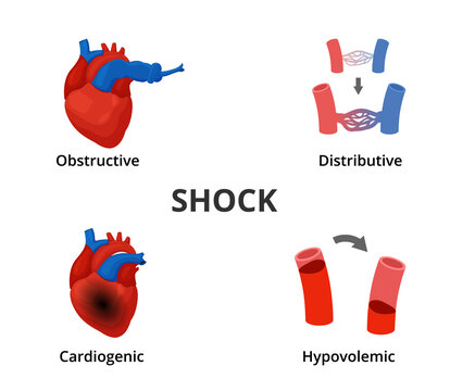
- Shock is a life-threatening condition. In which the body does not get enough blood supply, meaning that the cells and tissues do not get enough oxygen and nutrients, due to which they do not function properly and due to this, severe damage is seen.
Write types of shock
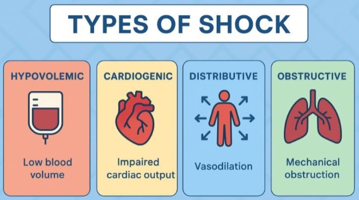
1) Hypovolemic shock :
- The condition of hypovolemia is observed due to significant blood or fluid loss and the shock seen due to it Known as hypovolemic shock.
2) Cardiogenic shock:
- Due to conditions like heart attack or heart failure, the heart is unable to pump enough blood and the shock seen due to this is known as cardiogenic shock.
3) Distributive Shock :
The blood vessels in distributive shock lose their tone and capacity to dilate. This is due to the following reasons.
- Septic shock: A condition of systemic inflammation is seen due to severe infection and due to this, septic shock is seen.
- Anaphylactic shock: Due to anaphylactic reaction, blood vessels constrict and a condition of anaphylactic shock is seen.
- Neurogenic shock: Due to injury to the spinal cord, vascular tone is lost, due to which a condition of neurogenic shock is seen.
4) Obstructive Shock :
- Due to physical obstruction in the blood flow, cells and tissues do not get adequate blood supply and due to this, the condition of obstructive shock is observed. Such as pulmonary embolism
Write sign and symptoms of shock
- Low blood pressure (hypotension)
- Rapid pulse (tachycardia)
- Rapid breathing (Tachypnea)
- Shortness of Breath
- Cyanosis
- Cold and clammy skin
- Ineffective Tissue Perfusion
- Thirst and Dry Mouth
- Confusion
- Loss of Consciousness
- Restlessness
- Weakness or Fatigue
- Decrease urine output
- Dilated pupils
- Nausea
- Vomiting
- Sweating
Write management of shock
- Vasoconstrictors: Vasoconstrictors contract the smooth muscles in the blood vessels. Due to which the blood vessels constrict and the blood pressure increases. For this, epinephrine, norepinephrine drugs should be used.
- Isotrope: In cases of cardiogenic shock, isotrope medicine should be used to increase cardiac output. Such as dobutamine
- Fluid resuscitation: Administer IV fluids in cases of hypovolemic and distributive shock. Such as normal saline, Ringer lactate
- Specific treatment: Know what type of shock is and what cause (cause) is responsible for the shock and treat that cause.
- Hemodynamic monitoring: Continuous ECG, blood pressure, central venous pressure monitoring.
Define anaphylactic reaction
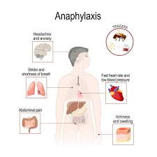
- Anaphylactic reaction is a severe and potentially life-threatening allergic reaction that occurs within a short time of exposure to an allergen.
Write sign and symptoms of anaphylactic reaction
✓ Skin reaction :
- itching
- hives
- Redness
- Swelling in face and eyes
✓ Respiratory symptoms :
- Breathing difficulty
- Cuffing
- Wheezing
- Chest tightness
✓ Cardiovascular symptoms :
- Rapid or irregular heartbeat
- Palpitation
- Low blood pressure
- Fancy
- Dizziness
- Lightheadedness
✓ Gastrointestinal symptoms :
- Abdominal pain
- Nausea
- Vomiting
- Diarrhea
✓ Other symptoms :
- Anxiety
- Confusion
- Slurred speech
- Unconsciousness
Write diagnostic evaluation of anaphylactic reaction (Write diagnostic evaluation of anaphylactic reaction) Reaction)
- History Collection
- Physical Examination
- Serum Tryptase
- Skin Prick Test
- Allergen Specific IgE Testing
- Complete Blood Count
Write management of anaphylactic reaction
Call for emergency:Call for emergency services.
Epinephrine administration: Epinephrine is the drug of choice for anaphylactic reactions. Therefore, epinephrine is administered to control the anaphylactic reaction.
Positioning: Place the patient on a flat surface and elevate the legs if possible and maintain airway patency.
Oxygen Therapy and Ventilation: Provide supplemental oxygen to the patient and provide mechanical ventilation if necessary.
Corticosteroid : Provide corticosteroid drug to reduce inflammation and prevent biphasic reaction.
Antihistamine : Provide antihistamine group medicine to relieve itching and hives.
Fluid : Administer intravenous iv fluids to manage hypotension.
Monitoring: Continuous vital sign monitoring as well as monitoring for treatment response.
Define abdominal trauma
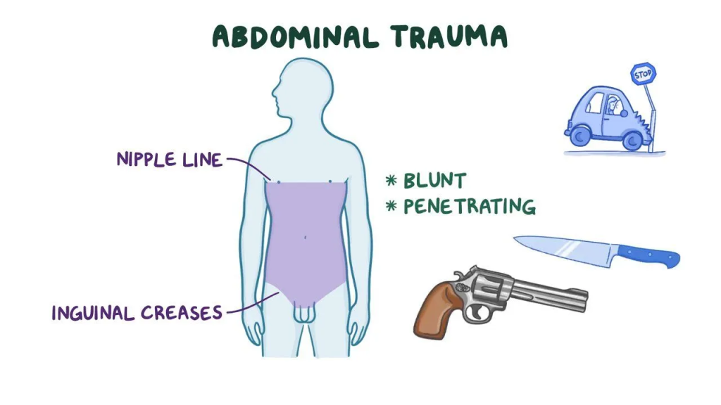
- Injuries found in the abdominal cavity or Trauma is known as abdominal trauma, in which the organs in the abdominal cavity are affected. Such as stomach, liver, spleen, intestine, kidney, bladder
Write types of abdominal trauma (Write type of abdominal trauma)
- Blunt trauma :
Blunt In trauma, force is applied to the abdomen, which causes damage to internal organs but does not break the skin. Such as falling down. Direct blow (kick)
- Penetrating trauma:
In penetrating trauma, the skin and abdominal wall break, penetrating it and damaging the internal organs. Such as knives, bullets.
Write sign and symptoms of abdominal trauma
- Abdominal pain and tenderness
- Distension or swelling
- Bruising
- Nausea and vomiting
- Rapid pulse
- Low blood pressure
- Palm and clammy skin
- Blood in stool and urine
- Difficulty in breathing
Write diagnostic evaluation of abdominal trauma
- History Collection
- Physical Examination
- Ultrasound
- CT scan
- MRI
- Blood Test (CBC)
Write management of abdominal trauma
- First assess the patient for ABCDE.
- A – Airway
- B – Breathing
- C – Circulation
- D – Disability
- E – Exposure
- If the airway is not clear, clear the airway.
- Then provide supplemental oxygen or mechanical ventilation if necessary.
- Secure the IV line.
- Apply pressure to the bleeding site To do.
- Check the severity of the injury using CT scan and X-ray.
- Laparotomy is performed in cases of intra-abdominal injury. With the help of which the injury to the organ can be repaired.
✓ Supportive Care :
- Fluid Resuscitation: Use crystalloids and blood products for fluid replacement.
- Pain Management: Use analgesic drugs to relieve pain.
- Antibiotics: In cases of penetrating injury, the chances of infection increase, so provide antibiotics group drugs to prevent infection.
Define multiple injury
- In multiple injuries, multiple injuries are seen due to any single traumatic event. Such as multiple bone fractures due to car accidents, fall down, lacerations, internal organ damage.
Write causes of multiple injury
- Vehicle accident (car, motorcycle and bicycle accident)
- Fall from height or sleep
- Sport injury
- Violence or assault
- Industrial accident
- Disaster (earthquake)
Write diagnostic evaluation of multiple injury
- History collection
- Physical examination
- X-ray
- CT scan
- MRI
- Ultrasound
- Complete blood count
- Coagulation study
- Kidney function test
- Liver function test
- Urine analysis
Write management of multiple injury
- First assess the patient for ‘ABCDE’ Do.
- A – Airway
- B – Breathing
- C – Circulation
- D – Disability
- E – Exposure
- If the airway is not clear, clear the airway.
- Then provide supplemental oxygen or mechanical ventilation if necessary.
- Secure the IV line.
- Apply pressure to the bleeding site.
- Manage head injury and spinal injury.
- Use splints if necessary.
- Transfer the patient to the nearest trauma center immediately.
- Administer IV fluids to the patient. If blood loss is severe, perform a blood transfusion.
- Treat life-threatening conditions immediately. Such as cardiac tamponade, pneumothorax, massive hemothorax.
- The area of injury If there is a chest injury, specific management should be done for the area that is damaged. For example, if there is a chest injury, rib fracture, hemothorax, pneumothorax should be managed.
- Administer tetanus toxoid vaccine to the patient.
- Administer analgesic medicine to the patient.
Define poisoning
- Poisoning occurs when a person comes into contact with or is exposed to a substance that is harmful to health.
- Poisoning occurs when a substance interferes with normal body functions.
- These harmful substances are introduced into the body through ingestion, inhalation, injection, or skin (absorption).
Types of positioning of poisoning)
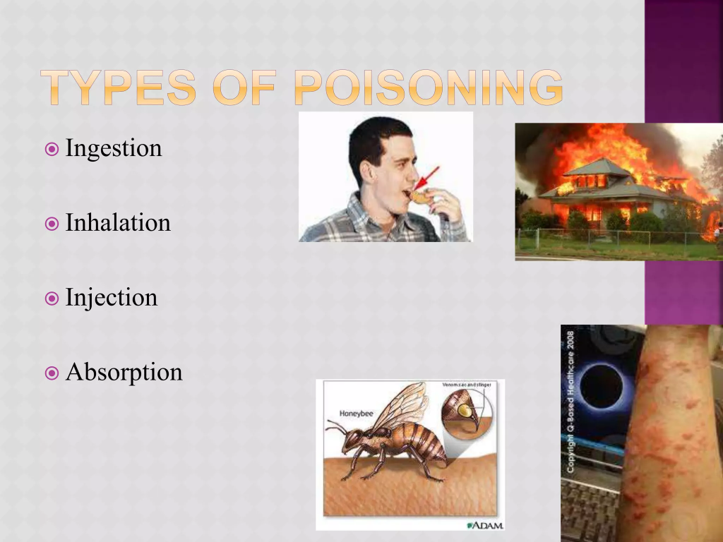
I) Chemical Poisoning :
- Chemical poisoning occurs due to exposure to household cleaners, pesticides, industrial chemicals or other toxic chemicals.
II) Drug Poisoning :
- Drug poisoning can occur due to overdose of prescription medicine, over the counter drops as well as illegal substances.
III) Food poisoning:
- Food poisoning occurs due to ingestion of contaminated food or beverages.
IV) Alcohol poisoning :
- Excessive alcohol consumption can cause alcohol poisoning.
V) Carbon monoxide poisoning :
- Carbon monoxide poisoning occurs due to inhaling carbon monoxide gas.
VI) Plant and Animal Poisoning:
- Poisoning conditions can also occur due to ingesting toxic plants or due to animal bites or stings.
Define inhaled poisoning

- Inhaled poisoning occurs due to the inhalation of toxic substances. Due to which, potential damage is seen in the respiratory system as well as other organs.
- Inhaled poisoning includes carbon monoxide poisoning, chemical fumes or vapor poisoning, smoke inhalation, ammonia and chlorine gas poisoning, asbestos poisoning, radon gas poisoning, hydrogen sulfide gas, nitrogen dioxide and sulfur dioxide poisoning.
Write sign and symptoms of inhaled poisioning (Write sign and symptoms of inhaled poisoning)
✓ Respiratory symptoms :
- Cuffing
- Shortness of breath
- Wheezing
- Chest Pain
- Rapid breathing
✓ Neurological symptoms :
- Headache
- Diarrhea
- Confusion
- Fainting
- Loss of Consciousness
- Seizures
✓ Cardiovascular Symptoms :
- Rapid or irregular heartbeat
- Palpitation
- Chest pain
- Hypotension
✓ Gastro-intestinal symptoms :
- Nausea
- Vomiting
✓ Eye and Skin Symptoms :
- Irritation or Redness in Eye
- Burning Sensation in Nose, Throat and Lung
- Skin irritation or rash
- Cyanosis
✓ General symptoms :
- Fever
- Fatigue
- Weakness
- Malaysia
Write first aid for inhaled poisoning
- Move to Fresh Air: Move the patient to an area with fresh air immediately.
- Call Emergency Services: Call emergency services immediately.
- CPR If Necessary: Provide CPR to the patient if breathing is not present.
- Provide Oxygen: Provide supplemental oxygen if oxygen is available.
- Remove contaminated clothing: If poisoning is suspected due to chemical exposure, remove contaminated clothing.
- Transfer to health center: Transfer the patient to the nearest health center area and provide medical care.
Define food poisoning
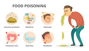
- Food poisoning is also known as ‘foodborne illness’. Which is caused by consuming contaminated food or beverages.
- Contaminants can be bacteria, viruses, parasites or toxins.
Write causes of food poisoning
Food poisoning is caused by consuming food or beverages contaminated with harmful microorganisms and toxins. is.
- Bacteria (Salmonella, E. coli, Listeria)
- Virus (Norovirus, Hepatitis A)
- Parasite (Toxoplasma, Cryptosporidium, Giardia)
- Toxin
- Chemical (Exposure to pesticide, Heavy Metal and Other Chemicals)
Write sign and symptoms of food poisoning
- Nausea
- Vomiting
- Diarrhea
- Abdominal pain and cramps
- Fever
- Headache
- Muscle pain
- Weakness or fatigue
- Dehydration
- Diarrhea
- Dry mouth
- Loss of appetite
Write diagnostic evaluation of food poisoning
- History collection
- Physical Examination
- Blood Test
- Stool Sample Analysis
- Urine Test
Write management of food poisoning
✓ Medication :
- Antidiarrheal drug: Provide over-the-counter medicine loperamide to control loose motions in mild cases.
- Antiemetic drug: Provide antiemetic drug to control nausea and vomiting. Such as Ondansetron
- Antibiotics : To provide drugs of the antibiotics group to treat bacterial infections.
- Antiparasitic : To provide drugs of the antiparasitic group to treat parasite infections.
✓ Hydration :
- Instruct the patient to drink plenty of fluids to prevent dehydration. Such as water, ORS, juice.
- In cases of severe dehydration, administer intravenous fluids.
✓ Rest :
- Provide adequate rest for the body to recover.
✓ Diet :
- If vomiting and diarrhea have subsided, provide the patient with easily digestible food. Such as rice, khichdi, curd
- If the patient can tolerate it, then reintroduce solid food.
- Avoid dairy products, alcohol, caffeine, nicotine and fatty foods.
Define chemical burns
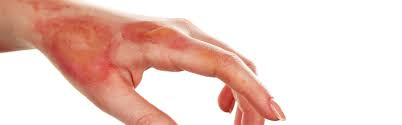
- Contact with harmful chemicals causes skin and tissue damage, which Known as chemical burns.
- Harmful chemicals include strong acids, alkalis, paint thinners, drain cleaners, gasoline, corrosive materials, etc.
Write sign and symptoms of chemical burns
The signs and symptoms of chemical burns depend on the concentration of the chemical and the duration of exposure.
- Redness and irritation
- Pain and burning
- Formation of blisters and peeling
- Discoloration of skin
- Swelling
- Numbness
- Ulceration
- Respiratory issues (cuffing, short ness of breath – in inhalation burns)
- Eye damage (pain, redness, vision loss – in case of eye burns)
- Systemic symptoms (nausea, diarrhea)
Write first aid for chemical burns
- Remove the chemical :
Quickly and carefully remove contaminated clothing and jewelry.
- Rinse the area:
Rinse the affected area with plenty of water for 20 minutes. Avoid hard sprays of water. Because it can cause tissue damage.
- Protect yourself:
Use gloves and cloths to avoid coming into contact with chemicals.
- Do not Apply Neutralizing Substance :
Do not use any other type of chemical to neutralize the chemical. Because it can cause a reaction.
- Cover the burn :
After flushing, cover the burn with a clean and dry cloth or sterile bandage.
- Medical attention :
In cases of severe chemical burns, breathing difficulties, and exposure to unknown chemicals, seek immediate medical help.
Write prevention of chemical burns
- Always wear protective clothing when handling chemicals.
- Chemicals should be stored properly and out of reach of children.
- Follow safety precautions and warnings associated with chemical products.
- Avoid prolonged contact with chemicals.
- Properly label the outside of chemical containers.
- Avoid storing chemical products in food or drink containers.
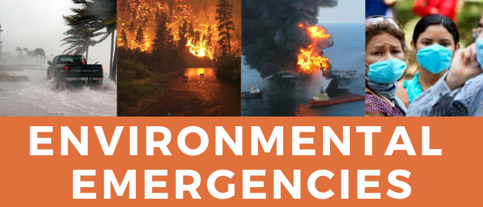
Environmental emergencies
Define heat stroke
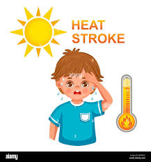
- Heat stroke is a serious and potentially life-threatening condition. In which the body temperature is found to be more than 41°C or 104°F.
- Which is mainly seen due to prolonged exposure to high temperatures or physical exertion during hot weather.
Write causes of heat stroke
- Prolonged exposure to high temperatures
- Dehydration
- Wearing heavy tight clothing
- Medication (diuretic, beta blocker, antihistamine)
- Medical conditions
- Drugs and alcohol Uses
- Sudden temperature changes
- Pure ventilation
Write sign and symptoms of heat stroke
- High body temperature (>104°F / 41°C)
- Alter mental status or behavior
- Confusion, agitation, slurred speech, irritability, delirium, seizures, coma
- Sweating
- Nausea and vomiting
- Flushed skin
- Rapid breathing
- Rapid heart rate
- Headache
Write first aid for heat stroke
- Call Emergency Services: If a person is suspected of having heat stroke, call 911 or your local emergency number.
- Move to Cooler Environment: Place the patient in an open area with plenty of fresh air.
- Cure the Person: Remove excess clothing and cool the patient using whatever is available. Such as placing the patient in a tub with plenty of water or using a fan with plenty of water.
- Monitor body temperature: Continuously monitor body temperature after cooling efforts.
- Rehydrate: Encourage the patient to drink plenty of water. Avoid drinking alcohol and caffeinated beverages.
Write prevention of heat stroke
- Stay hydrated: Drink plenty of fluids to stay hydrated. Avoid alcohol and caffeine.
- Avoid strenuous activity in the heat: Avoid working in areas with high heat. Work in a cooler area.
- Wear appropriate clothing: Wear loose-fitting, lightweight, and light-colored clothing.
- Protect against sunburn: Use sunscreen, sunglasses, and a hat to prevent sunburn.
- Acclimatize to the heat: Acclimatize means adapting. Gradual exposure to high temperatures over a period of days so that the plant can acclimate.
Define frost bite
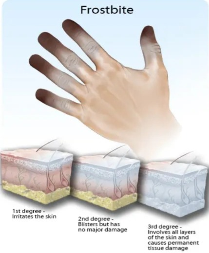
- Frost bite is a type of injury that occurs due to freezing of the skin and underlying tissue.
- A skin injury that occurs due to exposure to extremely cold temperatures is known as frost bite. Which mainly affects small parts of the body such as fingers, toes.
- Frost bite is mainly caused by contact with temperatures of 23° F or less.
Write causes of frost bite (Write causes of frost bite)
- Prolonged exposure to cold weather
- Wet clothing or skin
- Inadequate clothing
- Poor circulation
- Alcohol and drug use
- Fatigue and Dehydration
- High altitude
- Inactivity
- Medical conditions (diabetes, peripheral neuropathy, Raynaud’s phenomenon)
Write sign and symptoms of frost bite
- Cold skin
- Numbness (loss of sensation in affected area)
- Redness or pain
- Hard or waxy skin
- Blister formation in severe cases
- Discoloration of skin
- Skin may appear white, grayish yellow or blue
- Swelling at affected area
- Muscle and joint stiffness
- In severe cases tissue becomes dead (gangrene) leading to potential loss of limb
Write management of frost bite
- Get to a warm place: First, remove the patient from the cool environment and transfer him to a warm place.
- Remove wet clothing: Wet clothes can make frost bite worse, so replace the wet clothes with warm and dry clothes.
- Avoid Rubbing: Avoid rubbing the affected area as it can cause further damage.
- Warm the Area: Gently warm the frostbitten area with warm water. Avoid using direct heat sources. Such as fires, hot water, heating pad
- Protect the area: Gently wrap the affected area with a clean cloth or bandage.
Define near drowning

- This is a type of situation in which submersion or immersion in water Respiratory impairment is seen due to drowning. But if immediate care is provided, the patient can survive.
- This is a medical emergency. Due to which serious medical complications, respiratory distress and conditions like pneumonia can be seen.
Write causes and risk factors of near drowning
Near drowning is seen due to many factors. Which are as follows:
- Lake of Swimming Ability
- Alcohol and Drug Use
- Risky Behavior
- Attempted Suicide
- Lake of Safety Measures
- Environmental Factors (Current Tide, Waves and Weather Condition)
- Panic
- Accident (Boating accident, falling through ice)
Write sign and symptoms of near drowning
The signs and symptoms of near drowning depend on the severity of the incident and the person involved. Depends on how long you are submerged in water.
- Abdominal distension
- Breathing difficulty
- Chest pain
- Cuffing
- Cyanosis
- Cold skin
- Foaming at the mouth (white, frothy foam around the mouth
- Fatigue
- Confusion and disorientation
- Vomiting
- Unconsciousness
Write management of near drowning
✓ Immediate action:
- Ensure safety: Ensure that the area given for rescue is safe. Do not put yourself at risk.
- Call for help: Call for emergency services immediately.
- Rescue the person: Remove the person from the water safely.
- Check for responsiveness: Shake the person gently and check the person’s response.
✓ If the person is unresponsive:
- Open the Airway: Tilt the head back and lift the chin to open the airway.
- Check for Breathing: Assess the patient for breathing. If breathing is absent, provide CPR.
- Perform CPR: If breathing is not present, provide CPR.
- Use of AED: If an automated external defibrillator is present, use it.
✓ If the person is breathing:
- Positioning: Place the person in the recovery position to keep the airway open and prevent aspirating vomit.
- Keep warm: Cover the patient with a cloth or blanket to prevent hypothermia.
- Monitor: Monitor the patient for breathing and responsiveness until emergency services are available.
✓ Hospital Management:
- Oxygen therapy: Provide supplemental oxygen to maintain oxygen levels.
- Ventilation support: Provide mechanical ventilation if severe respiratory distress is present.
- Monitor for complications: Monitor the patient for secondary drowning, infection, and other complications.
- Chest X-ray: To check for water accumulation in the lungs and any other signs of Get a chest X-ray to check for injuries.
- Blood tests: Get blood tests done for electrolyte imbalances, oxygen levels, and other metabolic issues.
- Temperature management: Treat hypothermia if necessary.
Define hanging

- Hanging means the act of suspension by the neck.
- Hanging means the act of suspension by the neck.
- A hanging injury occurs when the neck is constricted by a ligature (such as a rope).
Write physical sign of hanging injury
- Ligature Mark
- Cyanosis
- Petechiae
- Protruding Tongue
- Saliva Dribbling
- Swallowed Face
- Congested eye
- Fracture of neck structure
Write mechanism of hanging injury
- During hanging, the ligature (rope, wire) compresses the soft tissue in the neck, which compresses the veins in the neck. The body is then hung on the rope, which causes more compression on the neck because the entire weight of the body is on the rope and due to this the weight increases, so that the artery in the neck (main coronary) is compressed and the blood supply to the brain is cut off, due to which death occurs.
Write causes of hanging
- Mental Health Issues (Depression, Bipolar Disorder, Anxiety)
- Acute Stress (Financial Problems, Loss of Loved One, Relationship Breakdown)
- Substance Abuse (Alcohol and Drug Addiction)
- Trauma and Abuse (Experience of Physical, Emotional and Sexual Abuse)
- Chronic pain or illness
- Social isolation (loneliness and lack of social support)
- Family history
- History of self-harm
- In case of execution (hanging is given as punishment in some countries)
Write management of hanging
✓ Initial assessment and resuscitation:
- Immediate response: Call for emergency medical services immediately. Carefully support the patient and lower him/her to a flat surface.
- Airway Management:If cervical spine injury is seen, provide thrust maneuvers to open the airway.
- Breathing:Check breathing and provide rescue breathing if necessary. Administer oxygen if oxygen is available.
- Circulation:Then check circulation, including checking pulse. If a pulse is not felt, provide cardiopulmonary resuscitation (CPR).
- Spinal precautions: Assume that there is a spinal injury until it is proven that there is no spinal injury. Therefore, immobilize the cervical spine.
✓ Emergency Department Care:
- Advanced Airway Management:Intubate to secure the airway.
- Breathing Support:Provide mechanical ventilation if respiratory failure is present To do.
- Circulation Support: Administer vasopressors and intravenous fluids to maintain blood pressure.
- Neurological Assessment: Perform a comprehensive neurological examination. Perform a CT scan and MRI of the head and neck to check for any injuries.
- Wound Care: Check for any injuries to the neck. Treat any injuries if present. Clean the wound properly to prevent infection.
- Supportive Care: Provide supportive care such as pain management and nutritional support.
- And this is mainly used to treat fractures in children.
nursing consideration
- To ensure that traction is effective or not.
- To see if the bandage applied over the traction is slipped or not.
- Maintain the proper position of the patient. The patient should be in the middle of the bed so that traction is more effective.
- The leg should be neutral to maintain the proper position.
- To maintain counter traction, there should be no wrinkling or slipping of the bandage.
- Avoid moving the patient so that the traction does not move.
- Limit the mobility of the patient to whom traction is applied.
- Provide proper psychological support to the patient.
- The color of the body part on which traction is applied, its temperature, its Check pulse or screen integrity and ED conditions.
- And check the patient for any other problems including pressure sores, constipation, urinary tract infection, loss of appetite, lung congestion, and screen breakdown.
- Closely monitor skin breakdown.
- Provide back care to the patient every two hours and use air and water mattresses to prevent pressure ulcers.
7)Skeletal traction:=
- Mainly used for fractures of the femur bone, tibia, and cervical spine.
- In this, traction is applied by an orthopedic surgeon while maintaining aseptic technique and wire pins and tongues are placed in the bone.
Nursing consideration:=
- When providing skeletal traction, an adequate amount of weight should be provided.
- When applying traction, the patient should have good body alignment and the patient should be in a proper position so that the foot does not drop.
- Anything on the side where traction is being applied should be Check for redness, swelling, warmth, drainage, etc.
- Continuously monitor the patient’s neurovascular status for signs of impaired blood flow.
- Check that the equipment is functioning properly.
- Check the skin integrity at the pressure points.
- Maintain traction properly.
- Nothing should be touched under the traction applied and it should be allowed to hang freely.
Traction apparatus:=
- Weight,ropes,
- Pulleys, spreader bars,footplate,
- Trapeze,hammocks,slings,and halter.
complication
- infection of skin.
- skin break down.
- stasis pneumonia.
- thromboplebitis.
- pressure ulcer.
- urinary infection.
- constipation.
explain the nursing management of patients with traction.
1)minimizing the effect of immobility.
- Advise the patient to do regular exercise to maintain joint strength and function.
- Ask the patient to do deep breathing exercises.
- Auscultate the lungs sound twice a day.
- Ask the patient to take fluid intake.
- Ask the patient to take a high-fiber diet.
- Provide the patient with stool softeners and enemas.
- Check whether the patient has thrombophlebitis.
2) Avoiding infection at pin site.
- Assess whether there is any pressure sore on the side of the bony prominence.
- Assess whether there is any skin irritation.
- Release pressure to a small extent.
- Check whether there is a burning sensation at the site where traction is applied.
- Check whether linen and clothing are wrinkle-free.
- Continuously check the patient’s vital signs.
- Immobilize the site where traction is applied.
- Check for any other signs of infection.
3) promoting tissues perfusion .
- Assess the patient’s motor sensory function.
- Check the patient’s sensation level.
- The patient’s neurovascular status assess.
- Prevent pressure ulcers by using protective devices.
- nurse should auscultate the patient’s lung sound.
General care of patient with traction.
- Assess the patient’s neurovascular status.
- Check the patient for pain sensation, active and passive range of motion exercises, skin colour, joint motion temperature, capillary refill time, numbness, and coldness.
- Check the patient’s skin area.
- Apply sufficient amount to the patient.
- Instruct the patient to do regular exercise, deep breathing, and wearing elastic stockings.
- Repeatedly assess the area where traction has been applied to see if there is any line in the tendon.
- Keep the patient in a neutral position.
- Provide the patient with a comfortable and functional position.
explain orthopedic splint
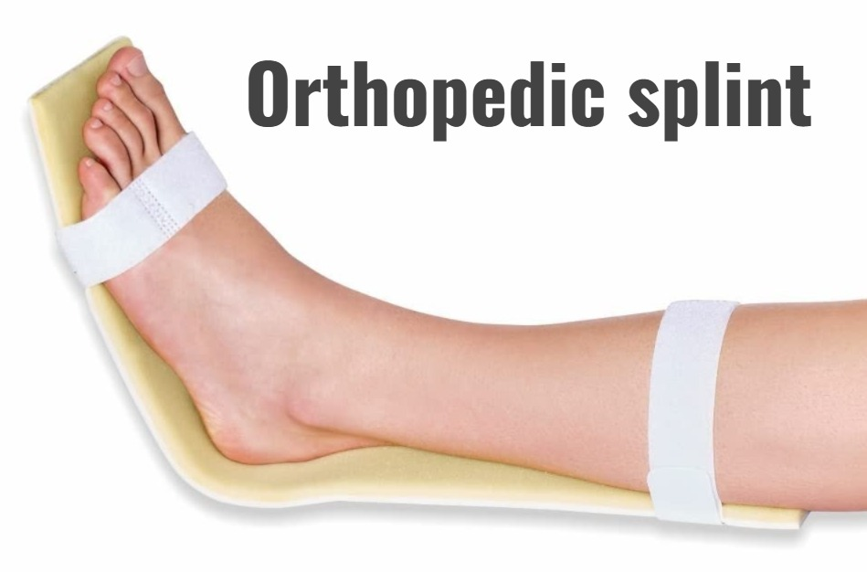
- A splint is a device that is used to support and immobilize an extremity or spinal cord.
- A splint is used in many situations, including temporary immobilization of a broken bone, damaged joint, and support for a joint during activities.
- A splint is a device that is used to immobilize an injured body part. It is used to provide support.
Ex:= broken bone, muscles sprain ,
Splint is used to provide support after surgery. - Splint is very padded due to which pressure can be prevented and skin break down can be prevented.
- Splint is mainly used in such conditions in which rigid immobilisation is not required.
explain the purpose of splint. (Describe the purpose of a splint.)
- To prevent movement of the injured extremity.
- To prevent further injury.
- To reduce oedema.
- To reduce pain.
- To maintain joint alignment For.
- To prevent contracture.
- A splint is used to support, protect, and immobilize the joint.
- To stabilize the joint if there is any ligament injury.
- To maintain the range of motion of the joint.
- To correct deformity For.
- To maintain tissue elongation (expansion).
- To stabilize and rest the joint if there is any ligamentous injury.
- To promote wound healing.
- To relieve pressure points.
- To protect a graft if it has been placed.
- To correct deformity.
- To strengthen the muscles.
- To support and immobilize the joint after an operation until healing is complete.
explain the indication for splinting.
- fracture, acute artheritis, suppuration,
- severe lacerations and abrasion,
- joint infection,
- skin A laceration,
- tenosynovitis (inflammation of tendon),
- puncture wound,
- laceration over joint puncture wound,
- reduced joint dislocation.
- Animal bites of hands or feet.
explain the contraindication of splint.
- compartment syndrome ( compartment syndrome := an increase pressure in side the muscles which restrict blood flow and cause pain).
- skin that is at high risk of infection.
- need for open reduction.
- open Fracture.
- Chronic neuropathy (nerve damage).
- Active infection.
explain the type of splint
Splints come in many shapes and sizes.
Splints come in many shapes and sizes.
Ex:= buddy taping ( for finger injury),
Some types of splints are large and are used to provide support to the hip and thigh.
Some splints are made of plastic and fabric.
These splints are fitted with hooks or buckles.
type of splint (type of splint) )
- 1)soft splint (soft splint).
- 2) hard splint (hard splint).
- 3)air/vacuum splint (air or vacuum splint).
- 4)Traction splint (traction splint).
1)Soft splint.
- This splint is mainly provided at home or by an emergency medical provider.
- This is a type of simple splint that is provided using a pillow or blanket.
- This splint is secured around the injured area and tied with tape. is.
- Soft splints are mainly provided for support and comfort of the injured extremity.
2) Hard splint.
- Hard splints are mainly used for injured extremities.
- Hard splints are provided using cardboard and padded boards.
- Some types of hard splints are made of fingerglass or plaster to support the patient’s extremities.
- The splint used for thumb injuries is called a thumb spica.
- Volar splint is used for wrist and forearm injuries.
- Boxer splint is used for hand and fist injuries.
- Pre-fabricated aluminum splints are mainly used to stabilize the finger.
3)air/vacuum splint (air or vacuum splint).
- Air splints are mainly used to provide support for orthopedic injuries.
- Air or vacuum splints are mainly used to provide comfort to injured extremities.
4)Traction splint (Traction splint) .
- Traction splints are used primarily to reduce the amount of deformity, align the bone, provide traction, and prevent the bone from moving.
- Track splints are used primarily to align the femur bone or the midshaft of the lower leg.
Explain the nursing care of a patient with a splint. Describe.)
- Check the skin where the splint is applied for any cracks, damage, swelling or soreness.
- Keep the area where the splint is applied dry and clean.
- Keep the area where the splint is applied properly padded to prevent cracks and skin breakdown.
- Keep the area where the splint is applied properly padded to prevent cracks and skin breakdown.
- When a splint is applied and bathing is to be done, wrap the splint properly with plastic. So that the splint can be prevented from getting wet.
- Apply a soft pad to the area where the splint is applied and if there are bony prominences, apply it to that area.
- Check that blood vessels or nerves are not compressed in the area where the splint is applied.
- Keep the splint properly clean.
- Provide support to the injured area.
- Elevate the affected limb and prevent swelling.
- Loosen the elastic bandage.
- Do not apply powder or deodorant to the skin, as this may increase itching.
- Do not try to crack the skin with a sharp object inside the hard splint, as this may cause cuts to the skin.
- Apply a pad to the skin to protect the skin.
- Perform range of motion exercises.
- And provide support to the patient.
explain pop application and removal
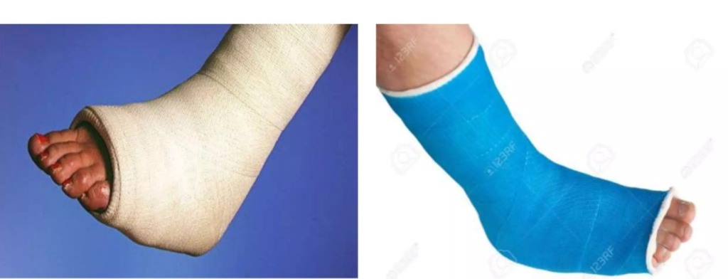
1) explain the plaster cast (Explain the plaster cast.)
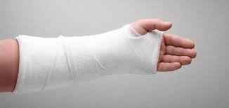
- A cast is a rigid device that is applied to immobilize injured bone and soft tissue and promote healing.
- A cast is primarily applied to immobilize the bone above and below a fractured bone.
- A cast is essentially a supporting bandage that is solid and is wrapped around an extremity.
explain the purpose of cast
- To support and protect bones and soft tissues.
- To reduce pain.
- To reduce swelling and muscle spasm.
- To immobilize a broken bone.
explain the casting material
1) plaster cast
- Plaster of Paris bandages are rolls of precut crinoline that come in a variety of sizes.
- And the plaster is also attached to it.
- The bandage rolls are easily applied to the body by dipping them in cold water.
- Then a crystallization reaction occurs and heat is generated.
- Then it is exposed to air to dry. is.
- Then it cools down after about 15 to 20 minutes.
- Plaster cast takes 24 to 72 hours to dry completely.
- Plaster cast is heavy and dries slowly.
2) fiberglass cast( Fiberglass Cast):=
- This water is made of activated polyurethane which has the versatility of plaster.
- It is light in weight, dries quickly, is strong, water resistant and durable.
- It has pores, so skin problems can also be prevented.
- If this gets wet, it must be dried to prevent skin breakdown.
- Fiberglass and plastic casts are generally expensive and also macerate the skin.
- Cotton and other synthetic materials are used to make the inside of the cast, which makes the inside soft and acts as padding that provides softness around bony areas, such as the wrist or elbow.
- Some types of special waterproof casts are used to prevent the plaster cast from getting wet and prevent screen problems.
explain the type of cast
1)short arm cast:=
- Short arm cast is mainly applied from elbow to thumb and is mainly used when there is a radius, humerus, carpal, metacarpal fracture.
2)long arm cast:=
- Long arm cast is mainly used from the axillary area to the palmar crease of the hand and is mainly used to treat fractures of the upper extremity.
3)short leg cast:=
- Short leg cast is applied from the bottom of the knee to the base of the toe.
- Short leg cast is mainly used to treat fractures of the tibia, fibula, ankle.
4)long leg cast:=
- Long leg cast is mainly applied from the base of the thigh. It is applied from the base of the toe.
- And this cast is mainly used to treat fractures of the femur, tibia and fibula.
5)walking cast:=
- Short and long leg casts are mainly used as weight bearing.
6) body cast:=
- This cast is applied around the trunk.
7) shoulder spica cast:=
- This cast is It is mainly used to enclose the trunk, ankle and elbow and is mainly used to treat ankle fractures.
8) Hip spica cast:=
- This cast is mainly applied from the middle of the trunk to the foot and is mainly used to treat hip fractures.
explain preparation of patients for plaster cast:=
- Explain the procedure to the patient.
- Assess the patient’s general health.
- Check the patient’s vital signs.
- Properly prepare the area where the plaster cast is to be applied. Shaving.
- Pay special attention to the skin and bony areas.
- Plaster bandage mainly depends on the size and number of bone fractures.
- Take proper consultation before applying plaster cast.
- Explain the procedure completely.
- Clean the affected area properly with a shop solution.
- Gentally scrub.
- Remove any tight clothes and ornaments.
- Ask the patient to sit comfortably.
- Apply pads properly to the pressure points.
explain the nursing management of patients explain the assessment of patient
- Check the extremity where the cast is applied.
- Check whether there is circulation in the area where the cast is applied.
- Check whether there is warmth in the area where the cast is applied.
- Check whether there is sensation in the area where the cast is applied. To do.
- Assess whether the affected limb has motor ability.
- Check whether the site on which the cast is applied is too tight.
- Assess skin integrity.
Explain the nursing interventions
- Elevate the affected extremity to stimulate circulation.
- Change the patient’s position frequently while the cast is being applied.
- Provide support to the foot to prevent foot drop while the splint is being applied.
- If the patient is experiencing pain over the bony prominence area, Check the area where the pressure bony part is.
- Assess the patient’s neurovascular function.
- Check the patient’s body circulation and movement of his extremities.
- Ask the patient to moment the body parts.
- Check the patient for any complications such as compartment syndrome, pressure ulcers or tissue damage.
- Instruct the patient to follow a well-balanced diet, including a high-fiber diet that prevents constipation.
- Instruct the patient to drink adequate amounts of fluid.
- Instruct the patient to avoid gas-forming foods while the cast is in place.
Nursing care
1)keep the cast dry:=
- Keep the cast completely dry and when washing the body part, protect it with plastic so that the cast is not exposed to water.
2) Watch the cast carefully:=
- Check the area where the cast is applied for any redness, skin breakdown, or bluish discolouration. To do.
3) elevate the cast:=
- Elevate the affected extremities to prevent swelling.
4)exercise the extremity:=
- Properly exercise the affected extremity.
5)Apply an ice bag to the cast:=
- Apply an ice pack to the area where there is swelling to reduce swelling.
6)Instruct the doctors if the following conditions occur:=
- When the body temperature is above 101°f.
- When the pain level increases.
- When the swelling increases.
- Numbness or tingling sensation.
- When there is any foul smell coming from the cast.
- When the toes are cool.
explain the nursing care after removal of plaster
- Plaster is mainly cut using electric plaster cutter and manual plaster cutter.
- After removing the plaster, wash it thoroughly and then dry it properly and do not wipe the affected extremity forcefully, due to which there is a possibility of peeling of the skin.
- How to wear the plaster so that oedema can be prevented.
- After removing the plaster, it Advise the patient not to rub or scratch the area.
- Instruct the patient to elevate the affected area to prevent swelling after the cast is removed.
- Instruct the patient to ambulate properly after the cast is removed.
- If the patient has been instructed by the physician to wear compression bandages and elastic stockings, wear them properly.
complication of cast
1)compartment syndrome
- Compartment syndrome is a syndrome in which the blood supply is impeded and the circulation is cut off at that place and swelling occurs on the affected area.
- And pain occurs on the affected area.
2)Pressure ulcer:=
- Ulcers develop on the bony prominences where the cast is applied.
- Conditions like skin breakdown, redness, warmth, swelling are also seen in the area where the cast is applied.
explain the health education to patient and relative. (Provide health education to patient and relative)
- Do not place any object on the cast that has been applied.
- If the cast is made of plaster of Paris, do not let it get wet.
- If fiberglass If the cast is applied, dry it properly if it is wet.
- Instruct the patient not to walk on wet and slippery surfaces.
- Do the prescribed exercises regularly.
- Elevate the area where the cast is applied to prevent swelling.
- Avoid scratching the skin inside the cast, as this can lead to skin breakdown.
- Do not insert any object inside the cast.
- If there is pain, swelling, or redness in the area where the cast is applied, report it to the physician immediately.
- If the cast breaks down for any reason, inform the physician immediately.
1)explain the definition of neurogenic bladder .
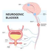
- Neurogenic bladder is a type of neurogenic dysfunction.
- Neurogenic bladder is a dysfunction of the urinary bladder, which is mainly caused by problems in the central and peripheral nervous system.
- Neurogenic bladder is mainly caused by impairment in the normal nerve pathways.
- Neurogenic bladder can cause urinary retention, incontinence of urine, urinary tract infection, stone formation, renal failure Conditions like this arise.
2) Explain the type of neurogenic bladder. (Write the type of neurogenic bladder.)
- 1) flassid neurogenic bladder ( flaccid neurogenic bladder).
- 2)spastic neurogenic bladder ( spastic neurogenic bladder.)
1) flassid neurogenic bladder ( flaccid neurogenic bladder).
- Flaccid bladder is mainly seen due to motor neuron lesion and any trauma.
- Due to the decrease in the sensation of bladder feeling, the bladder does not concentrate fully and due to this the bladder becomes full.
- And the bladder becomes distended due to which urinary incontinence is seen.
2)Spastic neurogenic bladder
- Spastic bladder is mainly characterized by uncontrolled and frequent expulsion of urine from the bladder.
- This is mainly seen due to brain damage and spinal cord damage.
- Due to this, urine emptying becomes incomplete.
provide Etiology (Give reasons)
- Neurogenic blood vessels are found in any age.
- Alzheimer’s disease.
- Alcohol neuropathy.
- Stroke.
- Meningomyelocele.
- Aids.
- Parkinson’s disease.
- Brain or spinal cord tumor.
- Diabetic neuropathy.
- Spina bifida.
- Multiples Sclerosis.
- nerve damage.
- due to any diabetes or alcoholic disorder.
- due to injury to the spinal cord.
- due to nerve damage.
- Vitamin B12 deficiency.
Clinical manifestations:
- Excessive urine production.
- Bladder overactivity.
- Urinary incontinence.
- Frequent urination.
- Urinary retention.
- Urinary frequency and urgency
- Problems in expelling the entire urine from the bladder.
- The bladder becomes full and urine leaks.
- Loss of bladder control.
- Loss of sensation of bladder fullness.
- Pain and burning during urination.
- Erectile Dysfunction.
- Urinary Tract Infection.
Diagnostic Evolution (Diagnostic Evaluation):
- history tacking and physical examination
- complete Neurological examination.
- post void residual volume.
- renal ultrasonography.
- Serum creatinine.
- cytography.
- cytoscopy.
- cytometrography.
- urodynamic testing.
explain the treatment (Explain the treatment)
- Provide medicine that relaxes the bladder.
- Treat and control urinary tract infections.
- Advise to drink plenty of water to reduce urinary tract infections.
- Ambulate the patient frequently.
- Change the patient’s position frequently.
- Tell the patient to take calcium in small amounts.
- Change the patient’s position frequently.
explain specific treatment
- 1)physical-psychological therapy.
- 2)bladder evacuation.
- 3) electrical stimulatory therapy.
explain surgery
- 1) Transurethral resection of the bladder neck.
- 2)urethral dilatation.
- 3)External sphincterotomy.
- 4)urinary diversional procedure.
- 5)implantation of artificial sphincter.
- 6)urethral stent.
Nursing Management:
- Monitor residual urine.
- Monitor for signs and symptoms of any renal calculi.
- Assess urinary stasis.
- Check for signs and symptoms of urinary tract infection, including urine color, order, volume, frequency, urgency.
- Patient’s Check intake output.
- Administer vitamin C to the patient to produce acidic urine and prevent bacterial growth.
- Assess the patient’s voiding pattern.
- Ask the patient to do kiggle exercises.
- Provide different methods to empty the patient’s bladder like Crede’s method, Valsalva’s maneuver etc.
- Use aseptic technique and sterile method when catheterizing the patient.
- Prescribe drugs to maintain continence in the patient.
- Instruct the patient to have regular follow-up.
explain the use of orthopedic aasist device
1) Explain the crutches.
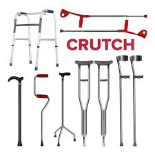
- Crutches are artificial devices that are used by patients who are unable to walk on their own.
- Crutches are mainly used to provide ambulation and independence to patients who have lower extremity injuries.
explain the indication
- disease, injury, birth defect.
explain the preparation for crutch walking
- Inform the client that he/she should wear well-fitted shoes.
- Before using crutches, ask the patient to stand on a chair and use the support of a chair to achieve balance. Can be done.
- The patient should stand against the wall and provide a neutral position.
- It is necessary to practice walking with crutches before starting.
- Explain the procedure to the patient.
- Ask the patient to wear full clothing and non-slippery shoes.
crouch walking gait
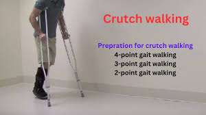
1)Explain four point gait:=
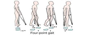
The four point gate is mainly used when a small amount of weight can be borne on both lower extremities.
- 1) First, forward the right crutches.
- 2) Then forward the left leg.
- 3) Then forward the left crutches. Do.
- 4) Then move the right foot forward.
- 5) Repeat in this way in a cruch-foot cruch-foot sequence.
2)three point gait:=
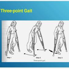
Three point gate is mainly used when some weight bearing is not possible.
- 1) In this, the affected leg (Non weight bearing) and both the crutches are moved forward.
- 2) Then the unaffected leg (weight bearing) to do it next.
- 3) Repeat in the same sequence.
3) Two point gait:=
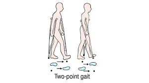
Two point gate is mainly used when a small amount of weight is to be borne above both lower extremities.
- 1) In this, the right leg and the left crutches are brought forward.
- 2) Then the left leg and the right crutches are brought forward.
- 3) Then repeat this sequence.
4)swing through gait:=
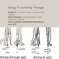
This is mainly used when the lower extremity is paralyzed.
- 1) In this, both the extremities are moved forward about six inches.
- 2) Then both the legs are moved forward six inches.
- 3) Then repeat in the same pattern.
explain walker and canes
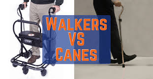
- Walkers and canes are primarily mobilisation aids that allow the patient to bear some weight on the affected leg but require some support.
- When a walker is used, the patient’s upper arm muscles and upper body are used for weight bearing.
- Then the following sequence should be used.
1) Grip the hand grip very tightly.
2) Then move the walker and the affected leg forward six inches.
3) Then bring the unaffected leg parallel to the affected leg.
4) Then repeat the same sequence.
explain the use of cane
Ask the patient to hold the unaffected side of the cane on the same side, six inches forward and six inches to the side of the foot, and then follow the sequence below.
1) Move the affected leg forward and move its parallel cane forward as well.
2)Then move the unaffected leg forward so that it is just a little behind the cane.
3) Then move the affected leg forward.
4) Then move the cane forward six inches.
5) Repeat this sequence.
If less support is needed, move both the cane and the affected leg forward together.
The walker and cane are used to provide support to the body part and to walk.