ENGLISH-MSN-2-SKIN (PART-6)(UPLOADED)
♣ What is abrasion ?

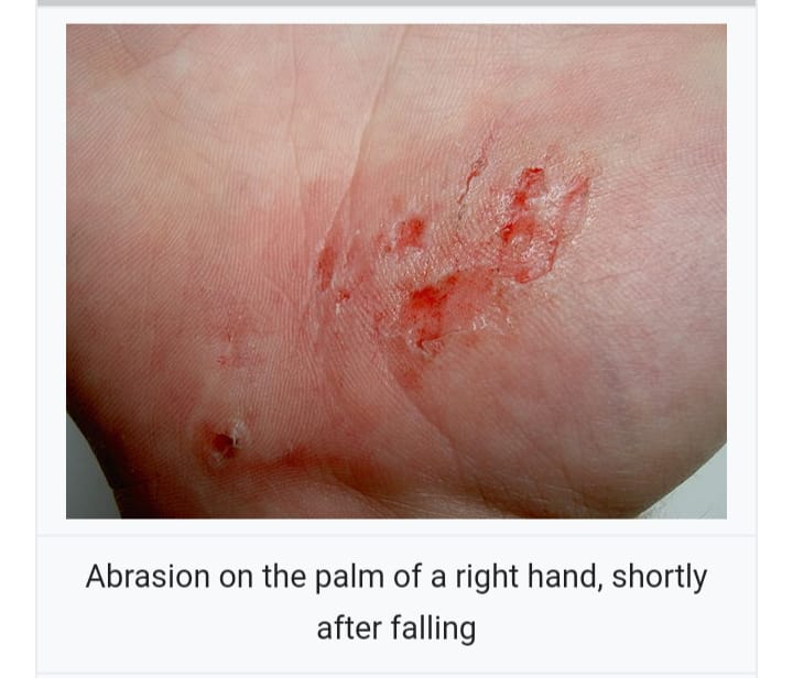
- Abrasion is a common injury. In which the skin is damaged due to rubbing with rough surfaces and the integrity of the tissue is broken.
- Skin injury due to rubbing, grinding and scraping of the skin due to contact with hard and rough surfaces. This is known as abrasion. For example falling on a rough surface.
- Abrasion occurs in a mild to severe form. Abrasions are mainly seen in palms, elbows, knees, ankles.
♦ Explain degree of abrasion (Explain degree of abrasion)
◙First degree abrasion
- First degree abrasion is mild abrasion. In which the epidermal layer of the skin is affected. Bleeding is not seen in first degree abrasion. First degree abrasion is also known as ‘graze’ or ‘scrapes’.
◙Second degree abrasion
- In second degree abrasion, epidermal as well as dermal layers are affected. Mild bleeding is seen in second degree abrasion.
◙Third degree abrasion (Third degree abrasion)
- Third degree abrasion is a more severe type of abrasion in which the subcutaneous layer is also affected. Also known as ‘avulsion’. A third degree abrasion involves heavy bleeding and requires medical care.
♦ Write classification of abrasion (Write classification of abrasion)
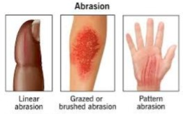
- Abrasions are classified into three types based on their clinical presentation and etiological factors.
i) Linear or scratch abrasion
- Linear abrasion is a simple injury. Linear abrasion is caused by sharp or pointed objects. Like nails, pins. Linear abrasion has greater length while less width. For example nail scratches on the skin. Linear abrasion also has medicolegal importance. For example, if a scratch is found in the throat, it indicates that an attempt has been made to strangle the person.
ii) Grazed or brush abrasion
- Grace abrasion results from friction on broad, hard and rough surfaces and has multiple presentations. For example, gray abrasion is seen in conditions like road traffic accidents, falls during sports activities. The depth of injury in gray abrasion varies depending on the surface irregularity and body speed.
iii) Patterned abrasion
- When a perpendicular force is applied to the skin by an offending object, the impression of that object is made on our skin and a patterned abrasion is seen.
♦ Write management of abrasion (write management abrasion)
- First and second degree abrasions can also be treated at home.
- Hand wash the affected area with warm water and soap to remove dust and other particles.
- Leave the wound open if there is mild abrasion and no bleeding.
- If the wound is bleeding, apply gentle pressure to the area using a clean cloth or bandage. So that the building is stopped and also to elevate that area.
- Apply topical antibacterial medicine on the affected area.
- Antibacterial medicine prevents infection and keeps the wound moist.
- Inform the doctor if signs of infection are present.
- If the wound is painful then use systemic analgesic drug.
- If there is a third degree abrasion, go to the hospital and get medical treatment.
♣ Define dermatitis
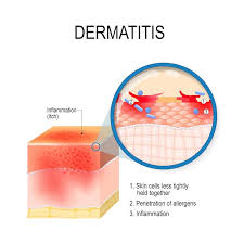
- Derm means skin and itis means inflammation. Dermatitis means skin inflammation. The term dermatitis is used to describe a group of skin disorders.
- The word eczema is similar to the word dermatitis. The term eczema is also used to describe a skin disorder. Including itchy, dry and inflamed skin conditions.
♦ Explain pathophysiology of dermatitis
Etiology or exposure to risk factor
|
Inflammatory reaction in the skin.
|
Resulting in skin eruption.
|
Local erythema, vesicles and itching.
|
Continuous irritation and scratching of the skin. is.
|
Due to which the skin becomes thick i.e. lichenification is seen.
Write type of dermatitis (Tite type of dermatitis)
There are four main types of dermatitis:
i) Contact dermatitis
ii) Atopic dermatitis
iii) Exfoliative dermatitis
iv) Seborrheic dermatitis
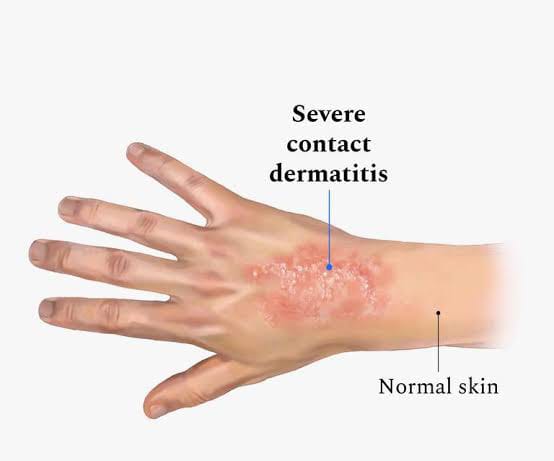
i) Contact dermatitis
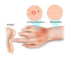
- Contact dermatitis is also known as ‘contact eczema’. Contact dermatitis is an inflammatory skin disorder. Physical, chemical and biological agents coming into contact with our skin cause a skin inflammatory reaction known as contact dermatitis.
There are two types of contact dermatitis:

1. Non allergic contact dermatitis or irritant dermatitis (Non allergic contact dermatitis or irritant contact dermatitis)
2. Allergic contact dermatitis (allergic contact dermatitis)
Non allergic contact dermatitis or irritant dermatitis
- Irritant contact dermatitis is a non-allergic reaction in the body that comes into contact with an irritating substance. Some types of soaps, detergents, cosmetic items and industrial chemicals are included as irritating substances.
Allergic contact dermatitis
- Allergic contact dermatitis is a sensitized person’s allergic reaction to the body coming in contact with the allergen. For example, some people are allergic to latex. So if such people wear latex gloves, allergic reactions are seen in them. Allergic substances are different in different people.
♦ Write sign & symptoms seen in contact dermatitis (Right sign and symptoms seen in contact dermatitis)
Acute phase:
- Initially there is itching, burning and erythema. Then there is edema, papule, vesicle and oozing.
Sub acute phase
- Vesicular changes are less common in the subacute phase and there is crusting, fissuring and peeling.
Chronic phase
- If there is frequent reaction and continuous scratching then the skin becomes thickened i.e. lichenification is seen and pigmentation is seen.
♦ How to diagnose contact dermatitis (How to diagnose contact dermatitis)
- History Collection
- Physical Examination
- Patch testing
♦ Write medical management of contact dermatitis (Write medical management of contact dermatitis)
- Apply topical corticosteroid and antipruritic medicine to the affected area.
- Apply a total and wet dressing that clears the oozing.
- Provide medicated bath (balenotherapy) if more area is affected.
- Provide systemic corticosteroids in severe cases.
♦ Write nursing management of contact dermatitis
- To assess vital signs.
- Checking the affected area for swelling, oozing, fissuring.
- Apply topical medicine on the affected area.
- Advise the patient to avoid contact with allergens and irritant substances.
- Change position every two hours.
- To administer the medicine prescribed by the doctor.
- To maintain records and reports.
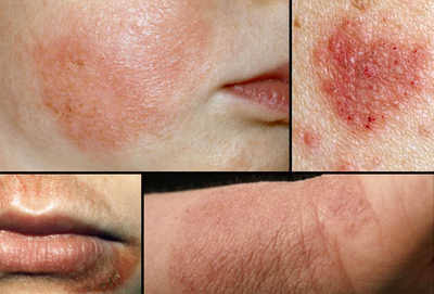
ii) Atopic dermatitis
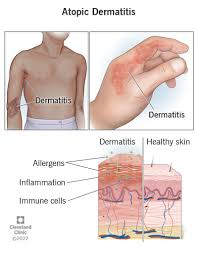
- Atopic dermatitis is a chronic pruritic inflammatory skin disease. The word atopic denotes three allergic diseases – asthma, allergic rhinitis and atopic dermatitis
- Atopic dermatitis is more common in early childhood. The exact cause of atopic dermatitis has not yet been discovered.
♦ Risk factor of atopic dermatitis
People who have atopic dermatitis also have asthma and seasonal allergies.
- Family History
- People working with chemicals, irritants and rough materials
- Weather Changes
- Stress
- Cold and dry air
♦ Explain pathophysiology of atopic dermatitis (Explain pathophysiology of atopic dermatitis)

Coming in contact with an allergen
|
The body’s immune system is activated and the body releases histamine, cytokines, bradykinin and other inflammatory mediators.
|
These released inflammatory mediators interact with the allergen.
|
This interaction causes an inflammatory reaction in the body.
|
The water binding capacity of the body decreases and water loss is observed.
|
Due to which the skin becomes dry and cracking.
|
And inflammatory symptoms on the skin include itching, scratching, redness, erythema and lesions.
♦Write sign & symptoms seen in atopic dermatitis (right sign and symptoms seen in atopic dermatitis)
- Symptoms can occur anywhere in the body and are different in different people.
- Skin becomes dry and cracked.
- A small raised bump is seen on the skin.
- Redness is seen around it.
- It has oozing and crusting.
- Etching is observed.
- The skin becomes thicker ie lichenification is seen.
- Changes in skin color are seen.
- The skin around the eye becomes dark in color.
- Discharge and bleeding are seen in the ear.
- The skin is seen to be raw due to scratching.
How to diagnose atopic dermatitis (How to diagnose atopic dermatitis)
- History Collection
- Physical Examination
- Skin biopsy
- Patch testing
♦ Write medical management of atopic dermatitis (Write medical management of atopic dermatitis)
- Apply topical corticosteroids to the affected area.
- Using oral corticosteroids to reduce inflammation and suppress immunity.
- Using topical immunomodulators.
- Providing therapeutic bath (Baleno therapy) to the patient.
- Using Ultra Violet Light Therapy.
♦ Write nursing management of atopic dermatitis
- To assess vital signs.
- Assess the affected area for wearing, cracking and lichenification.
- Apply topical medicine prescribed by the doctor.
- Provide cold moist compresses on the affected area.
- Advise the patient to avoid contact with strong shops and chemicals.
- Don’t take too much stress.
- Apply a lubricant to the skin after bathing to prevent the skin from drying out
- To administer the medicine prescribed by the doctor.
- To maintain records and reports.
iii) Exfoliative dermatitis
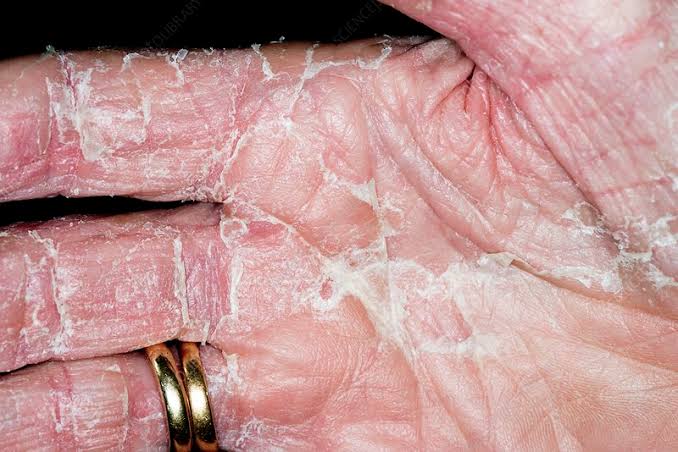
- Exfoliative dermatitis is a serious skin inflammatory disorder. In which progressive inflammation is seen and accompanied by erythema and scaling. It affects 90% of the skin surface.
- Exfoliative dermatitis occurs due to drug reactions, systemic diseases, preexciting conditions, and cancer. While in some people cause unknown.
♦ Write sign & symptoms seen in exfoliative dermatitis (right sign and symptoms of exfoliative dermatitis)
- Erythema and scaling are seen on the skin.
- Etching is observed.
- Peeling is observed on the skin.
- So that nail and hair loss is seen.
- Skin is pink to brown in color.
- Systemic symptoms like fever, malaise, chills are seen.
- Prostate and severe toxicity is observed.
- Water and protein are lost from the skin.
- Also heart failure, gastrointestinal disturbance, best enlargement and hyper uricemia are seen.
♦ How to diagnose exfoliative dermatitis (How to diagnose exfoliative dermatitis)
- History Collection
- Physical Examination
- Culture and sensitivity test Write management of exfoliative dermatitis (Write management of exfoliative dermatitis)
- Using oral and topical corticosteroids.
- Using antihistamine drug.
- Maintaining fluid and electrolyte balance.
- Administering a plasma volume expander.
- Provide iv fluids.
- Anchoring the patient for oral fluid intake.
- Providing bed rest.

iv) Seborrheic dermatitis
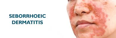
- Sebohric dermatitis is also known as ‘cradle cap’ or ‘dandruff’. Seborrheic dermatitis is a common and chronic skin inflammatory disorder. In which mainly the scalp is affected. Apart from this, the oily area of the body like face, eyebrow, ear, side of nose, axilla and upper chest are also affected.
♦ Sign & symptoms seen in seborrheic dermatitis (Signs and systems seen in seborrheic dermatitis)
- Dandruff occurs on the scalp, hair, eyebrows and beard i.e. flaking skin.
- Patches of gray color are seen around it.
- Peeling is observed on the skin.
- Etting is seen on the affected area.
- Small pustules are seen in the trunk.
How to diagnose seborrheic dermatitis (How to diagnose seborrheic dermatitis)
- History Collection
- Physical Examination
- Skin biopsy
♦ Write management of seborrheic dermatitis
- Apply topical corticosteroids and antifungal ointment to affected area.
- Wash hair with antidandruff shampoo.
- To wash hair twice a week.
- Maintain proper hygiene.
Apart from this, there are some types of dermatitis which are as follows.
Statis dermatitis (Statis dermatitis)
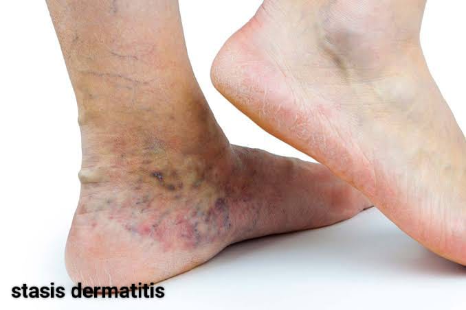
- Status dermatitis is also known as ‘gravitational dermatitis’ or ‘venous eczema’.Status dermatitis causes skin changes due to poor blood circulation. In which discoloration, itching and open sores are seen in the lower extremities and the skin becomes thick.
- Static dermatitis is caused by fluid buildup due to varicose veins, circulation issues, or heart disease.
Neuro dermatitis
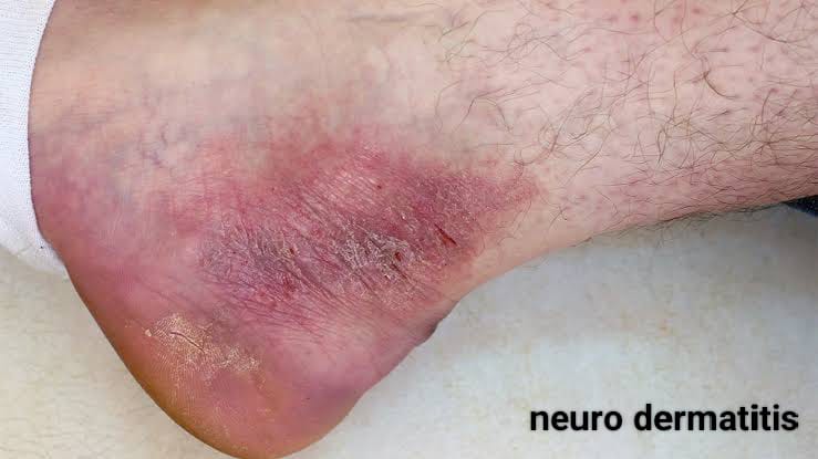
- Neuro dermatitis is also known as ‘lichen simplex chronicus’ .
- Neuro dermatitis is a neurological skin disorder. In which scaly patches and chronic itching are seen in the neck, scalp, wrist, ankle, and groin area. The cause of neurodermatitis is unknown.
Numular dermatitis

- Numular is a Latin word meaning coin. Nummular dermatitis is characterized by coin-shaped lesions and sores on the skin after injury.
- The skin surrounding the lesion becomes inflamed and appears red pinkish or brown in color when the ulcer is seen.
Dyshidrotic dermatitis
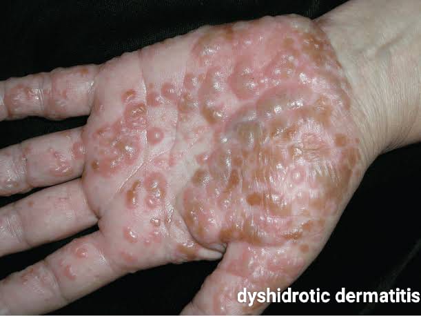
- Dyshidrotic dermatitis is also known as ‘pompholix’ or ‘palmoplantar eczema’ .
- Dyshidrotic dermatitis presents with tiny fluid-filled blisters on the palms and fingers. Also often seen on fit’s soles and toes.