ENGLISH-UNIT-4-MSN-II-OPTHALMOLOGY AND OPTHALMIC NURSING-DIAGNOSIS(PART-2)
Assessment of function of eyes Health history (subjective data):
- Health history includes present health history, past health history, past surgical history, personal history, family history and occupational history.
Present health history / cheif complain
- Ask the patient about their current complaints.
- Ask the patient about problems related to the eyes and vision.
- Ask the patient if there is pain, redness, inflammation in the eyes.
- If present, ask about its duration, intensity, and severity.
- To find out if the patient has any kind of vision problem.
- If there is a vision problem, get detailed information about it.
- Ask when the vision is impaired. If the problem occurs at night, it may be night blindness.
- Ask the patient if he has difficulty seeing near or far.
- Check for black spots during vision.
- Ask the patient if he has difficulty identifying colors.
- Ask the patient if he has sensitivity to light.
Past health history
- To obtain information about whether the patient has had any type of eye disease in the previous years.
- To know whether the patient has any systemic conditions like diabetes, hypertension, etc.
- To collect information about whether the patient has been hospitalized in the past.
- Collect information about childhood diseases and immunizations.
Past surgical history
- To know whether the patient has undergone any eye surgery in the previous years.
- In addition, whether the patient has undergone any other surgery or not To know.
Personal history
- Collect the patient’s personal history.
- To know whether the patient smokes or not.
- To know whether the patient consumes alcohol or not.
- Ask the patient if he has any allergies to any substance or medicine.
- Know about the patient’s visual habits.
- Know about the frequency and duration of the patient’s screen time. That is, how long the patient uses a laptop, computer, smart phone.
- In addition, know about the patient’s dietary pattern.
Family history
- Collect the patient’s family history.
- Ask the patient if any of their family members have had eye disease.
- Collect information about whether there is any hereditary condition in the family. (such as retinitis pigmentosa)
Occupational history
- To obtain information related to the patient’s occupation.
- To find out whether the patient does computer-related work.
- To find out whether the patient is exposed to radiation or chemicals during his work.
Physical examination / objective data
- Inspection method is used in physical examination of the eye.
- First inspect the external eye area, then Inspect the internal eye area.
- Examine the eyebrows for symmetry and inspect them for hair distribution.
- Then check the position and alignment of the eyes.
- Inspect the eyelids for redness, swelling discharge, ptosis.
- Inspect the eyelids for entropion, ectropion.
- Inspect the conjunctiva and sclera for color, texture, redness, lesions, secretions.
- Inspect the cornea for opacity and dullness.
- Then check the corneal reflex.
- Check the iris for obesity, cloudiness, and redness.
- Inspect the pupil for size, shape, and location.
- Check the pupillary reflex.
- Check the lens for opacity.
Diagnostic test for eye disorder
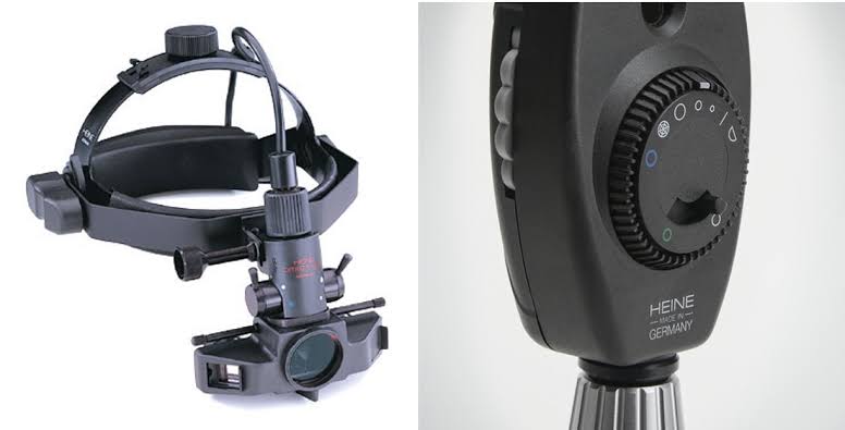
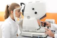
Opthalmoscopy (Ophthalmoscopy)
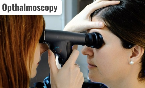
- Ophthalmoscopy is a diagnostic procedure in which an ophthalmoscope is used to examine the eye. The back part of the eye is examined with the help of. Such as the retina, choroid, optic disc and blood vessels are examined. There are two types of ophthalmoscopy:
- 1) Direct ophthalmoscopy
- 2) Indirect ophthalmoscopy
1) Direct ophthalmoscopy :
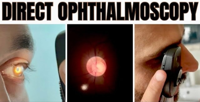
- Direct ophthalmoscopy is used to examine the anterior structures of the eye. Direct ophthalmoscopy uses a handheld (torch-like) instrument (ophthalmoscope) that is equipped with a light source and a lens. This ophthalmoscope is held close to the eye and the anterior structures of the eye are examined.
2) Indirect ophthalmoscopy:
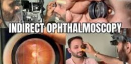
- Indirect ophthalmoscopy provides a wider view than direct ophthalmoscopy, i.e. it provides a complete view of the retina. In indirect ophthalmoscopy, the examiner wears a headband with a lens attached, and the lens is held close to the patient’s eye and examined. A bright light source (such as a slit lamp) is used for this.
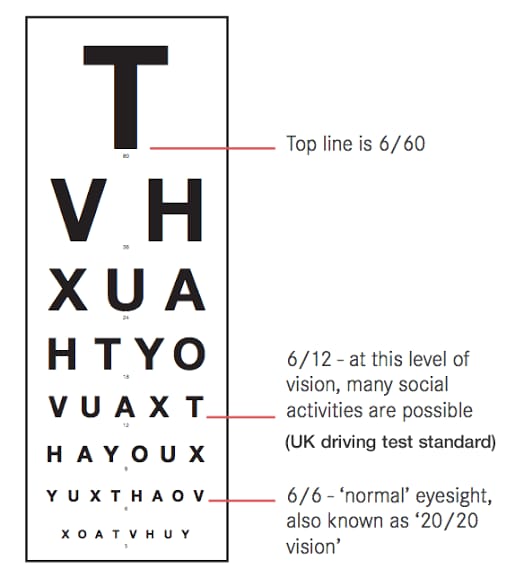
Visual acuity test
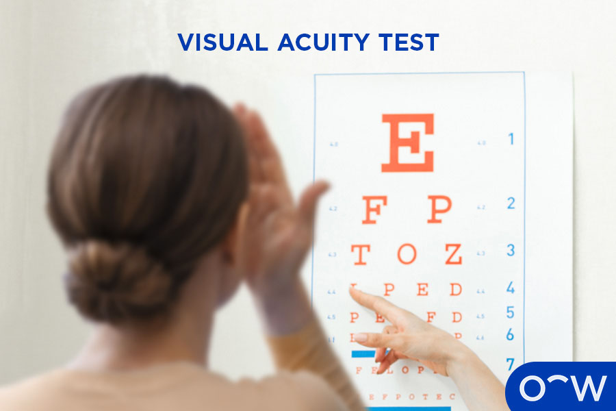
- Visual acuity test is a diagnostic test used to check the sharpness of vision. Method. For which the Snellen chart method is mainly used. In this method, the person is shown a row of words and symbols of different sizes in the Snellen chart as shown below from a distance of 20 feet (6 meter) and the person’s visual acuity is checked. During this test, one eye of the person is covered. During which row, when the person stops reading, the number written in that row and which eye is it is noted. Then this procedure is done in the other eye and its visual acuity is checked. 20/20 or 6/6 is considered normal vision.
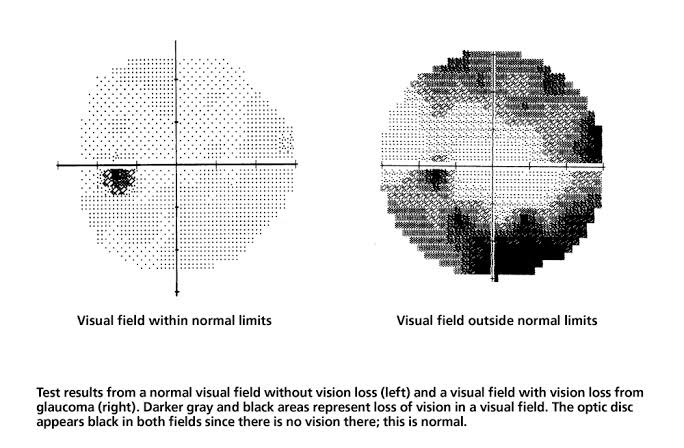
Visual field test
- Visual field test, also known as perimetry, is a diagnostic test used to check peripheral vision. During this test, the patient is placed in front of a machine and asked to focus on a target light. In addition, a light or other stimulus is placed in different areas of his visual field and the patient’s response to this stimulus is checked and the boundaries of the visual field are determined and areas of visual loss are detected. Visual field tests are used to detect blind spots and evaluate glaucoma, optic nerve damage, and neurological conditions that affect vision.
Refraction test
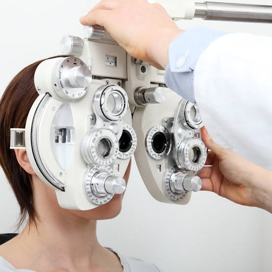
- A refraction test is a standard procedure used to determine a person’s eyeglass prescription, meaning it helps determine the number of lenses needed by people with vision problems. In this test, the doctor asks them to take readings from a chart using different lenses and identifies the lens for accurate vision. Refractive errors such as myopia, hypermetropia, presbyopia, and astigmatism can be detected with the help of a refraction test.
Gonioscopy
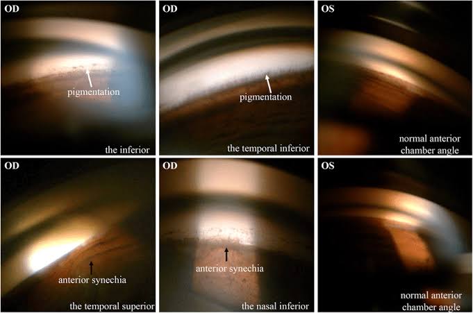
- Gonioscopy is a diagnostic method used to examine the angle formed between the iris and the cornea. This angle is located where the aqueous humor drains from the eye. In a gonioscopy test, numbing drops are instilled into the eye, after which a special lens called a gonioscope is placed in the eye. With the help of which the internal structures of the eye, especially the angle between the iris and the cornea, are examined. Which can be used to determine whether the angle is open, closed or narrow. Gonioscopy is used to examine the eye angle in conditions such as glaucoma.
Ultrasound biomicroscopy
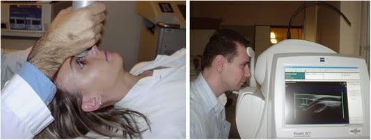
- Ultrasound biomicroscopy is a non-invasive imaging technique that uses high-frequency ultrasound waves to examine eye structures, providing detailed images of the anterior part of the eye. Ultrasound biomicroscopy is used to detect glaucoma, cataracts, tumors, and anterior segment abnormalities.
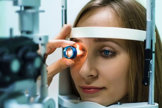
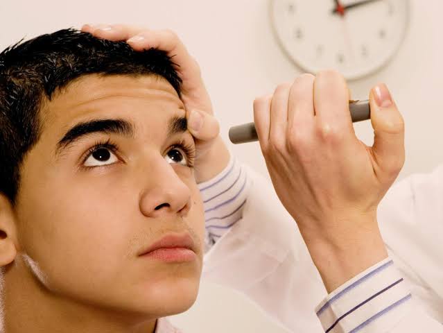
Slit lamp examination
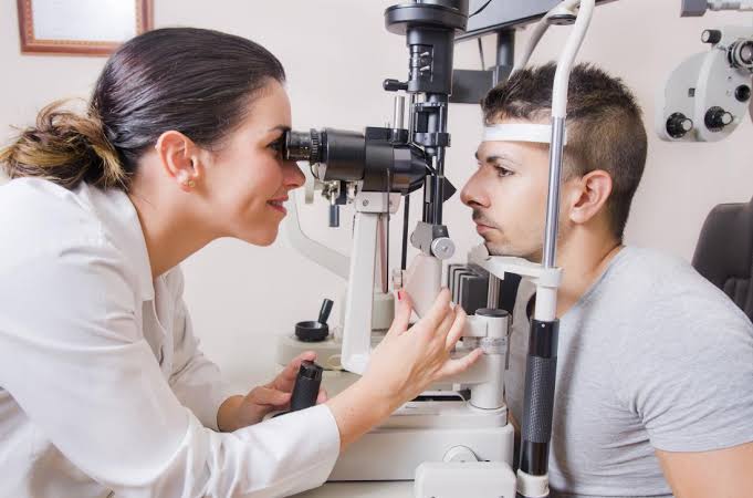
- A slit lamp examination is a common procedure used by eye care professionals to visualize eye structures. Slit lamp examination involves examining the eye structures in detail using a specialized microscope called a slit lamp. This slit lamp produces a thin focused light beam and uses it to examine the iris, cornea, retina, and lens. Slit lamp examination provides 10-25 times magnification.
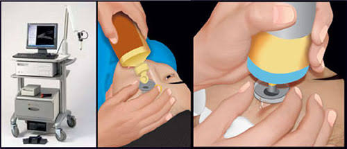
Tonometry
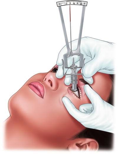
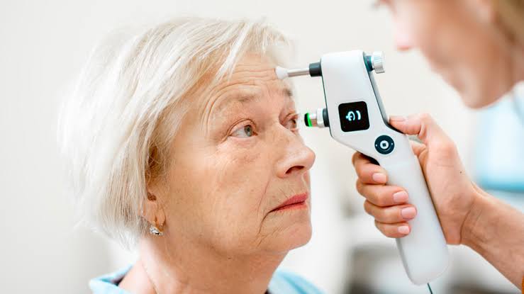
- Tonometry is a diagnostic test. It is used to measure the pressure in the eye, which is called intraocular pressure. In tonometry, intraocular pressure is measured with the help of a tonometer instrument. In tonometry, topical anesthetic eye drops are instilled in the lower conjunctival sac. Then, with the help of a tonometer instrument, gentle pressure is applied to the eye and a puff or air is sent on its surface and the intraocular pressure is measured. This pressure is measured in millimeters of mercury (mmHg). Tonometry is used to measure intraocular pressure in glaucoma and other eye conditions.
Pupillary dilation test
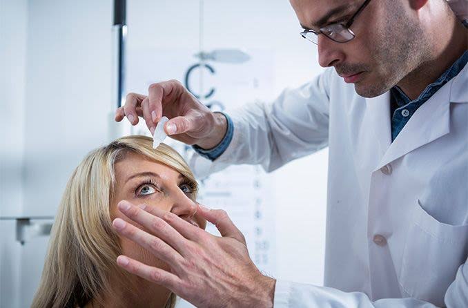
- This is a simple test in which special drops are instilled into the eye that dilate the pupil. The pupil dilates, allowing the retina to be examined systematically.
Electroretinography
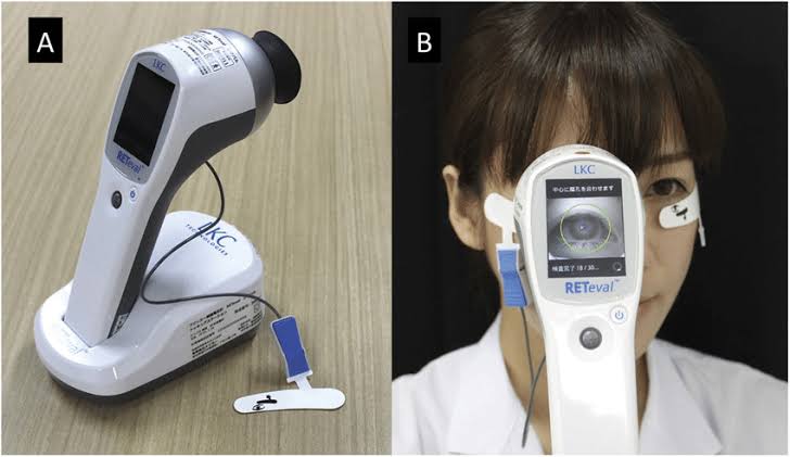
- Electroretinography is a type of diagnostic test that measures the response of the retina to light. Electroretinography measures the electrical response of different types of cells in the retina, such as photoreceptor cells, inner retinal cells, and ganglion cells. In electroretinography, electrodes are placed on the cornea or the skin around the eye and the electrical response generated by retinal cells when stimulated by light is checked. The patient’s eyes are then dilated and asked to focus on the light source, and the electrical signal is recorded and analyzed. Electroretinography is used to evaluate retinal function and diagnose various retinal disorders such as keratitis pigmentosa, macular degeneration, and diabetic retinopathy.
Colour fundus photography
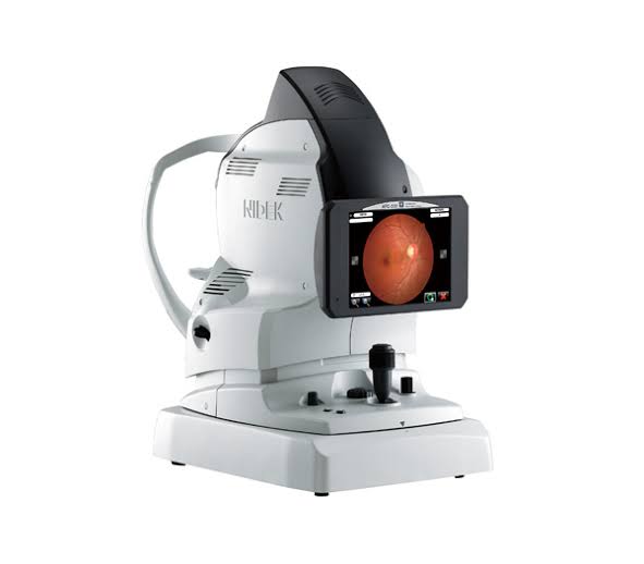
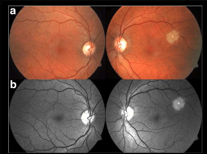
- Color Fundus Photography is a non-invasive diagnostic method that provides detailed images of the back part of the eye. Specifically, the retina, optic disc, macula, blood vessels. In color fundus photography, the patient’s eye is dilated, then a specialized camera with a lens and filter is placed on the patient’s front side and the camera is focused on the back part of the eye and photos are clicked from different angles and those photos are analyzed. Color fundus photography is used for early detection of retinal diseases. Such as diabetic retinopathy, retinal detachment, age related macular degeneration
Fluorescein angiography
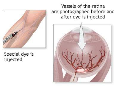
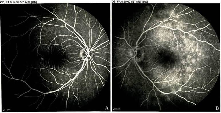
- Fluorescein angiography is an invasive imaging study. It is used to check the status of blood vessels in the retina and choroid. In fluorescein angiography, a small amount of fluorescein dye is injected into a vein, usually in the arm, which reaches the eye through the blood circulation. Then, a specialized camera is used to take pictures of the area and observe it. Fluorescein angiography can be used to detect conditions such as retinal vascular disease, macular degeneration, and diabetic retinopathy.
Corneal topography
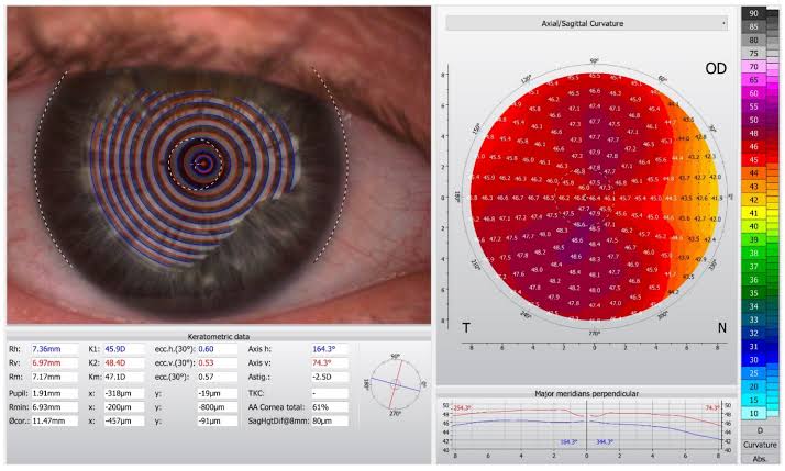
- Corneal topography is a non-invasive diagnostic method in which a map of the cornea is prepared, with the help of which information about the shape, curvature and elevation of the cornea is obtained.
- Corneal Topography is used to identify astigmatism, keratoconus, and irregular corneal shape. This method is also used before refractive surgery, before fitting contact lenses, and in corneal transplants.

Published
Categorized as Uncategorised