ENGLISH-Ent-part-4-NOSE AND THROAT
a)👃 Review of Anatomy and physiology of nose
👃 Anatomy of the Nose-(Anatomy of nose)
1. External Nose (outside of the nose)
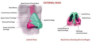
Structure:
- The External Nose is made up of Bones (nasal bones) and Cartilage (septal and alar cartilages). Which works to give shape and structure to the nose.
Parts:
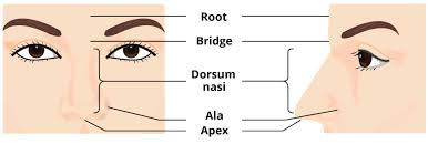
- Root: This is the upper part of the nose that meets the forehead.
- Dorsum: The Bridge of the nose. Which is the middle portion between the root of the nose (the part connected to the head) and the apex of the nose (Apex).
- Apex: The Tip of the nose (the part of the nose)
- Nostrils: These are the openings through which air enters.
Functions:
- Shapes the face.
- Facilitates airflow into the nasal cavity.
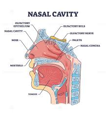
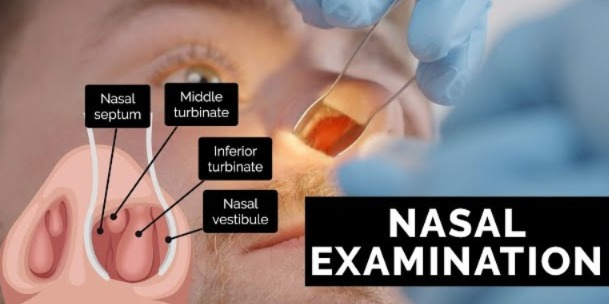
2. Nasal Cavity (Nasal Cavity)
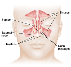
- Nasal Septum It divides the nose into two chambers.
- It is lined with Mucous membrane which works to filter and moisten the air.
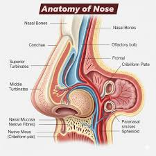
- Turbinates (conchae): Superior, middle, and inferior turbinates that increase the surface area for air filtration and humidification.
3. Paranasal Sinuses (Paranasal sinuses)
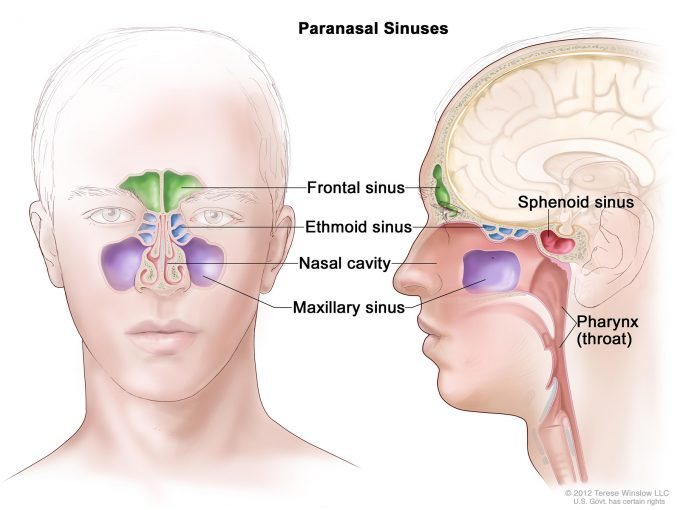
These are air-filled cavities that are located around the Nasal Cavity. They are as follows:
- Frontal
- Maxillary
- Ethmoidal
- Sphenoidal
Functions :
- Lighten the skull.
- Humidify and warm the air.
- Add resonance to the voice.
4. Blood Supply
-
- Richly supplied with blood by branches of the internal and external carotid arteries.
-
- Key Arteries include the sphenopalatine artery and anterior ethmoidal artery.
5. Innervation
-
- Sensory nerves: Supplied by the trigeminal nerve (cranial nerve V).
-
- Olfactory nerves: Responsible for the sense of smell.
Physiology of the Nose
1. Respiration
- The nose performs the primary function of filtering, warming, and humidifying the air before it reaches the lungs.
2. Olfaction (Sense of Smell)
- The olfactory region in the upper nasal cavity contains specialized receptor cells that detect odors and transmit signals to the brain through the olfactory nerve (cranial nerve I).
3. Filtration and Protection
- Mucociliary mechanism:
- Mucus traps dust, allergens, and pathogens.
- These trapped particles are moved by cilia towards the pharynx for swallowing or expulsion (sneeze or cough).
- It contains immune components, such as Lysozymes and Immunoglobulins, that fight infection
4. Speech Resonance
- The paranasal sinuses play an important role in the resonance and tone of the voice.
5. Airway Reflexes
- Sneeze reflex protects the nasal passage by expelling irritants.
Assessment of the Functions of Nose :-
👃 History taking and physical examination Doing physical examination ):=
1.Present Complaints History – (Present Complaint History)
- Ask whether respiratory tract infection (URTI or RTI) is frequent or not.
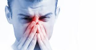
- Ask whether nose bleeding, sinus infection, hay fever, rhinitis, sneezing, pain radiating to the face or ear occurs or not.
- Airway obstruction due to polyps (nasal warts) and engorged mucous membrane.
- Loss of Sense Of Smell
- Snoring
- Itching
Past health history
Allergies:
-
- History of allergic rhinitis or sinus allergies.
Medication (Medication):-
-
- Patient has any type of nasal Ask if the person is taking nasal sprays, antihistamines, decongestants, over-the-counter drugs, herbal or allergic medicines.
Infections:
-
- Frequent sinusitis or upper respiratory tract infections.
Surgeries:
-
- Any nasal surgeries such as Septoplasty or sinus surgery.
Chronic conditions:
-
- Such as Asthma and other Respiratory Disorders.
Environmental and Lifestyle Factors
Ask about any situations in the environment that may aggravate the symptoms, such as
-
- High Humidity, Change in Weather, Seasonal Allergy, Change in Temperature ,such as going from air conditioning to hot weather, pollens, fumes, smoke, as well as dust particles Occupational hazards and chemicals or strong odors.etc. Take a history about them.
-
- Ask about foul taste in mouth and facial pain.
Examination of nose
- A full nose examination is performed to assess the function of the nose, airway resistance, sense of smell, as well as the mouth and pharynx. Common symptoms of nasal disease include:
- Airway obstruction,
- Rhinorrhea (runny nose),
- Sneezing,
- Loss of smell,
- Facial pain caused by sinusitis,
- Snoring associated with nasal obstruction.
inspection of 👃Nose
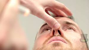
- First of all, inspect the external nose,
- Look at the nose from the front to the side for the following symptoms
- size and shape,
- obvious bend or deformity : a deviated nose is often best looked at from above,
- Swelling,
- scar or abnormal creases,
- Discharge or crusting,
- offensive smell,
Sinus assessment
- Palpation of the frontal and maxillary sinuses should be performed to detect any tenderness or To see if there is swelling or not.
Diagnostic test
- BLOOD TEST
- Full blood count and esr,
- electrolytes,
- growth hormone level,
- Allergy test,
- Wegener’s granulomatosis,
- syphilis serology, if indicated,
- coagulation profile,
- raised immunoglobulin levels and presence of certain autoantibodies.
Radiological investigation.
- Sinus X-ray,
- CT Scan of Nasal Cavity,
- Nasal Endoscopy,
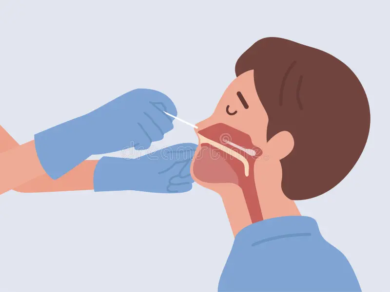
- Muzzle Smear and Culture for Bacteria and Fungi,
- Biopsy of 👃 Nose,
- skin smear from the skin or nasal lining,
- skin prick allergy testing,
- nasopharyngoscopy,
- Schirmer tear test
- If this test is a dry eye, it is done to assess
Disease and disorder of 👃 Nose
INTRODUCTION
Common Cold :
- Common cold (viral upper respiratory track infection, acute viral nasopharyngitis, acute viral rhinopharyngitis or acute coryza) is an acute viral contagious infection that Which occurs in the upper respiratory track and is caused by rhinoviruses, picoviruses or coronaviruses.
- A common cold lasts an average of one week.
- A mild cold lasts for two to three (2 to 3 days).
- A severe cold lasts for two weeks.
Transmission of infection
- The common cold is spread by direct hand-to-hand contact with infected secretions or from a contaminated surface. spread.
For example:=
- For example, if a person has a common cold and touches his nose and then touches something else, the common cold virus that is on that surface can be transferred to another person who touches that surface.
- In most cases, the common cold virus can survive on surfaces such as pens, books, telephones, computer keyboards, and coffee cups. The common cold virus can survive on these surfaces for a few hours.
clinical manifestation
- Clinical manifestation wp:list-item –>
- Nasal congestion occurs.
- sore or scratchy throat.
- sneezing 🤕.
- hoarseness.
- muscle weakness.
- mild fever.
- Uncontrollable shivering.
- Loss of appetite.
- rarely extreme 😩
- exhaustion.
- fatigue.
treatment
- There is no best treatment for common cold.
- Since common cold is mainly caused by viral infection and no type of antibiotic is used in it.
- Home treatment is mainly used to reduce the symptoms of common cold, that is, there is no specific treatment for common cold.
- Proper rest and drinking sufficient water are used as supportive measures in common cold.
- Over-the-counter medicines such as throat lozenges, throat spray, cough syrup can help relieve symptoms.
- Decongestants such as pseudoephedrine or antihistamines Used to relieve nasal symptoms.
- Saline spray or humidifiers are also beneficial.
- Acetaminophen (acetaminophen) or ibuprofen
- ( ibruprofen) is a sore It is used for sore throat, mild fever or body ache.
- Aspirin or aspirin group medicines should not be used in children and
- teenagers because it can cause Reye’s Syndrome-like conditions may occur.
(Reye’s syndrome :=
- A rare but serious condition in which confusion,Brain Swelling 🧠and liver damage
- Such There are other steps that can help relieve nasal congestion, such as:
- Instilling salt water drops into the nostrils can help relieve nasal congestion.
- A cool mist humidifier increases air moisture.
- Applying petroleum jelly to the skin of the nose can help soothe the rawness.
- Hard candy or cough drops can help soothe sore and irritated nasal passages. Helps relieve sore throat. (For children over three years of age.)
- A warm bath or heating pad is used to relieve pain.
- Inhaling steam from hot water also helps relieve nasal congestion.
prevention
- Prevention is more important than curing the common cold.
- The common cold virus can be transmitted from one person to another up to 12 feet, so it is very important to take care of children to prevent it.
- To prevent the common cold, keep children away from people who have the common cold for two to three days.
- Not washing children’s hands frequently and properly.
- Antibacterial or alcohol-based Hand sanitiser is used to prevent the transmission of any respiratory illness.
- A person who has a respiratory tract infection should cover their nose and mouth.
- If any family member has an infection, i.e. a common cold, keep their towels and all their utensils separate and use disposable items so that the infection cannot be transmitted from one person to another.
- Lifestyle modifications such as avoiding smoking Doing this, stress management reduces the chances of getting the common cold
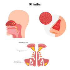
Rhinitis
introduction
- Rhinitis is inflammation and swelling of the mucous membrane of the nose. And it causes runny nose and stiffness of the nose.
- Rhinitis is mainly caused by the common cold or allergies.
classification
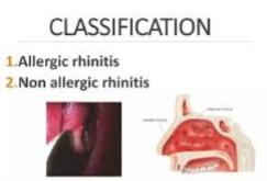
1) Allergic Rhinitis ( Allergic Rhinitis), 2) Non Allergic Rhinitis 🤧 (Non Allergic Rhinitis)
1) Allergic Rhinitis 🤧:=
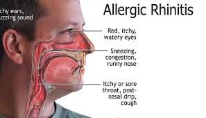
- Allergic rhinitis is caused by seasonal changes and involves inflammation of the mucous membrane of the nose.
- Symptoms:=
- runny nose,
- Irritate eye,
- itching of nose and eye,
- These symptoms are caused by coming into contact with any dust, dander or certain types of seasonal pollen that people are allergic to.
2) Non Allergic Rhinitis:=
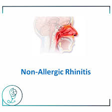
- Non-allergic rhinitis is caused by bacteria and viruses.
- Occupation ( occupation),
- Hormonal rhinitis ( Hormonal Rhinitis),
- drage induced,
- Gustenary Rhinitis occurs after contact with any heat or spicy food or alcohol.
risk factore
- Viruses such as rhinovirus, adenovirus, or entero virus.
- Environmental factors,
- Health changes,
- Infections,
- Food and beverages,
- Certain types of medications,
- Health problems,
- Animals,
- Any other disease condition,
- Weak immune system.
clinical manifestation
- Dry eyes, nose and soft palate,
- Fever and chills,
- Headache,
- Swelling in the mucous membrane of the nose makes it difficult to breathe through the nose.,
- A watery discharge comes out of the nose,
- 🤕 sneezing ,
- tears fall from the eyes,
- nasal irritation,
- restlessness,
- discomfort.
management
- Provide adequate bed rest to the patient.
- Instruct the patient to drink adequate amounts of water,
- Provide steam
- Provide inhalation.
- Provide antihistamines and decongestants to the patient to prevent sneezing and tearing from the eyes.
- Advise the patient to take antihistamines before driving or operating machinery
- Do not take antihistamines as they can cause excessive drowsiness.
- Advise the patient not to rub the nose excessively.
- If there is nasal obstruction, ask them to insert nasal drops every four hours to remove nasal decongestants For.
- Teach the patient how to insert nasal drops.
- If nasal discharge continues for 7 to 10 days and the temperature increases, seek medical advice.
- If the patient has a continuous common cold, seek medical advice as it may be a sign of a nose May also be due to deformity.
Allergic Rhinitis 🤧 introduction
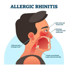
- Allergic rhinitis can be acute or seasonal.
- And it can be caused mainly by pollen and flower pollen, while it can be chronic when a person comes into contact with dust, animal dander, wool or certain types of food and is allergic to it. Can happen.
Etiology
- seasonal allergy 🤧,
- pollens,
- outdoor molds,
- trees,
- grass,
- ragweed pollens,
- occupational Allergic Rhinitis,
- in occupation exposure to allergen in workplace,
- dust mites,
- cockroaches,
- molds
- animal danger,
clinical manifestation
- Sneezing,
- Nasal obstruction,
- Watery discharge from the nose.
- Headache.
- Itchy eyes and nose Coming.
- Nasal congestion.
- Generalized fatigue,
- Dark circles around the eyes,
- Sore throat,
- Reduced sense of smell,
- Mental changes,
- slower thinking.
Management
- Keep the client away from the allergen.
- Provide the patient with antihistamine medicine.
- Avoid allergens.
- Cover pillows and mattresses with plastic covers.
- Animal products such as dust and pollen To separate.
- If you have mild allergic rhinitis, do nasal wash.
- Nasal wash
- It helps to remove mucous from the nose.
- Keep your head down in a Gandi So that the mucous can be removed properly.
- Some kind of solution or take it in your hand and then inhale it through the nose so that the mucus can come out of the nose properly.
- Immunotherapy.
Pharmacological therapy
- 1. Antihistamine ( antihistamine),
- 2) nasal corticosteroids ( nasal corticosteroids),
- 3)oral decongestant ( oral decongestant),
- 4)intranasal cromolyns ( intranasal cromolyns),
- 5) intranasal anticholinergic agent ( intranasal anticholinergic agent).
Sinusitis (Sinusitis)
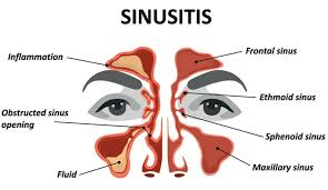
- introduction
- Sinus is an air-filled space located within the bones of the face and nose. It is called sinus. Infection and inflammation in it is called sinusitis.
- Sinusitis may or may not be caused by infection, bacteria, fungi, viruses, allergies, or autoimmune diseases.
Etiology
- bacteria :=
- Streptococcus pneumoniae ( Streptococcus pneumoniae),
- staphylococcus aureus ( Staphylococcus aureus),
- Haemophillus influenzae ( Haemophilus influenzae),
- morexella catarrhalis ( Moraxella catarrhalis ),
- viral,
- Repeated contact with allergens such as dust, mites, molds, cockroaches,
- It causes infection and inflammation of the nose or paranasal sinuses, i.e. sinusitis.
Risk factore
- Due to changes in the environment,
- Due to excessive use of decongestant sprays,
- Smoking,
- Due to excessive swimming,
- Deviated nasal septum Due to.
- Adenoids,
- Infected tonsils,
- Dental infection,
- small sinus ostia,
- concha bullosa ( Concha Bullosa),
- Nasal polyps,
- Cystic fibrosis.
type
1) Acute sinusitis:= If the infection lasts for less than four weeks, it is called Acute sinusitis.
2) Chronic sinusitis:= If the infection and inflammation last for more than three months, it is called Chronic sinusitis.
1) Acute sinusitis:= Acute sinusitis is usually less than four weeks in duration.
It is mainly caused by viruses or bacteria.
Etiology
- bacteria,
- viral,
- blocked sinus,
- deviated nasal septum ,
- allergens.
clinical manifestation
- 1) maxillary sinusitis:=
pain, pressure at the maxillary region (cheek, jaw, gums, and teeth),
- 2) frontal sinus:=
Pain and pressure in the area,
headache,
pain is worse in the morning and gradually decreases.
- 3) ethmoid bone:=
In ethmoid sinusitis pain and pressure occurs in the back of the eye
- 4) Sphenoid sinusitis:=
This causes pain behind the eyes and mainly in the occiput area.
Others symptoms: =
- Feeling tired,
- Weakness,
- Sense of smell decreases,
- Excessive phlegm is produced at night,
- Tenderness is felt when pressure is applied,
- sore throat,
- bad breath,
- fever,
- Discharge comes out of the nose,
- Swelling of the eye lid,
- Bed breath,
- Headache,
- Pain behind the eye,
- Toothache and tenderness in the face.
- Natural stiffness and nasal discharge,
Diagnostic evaluation
- History taking and physical examination
- Looking at the nose to see if there are any polyps,
- Inflammation of Nose,
- Taping over a sinus to find infection,
- X Ray,
- endoscopy,
imagine test like :=
- X Ray,
- Ct scan,
- MRI,
- allergy testing,
- blood test,
- ciliary function test,
- nasal culture,
- nasal cytology,
- sweat chloride test for cystic fibrosis.
management
- antibiotics,
- antihistamine,
- steam inhalation,
- nasal decongestant.
- self care:=
- warm Apply a washcloth to the face for a few days.
- Drink plenty of water to prevent dehydration.
- Take steam inhalation two to four times throughout the day.
- Use nasal saline spray.
- Use a humidifier.
- When over-the-counter drugs or nasal sprays or decongestants are not Do not overuse as it causes nasal stiffness and this leads to pressure and pressure.
complication
- Abscess,
- Bone infection,
- Meningitis,
- Skin infection around the eyes.
prevention
- Eat more fruits and vegetables.
- Fruits and vegetables contain antioxidant agents that boost the body’s immune system, which can fight infections.
- Provide an influenza vaccine every year To do.
- Take less trace.
- Wash your hands properly.
- Reduce smoke and pollution.
- Drink plenty of water so that the water provides moisture to the body.
- Use decongestants when there is an infection of the upper respiratory tract.
- If there is an allergic condition, it should be treated immediately.
Chronic Sinusitis (Chronic Sinusitis):

introduction
- Chronic sinusitis causes infection and inflammation in the mucous membrane surrounding the nasal passages, causing them to swell. is.
- In chronic sinusitis, the membrane of both paranasal sinuses becomes thick because of constant infection and inflammation, which can last up to eight (8) weeks if the person is an adult and up to two (2) weeks if the person is a child.
- If acute sinusitis is not cured properly, it results in chronic sinusitis.
- If chronic sinusitis is present, the nose The mucus becomes quite thick and due to this the person may experience: Cannot breathe through the nose.
- Chronic sinusitis causes swelling of the area around the eyes and face, causing conditions like facial pain and headache.
Etiology
- nasal polyps,
- nasal Tumor,
- Allergic conditions,
- Deviated nasal Septum,
- Trauma to the face,
- Due to any other medical condition,
- Respiratory tract infection,
- Any type of allergy,
- Immune system down.
clinical manifestation
- Nasal obstruction,
- Nasal congestion,
- Difficulty breathing through the nose,
- Drainage becomes very thick,
- Pain, tenderness, and swelling in the area around the eyes, and all these symptoms are also seen on the cheeks, nose, and forehead.
- Loss of sense of smell and taste,
- sore throat,
- dryness of throat,
- coughing,
- genital malaise,
- difficulty in assessing smell,
- dental infection,
- vertigo,
- lightheadness ,
- Looking pale,
- Earache,
- Teeth and jaw pain,
- Coughing a lot at night,
- Bad breath,
- Fatigue or irritability,
- nausea,
complication
- asthma flare up, (asthma)
- meningitis, ( Menin Jaitis)
- vision problems, ( Vision Problem)
- aneurysm or blood clot (aneurysm or blood clot).
Diagnostic evaluation
- history taking and physical examination,
- Nasal endoscopy,
- Imaging studies,
- Nasal and sinus culture,
- An allergy test.
treatment and drugs the goal of treating Chronic sinusitis is:=
- To reduce inflammation.
- Sinusitis To reduce inflammation.
- To ensure proper drainage of the nasal passages.
- To improve breathing.
medical management
- antibiotics,
- saline nasal sprays,
- nasal corticosteroids,
- oral or injected corticosteroids,
- decongestant,
- over the counter drug.
- antihistamine drug,
- angelic medicine,
- steam inhalation.
Surgical management
1) Antrum puncture (Antrum Puncture):=
- In this procedure, a trocar and cannula are placed under the inferior turbinate, about half an inch below the turbinate.
- After incising the nasoantral wall, the trochar is inserted into the sinus cavity. The trochar is then removed and the cannula is placed properly into the sinus cavity.
- This procedure involves inserting a cannula into the sinus cavity. It is done to irrigate and their normal saline is used.
2) Intranasal antrostomy (Intranasal enterostomy):=
- This is a drainage operation.
- Which is performed to create a permanent window near the floor of the antrum to facilitate drainage of secretions from the maxillary sinus.
3) Caldwell luc radical antrostomy :=
- In this, an incision is made in the upper gum.
- Which opens in the front wall of the intertum.
- This entire affected The maxillary sinus is removed and a large window is made next to it, creating a window in the lower and middle space.
- Which allows for proper drainage.
4) Balloon sinoplasty:=
- This method is similar to balloon angioplasty in that it dilates the arteries of the heart. Used to unclog.
Nursing management
- Tell the patient to relax a lot.
- When the line is down, the sinuses expand and due to this, proper breathing can be done.
- And by placing a small pillow under the head, good breathing can be done.
- The nurse should provide education to the patient about steam inhalation.
- To drink sufficient water Say.
- Ask the patient to apply a heat pack.
- Make a warm cloth and then place it over the nose and do a heat inhalation. Doing this will thin the sinuses
- The mucus in the cavity will be thinned and the sinuses will have a place to open.
- Tell the patient not to bend over too much and not to lift very heavy objects.
- When over-the-counter drugs Consult a doctor before using decongestants as they can also cause harm to the body.
- Nasal decongestants should not be used for more than three days as they will cause swelling of the nasal sinuses and cause congestion. symptoms will be much worse.
prevention
- Keep the nasal cavity moist as much as possible.
- Take care not to let an upper respiratory infection develop.
- Stay away from people who have the common cold as much as possible.
- Wash your hands frequently with soap and water before eating and before doing any procedures.
- Use a humidifier if necessary. Use.
- If your room has dry air, add a muster to prevent sinusitis.
- The outside environment should not be too dry.
- Keep in mind that any allergens such as mold, house dust, mites, cockroaches, such an environment creates a problem.
- Things that irritate like cigarettes or any cigarette smoke can also cause problems. smokers, stay away from chemicals if you have an order.
- Tobacco smokers and irritants The material damages the lungs, so it is important to stay away from it and alcohol, which conditions and worsens this sinusitis, should also be avoided.
- If you are allergic to any kind of thing, then you should stay away from it.
- You should not swim in the swimming pool for too long.
- And you should stay away from people who smoke cigarettes.
- Using minimally invasive methods, a balloon is used to expand the sinus cavity. It has arrived.
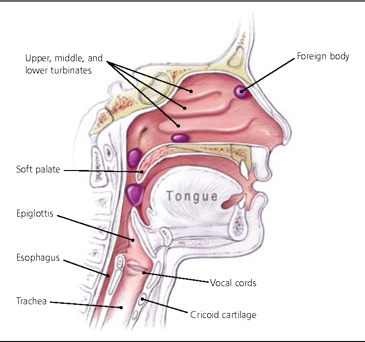
forien body (foreign body)
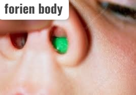
Introduction
- The nose becomes accidentally blocked by a substance that is not normally found there.
- These The object or material is called a foreign body.
- Some objects of concern such as beads are inserted into the nose and get stuck there.
- This is mainly seen in children.
- It is very important to remove it to prevent the problem.
- This mainly happens in young children who put small objects such as pebbles and marbles in their Can be put in the nose.
- Smooth objects get stuck in the nose and rough objects damage the nasal tissue.
- It is difficult to remove objects inside the nose.
causes/ Etiology
- Any small object should be removed immediately Put it in your nose like a bean.
- having the Airway packed in healthcare settings.
- playing or rough housing.
- Children, especially boys, are at higher risk of putting small objects in their noses.
clinical manifestation
- Discharge from the nose is.
- bloody nose,
- When the patient breathes, a whistling sound is heard.
- There is irritation in the nose.
- There is swelling.
- There is sneezing.
- There is difficulty in breathing.
- The discharge from the nose is mainly blood. It is.
Diagnosis
- History tacking and physical examination.
- A foreign body in the nose is usually diagnosed when the child has foul-smelling drainage from one of the nostrils. is.
- Some instruments with specific lights help in visualizing the nose.
treatment
- Encourage the patient to breathe slowly through the mouth. If the patient breathes too suddenly, there is a possibility of small objects being immediately pushed deep into the nose.
- Slowly close one nostril and gently move that nostril to see if any foreign object is present in that nostril.
- Avoid blowing the nose excessively and do not move it repeatedly.
- If the above method fails, seek medical advice Take.
- Do not try to remove any objects using tweezers.
- Even if it appears deep in the nose.
- Do not try to remove objects that cannot be easily grasped or seen.
- Follow the steps below Do not attempt to remove the foreign body.
- Do not grasp any foreign object with tweezers that Harms the nose.
- Do not push any foreign object with cotton or other objects so that any foreign object flows further into the nose.
- Press or move the nose.
- Ask the patient to breathe through the mouth.
- Calm the patient and provide him with reassurance. –>
- Take the patient to the hospital immediately if possible so that he can receive medical treatment and prevent any other complications.
- The aim of treatment is to remove any foreign body so that no situation arises.
- If first aid If that fails, consult a health provider.
- And this is mainly done in the operation room by providing general anesthesia.
- If any infection has occurred, it is treated by providing antibiotic medicine.
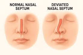
Deviated nasal septum
introduction
- Septum which is mainly thin and straight and is located between the two nostrils.
- And if this septum deviates from its place and flows inward into the nostril or becomes to one side, it is called deviated nasal septum.
- Deviated nasal septum It is a common physical disorder of the nose and it involves displacement of the nasal septum.
- The septum mainly separates the right and left nasal cavities and is mainly located in the middle and divides them equally into two parts.
- A deviated nasal septum mainly causes nasal obstruction and this can mainly occur in infections or allergic reactions.
- When obstruction is seen, the nose Difficulty in breathing occurs.
- This is mainly seen in children and adults and is mainly congenital and also due to any injury.
cuses
- trauma,
- birth defect,
- congenital disorder,
- compression of the nose,
- automobile accident,
Type Of Deviated Nasal Septum
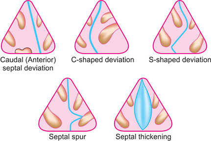
1) colluminar deviation or anterior dislocation (Column deviation or anterior dislocation.)
- In this, the septum is displaced from the front to the bottom and it goes to one side and it blocks the nostril completely or partially.
2) c – shaped deviation (C shape deviation).
- In this, the septum is deviated on one side and its shape is like the English letter ” c”.
3) s – shape deviation (S sep deviation)
- In this, the septum is deviated ( s – shape deviation) on both sides or above and below like the English letter S.
4) bony spurs (Bonny spurs)
- In this, the vomer bone of the nose is sharply deviated to one side of the nostril and forms a bony projection and it also causes nasal obstruction.
5) thickened septam (Thickened septum)
- If the nasal septum is normal, it is thin.
- But due to any infection or trauma, there is a connection of blood and Due to hematoma, the septum thickens and causes difficulty in breathing.
clinical manifestation
- If the patient has a deviated nasal septum in a minor amount, then there are no symptoms.
- If the deviated nasal septum is severe, then the following types of signs and symptoms are seen.
- Obstruction is seen in one or both nostrils.
- Nasal congestion occurs.
- Blood comes out of the nose.
- Frequent sinus infections occur.
- A whistling sound is heard while breathing while sleeping.
- The mucosa gets dry.
medical management
1)Decongestant.
2)antihistamine.
3)nasal cortisone spray.
surgical management
- septoplasty,
- submucous resection.
- rhinoplasty.
management
- If the deformity causes nasal obstruction, sub mucosal resection is performed and is done under general local anesthesia.
- If a large part of the septum is resected, plastic surgery becomes necessary.
- If bleeding occurs, packing is placed is.
- After the operation, the person is sent back to his home immediately, meaning the hospital stay is not long.
- Provide cold compression to the patient.
- Dress the wound properly.
- Tell the patient to use humidified fire.
- Perform saline irrigation.
- Tell the patient not to blow their nose too much.
- Teach the patient relaxation and deep breathing techniques Teaching.
- Ask the patient to do some daily routine activities.
fracture of nasal bone
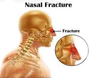
introduction
- nasal A bone fracture is a It is the most common bone fracture of the body.
- Fracture of the nasal bone can occur due to any trauma or assault.
- Nasal bone fracture can cause nasal deformity and due to it The condition of nasal obstruction arises.
clinical manifestation
- Bleeding from the nose,
- Bleeding from inside or outside the nose,
- Swelling in the nose,
- Be hurt.
Diagnostic evaluation
- History taking and physical examination
- inspection,
- palpation,
- X Ray,
medical management
- nasal Cold compression is provided to remove the build-up.
- The nasal cavity is assessed for swelling or any deformity.
- If there is any injury to the nasal cavity, immediately consult a doctor and nasal fractures are usually surgically reduced within 7 to 10 days.
Nursing management
- Apply ice packs to the nose for 20 minutes four times a day to reduce swelling.
- If there is bleeding from the nose, apply nasal packing and instruct the patient to breathe through the mouth if they cannot breathe through the nose.
- If breathing through the mouth, it can dry out the oral cavity mucus membrane or tell the patient to rinse the mouth frequently to keep the mouth moist. Mucous membrane should be moistened
Epistaxis
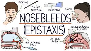
Introduction
- Epistaxis is also called Nose Bleed.
- This is a condition in which there is active bleeding from the nose. It occurs from the nostril, nasal cavity, or nasopharynx.
- This occurs when the blood vessels inside the nose are damaged or injured.
- Bleeding from the front or back of the nose is called a nose bleed or epistaxis.
Type
1) anterior nose bleed (Anterior Nose Bleed), 2) posterior nose bleed (Posterior Nose Bleed)
1) anterior nose bleed (Anterior Nose Bleed):
- Anterior Nose bleeds occur in 90% of people.
- Anterior nose bleeds occur from the front of the nose and the lower part of the nose.
- And the most common side of nose bleed is the nasal septum.
2) posterior nose bleed (posterior nose bleed) Bleed):
- Posterior nose bleeds are less common than anterior nose bleeds.
- In this, the bleeding mainly occurs from the artery at the back of the nose.
- In this posterior nose bleed, the blood mainly comes to the throat.
Etiology/ cause
1) Local Causes:
- Bleeding occurs due to excessive shaking of the nose or nail biting in the nose, which is one of the most common causes of nose bleed.
- Trauma to the nose or face.
- Falls or road traffic accidents can cause nose injuries, which can lead to conditions like epistaxis.
- Fracture of the Nasal Bone (Fracture) .
- Iatrogenic Cause.
2) Infection:
- Epistaxis can occur due to upper respiratory infection in children.
- Viral Rhinitis.
- Acute Rhinitis.
Other Causes:
- Due to any foreign body in the nose.
- Nasal septum Due to deviation (Nasal Septum Deviation).
- Due to changes in the environment.
- Due to any growth in the nasal cavity, such as polyps or benign and malignant tumors.
General cause:=
- Causes of Epistaxis:
Idiopathic:
- Bleeding of the nose without any apparent cause.
Systemic Causes:
- Hypertension.
- Heart Disease.
- Pregnancy.
- Bleeding Disorder.
- Drugs such as Oral Anticoagulant.
- Vitamin K Deficiency.
Infections:
- Acute Infections such as Typhoid, Pneumonia, Malaria.
- Dengue Fever.
- Measles.
Local Causes:
- Violent Sneezing.
- Nose Blowing.
- Injury in the Nose (Trauma to the Nose).
- Picking of the Nose.
Hematological Causes:
- Leukemia.
Symptoms of Epistaxis:
Symptoms related to the nose:
- Blood from the nose Bleeding from the Nose.
- Nasal Blockage.
Respiratory Problems:
- Difficulty in Breathing.
- Difficulty in Swallowing).
Head and body symptoms:
- Headache.
- Dizziness.
- Confusion.
- Fainting.
Physical Weakness:
- Weakness.
- Vomiting.
Medical management
1) Anterior nose bleed:=
Treatment for Epistaxis:
Minor Bleeding:
- If the bleeding is minor, it stops on its own and does not require any treatment.
- A blood clot forms at the site of the nosebleed, which stops the bleeding.
Cauterization:
- If the bleeding is from a blood vessel and is easily visible, the doctor may perform cauterization.
- Cauterization seals the blood vessel, which stops the bleeding.
Nasal Packing:
- If the case is more complicated, then nasal packing should be done.
- Nasal packing creates pressure in the nose, which causes a blood clot and stops bleeding.
- Types of nasal packing: Petrolatum Gauze.
- Balloon Nasal Packs.
- Synthetic Sponge Packs.
Local Application:
- Medical personnel apply medications to the nose, which relieve congestion, reduce pain, and stop bleeding.
- Bleeding can also be stopped by applying vasoconstrictor medications locally.
Sinus Infection Prevention:
- People who undergo nasal packing are at risk of sinus blockage and sinus infection.
- Starting antibiotic treatment.
- Nasal packing is usually left on for 48 to 72 hours.
- A doctor’s advice is very important for these treatment methods.
2)Posterior Nose Bleed:
Severity and Need for Medical Attention:
Posterior Nose Bleed usually does not stop on its own.
This condition is considered very serious and immediate medical treatment is required to stop it.
Treatment with Nasal Packing:
The doctor places a packing in the nose to control the posterior nose bleed.
The most common nasal packing is a balloon nasal pack.
Posterior nasal packing is more uncomfortable than anterior nasal packing.
Medication and Monitoring:
The patient often requires sedative and painkiller medicine.
There is a risk of infection and breathing passage blockage during packing.
The patient needs to be kept under close monitoring.
Duration of Nasal Packing:
Posterior nasal packing is usually kept in the nose for 48 to 72 hours.
Surgical Intervention (Surgical Procedure):
If the bleeding does not stop with nasal packing, then a surgical procedure is required.
It is necessary to provide appropriate treatment along with advice and monitoring from a health care professional.
Nursing management
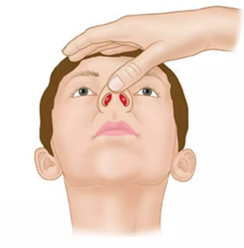
Self-Management of Nose Bleed :
Immediate Steps During a Nose Bleed:
- When the nose bleeds, lean forward slightly. This can prevent blood from flowing into the throat or windpipe.
- Press the hollow part of the lower nose with the thumb and first finger for 5-20 minutes.
- Sit quietly keep your head slightly higher than the level of your heart and do not lie down.
- Apply an ice pack to the nose. Using ice constricts blood vessels and stops bleeding.
- If bleeding continues, pinch the nose again and apply an eye pack.
Prevention of Re-Bleeding (to prevent nosebleeds from recurring):
- Rest at home and keep your head elevated 30-45 degrees.
- Do not pick or blow your nose.
- Keep your mouth open when sneezing, so that the air does not damage your nose.
- Do not lift any heavy objects.
- Avoid straining during bowel movements (stool passing). Use stool softeners.
- Keep your head elevated above heart level.
- Avoid smoking.
- Avoid hot liquids for 24 hours and eat plain cold foods.
- Avoid blood thinners (Aspirin, Ibuprofen, Clopidogrel, Warfarin).
- If the nosebleed starts again, So:
- Use a decongestant spray.
- Place a packing in the nose.
- Check the bleeding site.
Medical Management of Severe Nose Bleed Treatment):
Close Monitoring:
- When there is excessive bleeding from the nose, keep the patient under close monitoring.
- Watch for signs and symptoms of shock.
Vital Signs and Oxygenation:
- Check vital signs and provide adequate oxygen.
Medication:
- Give the patient Analgesic and Antibiotic.
- Provide liquid food if there is difficulty in eating or swallowing due to nasal congestion.
Surgical Intervention (Surgical Procedure):
- If there is frequent bleeding Or if packing does not control it, Surgical Ligation becomes necessary.
- In the process of ligation, the External Carotid Artery, Ethmoid Artery, or Internal Maxillary Artery is tied.
Prevention of Nose Bleeds
- Humidifier: Use a humidifier especially in the winter season, so that there is no dryness in the nose.
- Moisturizing the Nasal Passage (Nasnus Moist (Maintenance): Use petroleum jelly, antibiotic ointment, or saline nasal spray.
- Avoid Irritation (Maintenance): Do not pick or blow your nose too much.
- Medical Condition Management: If you have problems like liver disease, sinus infection, then consult a doctor and get it controlled.
- Avoid Straining: Do not strain during bowel movements.
- Avoid Heavy Lifting: Avoid activities that cause strain.
- Smoking: Avoid smoking.
Following these instructions will help prevent the recurrence of nosebleeds.
tonsilitis
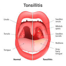
introduction
- Tonsillitis Infection and inflammation of the palatine tonsils is called tonsillitis and attacks of tonsillitis are mainly seen between the ages of 10 and 40.
- And this mainly happens when the immunity of any person is reduced and it starts very suddenly. And it is mainly caused by Streptococcus bacteria.
Etiology
- Acute:= can either be bacteria or virus.
- recurrent:=caused by bacteria.
- chronic:=caused by bacteria infection.
- Streptococcus ( Streptococcus ).
- glandular fever ( glandular fever).
- most common bacteria is group a beta Streptococci.
other virous are:=
- Herpes simplex virus.
- Streptococcus pyogenes.
- Epstein Barr virus.
- Cytomegalovirus.
- Adenovirus.
- Miscellaneous virus.
clinical manifestation
- sore throat.
- pain.
- difficulty swallowing.
- fever.
- feeling cold.
- malaise.
- Redness.
- irritation.
- Discomfort.
- Red swollen tonsil which may have a purulent Exudative coating of yellow or white patches.
- Sore throat.
- Earache.
- Fever.
- Headache.
- Bad breath.
- Muscle pain To be.
- Stiff throat.
- Swelling in the lymph nodes of the neck.
- Swelling of the eyes, mouth and throat.
- If the case is very severe, nausea may occur.
- Difficulty sleeping.
- Loss of appetite.
- Vomiting.
- Stomach ache.
- Constipation.
- Tongue that feels furry or fuzzy.
- Difficulty opening your mouth.
- Headache.
- Irritation and Discomfort
complications
- Heart and kidney failure.
- Pneumonia and chorea.
management
- Analgesic medicine,
- lozenges,
- Tell the patient to take bed rest.
- Tell the patient to drink plenty of water.
- Use warm saline solution if there is throat irritation.
- The patient’s antibiotic medicine Provide.
- Use acetylsalicylic acid and often codeine sulfate to manage pain and discomfort.
- Ice on tonsils Apply a pack.
- Instruct the patient to drink plenty of water.
- Provide antibiotic therapy to the patient.
- Provide antiviral therapy to the patient.
surgical management
Tonsillectomy (Tonsillectomy).
Most physicians believe that a person who has recurrent attacks of tonsillitis should undergo a tonsillectomy, and this procedure is mainly performed when the acute attack of tonsillitis subsides after four to six weeks.
preoperative care
- All the patient’s laboratory tests should be done such as:=
- Hb,
- Esr,
- B.T,C.T.
- Blood groping,
- Urine for sugar and Albumin,
- X-ray chest,
- Throat culture.
- Check vital signs every 4 hours.
- Do a general physical examination of the patient.
- Consult the patient and their family members for the operation.
- The operation is performed under general anesthesia if it is a child and under anesthesia if it is an adult. Prepare the patient in this way for the operation to be performed under local anesthesia.
- Give the patient a mild sedative at night so that he can get proper sleep and relieve anxiety.
- If the operation is to be performed under general anesthesia, then
- Preanesthesia which It is done in the evening before the operation and medicine is provided half an hour before and if the operation is done under local
- Xylocaine gargles are done 15 minutes before the operation.
- The patient is dressed in a clean operative cloth and sent to the operation area.
post operative care in 1st 24 hourse .
- After the operation, the patient should be immediately taken to the post-operative bed and placed in a semi-prone position. provide.
- Turn the patient to one side and place a kidney tray under him and a small pillow so that all the secretions can drain properly.
- Check the vital signs every 15-15 minutes.
- If the pulse is weak and fast or the BP is low, then inform the doctor immediately.
- When blood is coming out of the throat, if it is completely dark, it is considered normal and if it is fresh red, then inform the doctor immediately.
- Provide proper IV fluid to the patient.
- Maintain the patient’s intake output chart.
routine care
- If the patient is fully conscious, place him in the supine position.
- Tell the patient to spit out any saliva if he has any.
- Tell the patient to eat cold things like ice cream, jelly, pudding, etc.
- Provide soft-boiled eggs, milk, bread, milk shakes, etc. from the second day and bring the patient on a full diet on the third to fourth day.
- Do not give the patient very hot food and do not give him hard things like bread, raw, salad, spicy and fried and do not give fruits, vegetables, juice, soup for a few days.
- Maintain the patient’s oral hygiene.
- Provide the patient with analgesic and antibiotic medicine To do.
- If there is a sore throat, provide aspirin spray to relieve it.
- And gargle 15 minutes before eating.
- If everything is normal, provide discharge to the patient on the third to fifth day and Advise on follow up.
advice on discharge
- Avoid exposure to the sun.
- Vigorous games, shouting, and Vigorous cleaning of throat, blowing nose.
- avoid respiratory tract infection.
- report for follow up
periltonsillar abscess
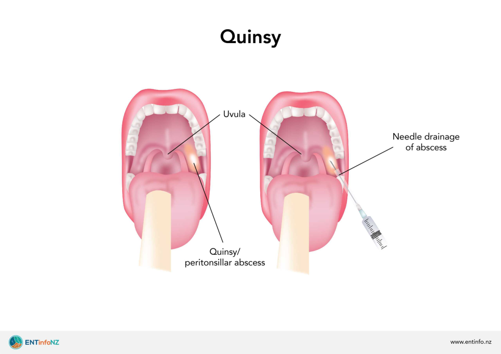
introduction
- A peritonsillar abscess or it is known as a complication of tonsillitis in which the collection of pus occurs around the tonsils i.e. the collection of pus occurs in the peritonsillar space. Thus the collection of pus in the area around the peritonsillar space is called a peritonsillar abscess.
Etiology
- most common complications of tonsillitis.
- Streptococcus bacteria.
- dental infection,
- Smoking,
- chronic lymphatic leukemia,
- stones or calcium deposits in the tonsil,
- aerobic and anaerobic bacteria,
- Streptococcus,
- staphylococcus.
clinical manifestation
- the first symptoms of peritonsillar abscess.
- Inflammation occurs in the throat area.
- contralateral deviation of the uvula.
- hot potato voice ( hot potato voice).
- swelling occurs in the soft palate.
- lymph nodes in enlargement occurs.
- severe sore throat.
- Difficulty swallowing.
- Be hurt.
- drooling,
- salivation,
- trouble handling oral secretion,
- Fever,
- fever,
- muscle spasm,
- hot potato/ muffled voice.
- Ear ache,.
- pain in the neck.
- Redness and Edema in the Tonsillectomy area.
Diagnostic Evaluation
- history tacking and physical examination.
- X Ray of neck.
- cbc.
treatment medical management
- antibiotic.
- Antipyretic.
- Analgesic.
- Immediate endotracheal intubation.
surgical management
1) Needle aspiration ( Needle aspiration)
- In needle aspiration, a needle is inserted into the abscess and slowly sucked out to drain the abscess. is done.
2) Incision and drainage ( Incision and drainage)
- If there is a large abscess, a large incision is made and the pus is drained from it.
3) Tonsillectomy (Tonsillectomy)
- In tonsillectomy, the entire tonsil is removed and treated.
Nursing management preoperative care:=
- Pathophysiology of tonsillitis and its surgery with the patient Explain all the procedures to the patient and his family members.
- Obtain consent from the patient and his family members.
- Properly anesthetize the patient
- Have the anesthesiologist provide anesthesia.
- If the patient has airway obstruction, then properly Patent the airway.
post operative management :
- If there is improvement in the pain level, the patient is discharged from the hospital immediately.
- Provide IV fluid to the patient.
- The patient is given 24 hours of rest. Only per hour Ask the patient to drink only fluids. Provide the patient with analgesic medicine. Do not give the patient hot drinks. Provide the patient with continuous antibiotic medicine. Maintain proper hydration status. Do not give the patient spicy food. Give.
- When the patient is ready to take oral fluid , give him oral antibiotic medicine.
- Provide the patient with analgesic medicine.
- Rinse the mouth with mouthwash. Mouthwash is 105 Degrees Fahrenheit to 110 degrees Fahrenheit ( 40.6 °cto 43.3°c).
- Ask the patient to apply cold.
- Ask the patient to gargle 3 to 4 times in 24 to 48 hours
Pharyngitis (Pharyngitis) introduction
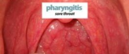
- Pharyngitis-often simply referred to as a “sore throat”.
- Pharyngitis is also called sore throat.
- Pharyngitis The infection and inflammation of the pharynx, which is located at the bottom of the mouth, is called sore throat.
- This is mainly caused by certain types of viruses and bacteria that enter the pharynx due to cold, flu or sinus infection and cause the condition of pharyngitis.
type of pharyngitis
1) Acute pharyngitis :
- Acute pharyngitis is mainly caused by viral infections and is caused by viral infections Acute pharyngitis is caused by the common cold.
2) Chronic pharyngitis:
- Is a persistent sore throat. Chronic pharyngitis is a long-term condition and lasts much longer than acute pharyngitis.
Etiology
bacterial Pharyngitis ( Bacterial Pharyngitis ):=
- Streptococcus Bacteria,
- Corynebacterium diphtheriae,
- Nigeria gonorrhoeae,
- Arctic bacterium hemolyticus (Corynebacterium),
- Chlamydophila pneumoniae,
- Mycoplasma pneumoniae,
- Fusobacterium necrophorum,
viral pharyngitis ( viral pharyngitis):=
- Rhinovirus,
- Coronavirus,
- Respiratory syncytial virus Virus,
- Parainfluenza virus,
- Adeno virus,
- Orthomyxo viridae,
- Herpes simplex virus,
- Miscellaneous virus,
- Coxsackie virus A & B,
- Epstein Barr Virus,
- Cytomegalovirus,
- H. I. V. Infection .
- Infection Mononucleosis.
- Adeno virus.
other cause
- Candida albicans,
- Immunocompromised
- Patient ,
- whites plaque in Oropharynx,
- cold and flu seasons,
- dry air,
- Allergies,
- Being around someone who has a cold 🥶 or sore throat.
- Being around people who smoke.
- Exposure to chemical irritants.
Clinical manifestation
- Dry throat.
- Redness in the throat.
- soreness.
- Extreme pain in the throat.
- Difficulty swallowing.
- Coughing a lot.
- High fever.
- Irritation.
- Discomfort.
- Lump in throat feeling.
- Headache.
- Earache.
- Swelling of the lymph nodes in the throat.
- Difficulty breathing.
- Difficulty speaking.
- Pain when swallowing.
- Hoarseness.
Diagnostic evaluation
- History taking and physical examination,
- Throat swab culture,
- ct scan,
- X-ray.
management
- antibiotics,
- corticosteroids,
- antifungal,
- pain medication (Analgesic),
Nursing management
- Provide adequate bed rest to the patient.
- Gargle with warm salt water.
- An ice pack may make the patient feel more comfortable .
- Provide acetylsalicylic acid for gargling.
- If there is local soreness, lozenges such as mild Anesthetics and lozenges are used to relieve local soreness.
- Do moist inhalation to relieve throat dryness.
- Take a liquid diet.
- Ask the patient to drink plenty of water, about 2500 ml of water throughout the day.
- Ask the patient to get enough rest and provide antipyretic medicine if there is fever.
- Maintain hygienic condition of oral cavity.
- If there is a lot of infection, then use antibiotics such as penicillin and erythromycin.
- Avoid smoking.
- Keep your mouth shut.
- Stay away from people who have infections.
prevention
- One of the best ways to prevent infection is to wash your hands properly.
- Use soap and water or an alcohol-based hand sanitizer.
- If someone has an infection, keep all their belongings separate.
- Wash your hands frequently.
- Do not touch public phones.
- Clean the telephone every day and clean the TV remote, computer keyboard, etc.
- Avoid close contact with a person who is sick.
- Do not smoke or be around people who smoke.
- Umidify the air.
- When sneezing or having a cold, cover your mouth and nose with a tissue or use a mask.
- And maintain proper hygiene
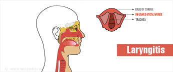
laryngitis (laryngitis) introduction
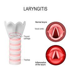
- Laryngitis is an infection and inflammation of the larynx It is called laryngitis.
- Infection and inflammation of the larynx, which is situated in the upper part of the respiratory track, is called laryngitis.
- Hoarseness is heard and speech is also lost.
Classification
1) Acute laryngitis :
- Acute laryngitis is short-lived and its causes are as follows There are causes.
- Viral infection such as those that cause a cold.
- Vocal strain, caused by yelling or overusing of voice.
- Virus such as measles or mumps.
- bacterial infection such as diphtheria.
- cold,
- sudden changes in temperature,
- irritating fumes,
- use their voice excessively,
- Smoking.
2) Chronic laryngitis ( chronic laryngitis):
- Laryngitis that lasts for more than three weeks is called chronic It is called laryngitis.
- If acute laryngitis
- recurs repeatedly, it can lead to chronic laryngitis.
- It causes irritation, injury, and strain to the vocal cords.
- It is caused by:=
- inhaled irritation such as chemical fumes, allergens or smoking.
- acid reflux also called gasteroesophageal reflux disease.
- chronic sinusitis,
- Bronchitis,
- excessive alcohol use,
- Habitual over use of voice.
- Smoking.
- infection such as tuberculosis, syphilis,
- Fungal infection.
- infection with certain parasite,
- cancer.
- vocal cord paralysis which can result from injury, stroke or lung tumor.
- chronic sinusitis.
- bronchitis.
- vocal cord strain.
- gasteroesophageal reflux disease.
- vocal cord strain.
- Smoking.
- infection such as tuberculosis or any fungal infection.
Diagnostic evaluation
- History taking and physical examination.
- X Ray,
- laryngoscopy,
- Biopsy.
clinical manifestation
- dryness,
- coughing,
- hoarseness,
- difficulty in making a voice.
- sore throat.
- fever.
- irritation.
- Discomfort.
- respiratory distress.
- Eruption of dry cough.
- cold or flu like symptoms.
- Swelling of the lymph nodes.
- Pain when lying down.
- Difficulty breathing.
management
- If laryngitis is caused by any bacteria or fungus, then provide antibiotic and anti-fungal treatment to the patient.
- If the patient has laryngitis, then provide steroid medicine.
- If the patient has gastroesophageal reflux disease, then provide antacid medicine.
- If the infection is caused by any virus, then provide antiviral medicine.
- Do not smoke and people who Don’t be around someone who smokes.
- Drink plenty of water.
- If you have a mild upper respiratory tract infection, treat it immediately.
- Humidifier Use.
- Drink proper fluids to prevent dehydration.
- Don’t talk too much until you’re well.
- Don’t use decongestants because they dry out your throat.
- Don’t talk too loudly.
- Keep your throat moist until you’re well.
- Lozenges or salt water gargles will help moisten the throat.
- Tell the patient to keep the room temperature normal.
- Tell the patient to take steam inhalation to moisten the throat.
- Tell the patient to take cough syrup.
- Tell the patient not to smoke.
- Provide antipyretic medicine to control fever.
- Provide antibiotic medicine to control infection.
- Keep things that cause irritation away.
- Tell the patient to sit in a quiet position to avoid stress.
prevention
- The main cause of laryngitis is viral infection, so to prevent it, wash your hands properly and especially when touching your mouth.
- And in children, mainly Haemophilus influenzae Get vaccinated against life-threatening bacterial infections.
- To prevent laryngitis, avoid talking loudly.
- And don’t talk too loudly.
- Take steam inhalation.
- Use a humidifier.
- Drink plenty of water.
- Keep your throat moist.
- Don’t smoke
laryngeal cancer introduction
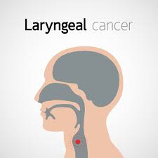
- Laryngeal cancer is also called cancer of the larynx or laryngeal carcinoma.
- The inner wall of the larynx is made up of squamous cells.
- Almost all laryngeal cancers are It is produced by squamous cells.
- Therefore, laryngeal cancer is called squamous cell carcinoma.
- Laryngitis is an infection of the vocal cords. and inflammation.
- Laryngitis occurs in people under the age of 50.
- Men are ten percent (10%) more likely to get it than women.
Risk factors
- Smoking,
- alcohol,
- a personal history of head and neck cancer.
- occupation,
- Smoking.
clinical manifestation
- abnormal high pitch breathing sounds.
- Coughing up.
- Blood in the phlegm.
- Difficulty swallowing.
- Hoarseness.
- Neck Pain.
- sore throat.
- Swelling in the throat.
- Weight loss.
- Change in voice.
- Pain.
- Difficulty swallowing.
- hot changes ” hot potato voice “.
- Airway obstruction occurs.
- Strider or dysphonia.
- Loss of voice occurs.
- Irritation.
- Discomfort.
- weight loss.
Diagnostic evaluation
- History taking and physical examination.
- radiation therapy.
- chemotherapy.
- Biopsy.
- X Ray.
- ct scan.
- MRI.
management assessment
- Ask the patient about their smoking history, alcohol intake history, and chronic illness.
- Ask about the patient’s breathing pattern.
- Ask about the patient’s ability to swallow.
- Check the patient’s vital signs.
- Ask the patient about their breathing pattern. Check fluid electrolyte balance.
- Take a complete nutritional history of the patient.
- Do all laboratory investigations of the patient.
- Check the patient’s weight.
- Assess the patient’s communicative ability.
- Check the patient’s anxiety level.
- To check the knowledge level of the patient and his family member.
Nursing diagnosis
1) ineffective breathing pattern related to obstructive and restrictive respiratory process associated with cancer of larynx.
interventions
- Assess the extent of the patient’s breathing difficulty.
- See if there are any changes in the patient’s breathing pattern.
- Listen to the patient’s lung sounds to see if there are any abnormalities.
- The patient’s hydration status assess.
- Ask the patient to do deep breathing exercises.
2) Ulttered nutrition less than body requirement related to hyper metabolism state , taste aversion, Anorexia secondary to radiation therapy and chemotherapy.
interventions
- Assess the patient’s nutritional status.
- Tell the patient to eat some high-calorie and high-protein food at regular intervals.
- Medicine as a supplement to prevent the patient from deficiency diseases Giving.
- Patient’s oral Maintain hygiene.
- Ask the patient to take a liquid diet.
- Ask the patient to drink plenty of water which will soften the stool and relieve the condition of constipation.
- Provide anti-emetic medicine to the patient.
3) Impaired verbal communication related to presence of trachiostomy tube.
interventions
- Asking the patient to write down the conversation improves communication.
- Do not speak too slowly and loudly.
- Asking the patient to take care of the tracheostomy tube and prevent infection.
- Associating the patient with a speech therapist.
4) Anxiety related to uncertain outcome and fear of recurrence.
interventions :=
- Check the patient’s anxiety level.
- Encourage the patient to describe their feelings.
- Tell the patient how to get out of a difficult situation.
- Tell the patient about stress reduction techniques.
- Ask the patient to describe their feelings.
- The patient has cancer Provide education about treatment.
- The goal of treatment is to completely remove the cancer and prevent it from spreading to other parts of the body.
- If the tumor is small, it is removed surgically or by providing radiation therapy.
- If the tumor is large and has spread to the lymph nodes in the neck, then such tumors are provided with a combination of both chemotherapy and radiation therapy.
- Many patients Swallowing therapy is also performed.
complication
- Airway obstruction.
- Difficulty swallowing.
- Disfigurement of the neck or face.
- hardening of the skin of the neck.
- Loss of voice.
- Cancer can also spread to other parts of the body.
prevention
- Avoid smoking Do.
- Do not sit next to people who are smoking.
- Do not consume alcohol.
Nasopharyngeal carcinoma (in Nasopharyngeal carcinoma) introduction

- Nasopharyngeal carcinoma is a cancer that primarily affects the nasopharynx. Which is located at the back of the nose and slightly above the throat.
Etiology
- Gene mutation.
- Epstein Barr virus.
Clinical manifestation
- In the early stages, nasopharyngeal carcinoma does not create any symptoms, but gradually and slowly, symptoms begin to appear.
- A lump in the throat is felt due to swelling of the lymph nodes.
- Blood with saliva.
- Bloody discharge from the nose Discharge.
- Nasal congestion.
- Hard hearing.
- Frequent ear infections.
- Headache.
Diagnostic evaluation
- history tacking and physical examination.
- Nasal endoscopy,
- Biopsy,
- Computerized tomography (CT scan),
- Magnetic Resonance Imaging (MRI),
- Positron Emissive Tomography (PET).
- X-ray.
management
1) Radiation therapy :=
- Nizopharyngeal External beam radiation is used in carcinoma.
- If there is a small Nasopharyngeal tumor, then treatment can be done with radiation therapy alone.
- And if there is any other situation, then radiation therapy and chemotherapy are provided in combination.
2) Chemotherapy :=
- Chemotherapy is used to kill cancer cells using chemicals.
- Chemotherapy is given in the form of pills or intravenously.
3) surgery :=
- Surgery is Nasopharyngeal cancer is not used to remove the head but is used to remove the lymph nodes in the neck.
- And to remove these cancer lymph nodes, the surgeon makes a cut in the roof of the mouth An incision is placed above.
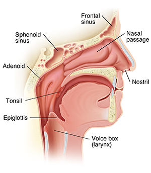
Adenoiditis:= introduction
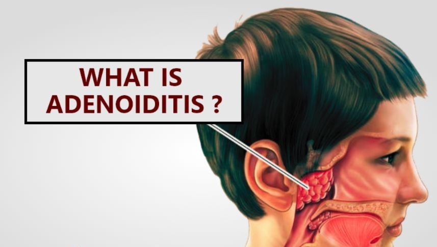
- In this, there is abnormal growth of lymph nodes and they grow in the area of the nasopharynx.
- Tonsils and adenoids are lymphoid structures and is located mainly in the oropharynx and nasopharynx and is at its full size in childhood and decreases in size as we move into adulthood.
Etiology
- The exact cause is unknown.
- Streptococcus bacteria,
- Staphylococcus bacteria,
virus:=
- Adeno virus,
- Enterovirus,
- Due to repeated upper respiratory tract infections.
CLINICAL MANIFESTATION
- Nasal obstruction.
- Facial expression becomes dull.
- Nasopharynges become blocked.
- Difficulty swallowing.
- Difficulty hearing.
- Difficulty speaking.
- pulmonary hypertension.
surgical management
- Most tonsils and adenoids are surgically removed.
pre operative nursing management
- Admit the patient to the hospital for surgery.
- Do a complete examination of the patient.
- If there are children, carefully prepare them for surgery and keep their parents with them.
- Do a general physical examination of the patient.
- Patient’s routine To conduct laboratory investigations. Such as urine, blood count, clotting time etc.
- If the patient has any condition that may create complications during anesthesia and post-operative course such as fever, upper respiratory tract infection, then the operation should be postponed.
- Tonsillectomy is performed under general or local anesthesia.
- It is performed under anesthesia.
post operative management
- After the operation, the patient should be placed in a side lying position or prone position and a pillow should be placed under his chest.
- When the patient is awake, he should be placed in the Fowler position provide.
- A patient who has had a tonsillectomy has dark brown blood coming out of the area.
- Instruct the patient to take adequate rest.
- Watch the patient for any type of hemorrhage.
- Ask the patient to sit quietly for 24 to 48 hours.
- Apply an ice pack to the patient Tell.
- Tell the patient not to cough because it can also cause bleeding.
- Give the patient plenty of fluids.
- Maintain the patient’s hydration status.
- Give the patient ice cream.
- Give the patient analgesic medicine.
- Give the patient antipyretic medicine.
- Avoid sour juices, hot food for a week as they can irritate the operative area.
discharge teaching
- Tell the patient that if any bleeding is observed after discharge, they should immediately report it to the physician.
- If there are children, ask them to take complete bed rest for two to three days.
- Advise the patient not to go out in the sun, not to exercise too much, not to cough, not to sneeze, not to try to clear the throat, or not to blow the nose too much, otherwise it may cause an erection.
- Any adult If the patient is present, ask him to take a specific type of treatment.
- Give him food as the patient can tolerate.
- Ask the patient to drink plenty of water.
- Ask the patient to eat plenty of food.
- Inform the patient that the stool will be dark or black because blood was swallowed during the surgery.
- If consistent If the body temperature remains high and there is discomfort in the ear, report it to the physician immediately.
- Tell the patient to have regular follow-up and to come back after a week after the operation
throat injury/ trauma ( throat injury) introduction

- The main structures of the throat include the esophagus, voice box, (i.e. The larynx (larynx) is a cartilage, vocal cords and trachea and all these structures play a very important role in daily living and all these parts are injured due to direct trauma such as a ball, any stick, elbow, pipe, or fall and all these structures are damaged.
cause and risk factore
- Due to trauma.
- Due to a bullet wound.
- Due to injury by any heavy machinery.
- Due to injury to the throat by a stick.
- Throat compression.
- Fall Due to.
- Due to an accident.
sign and symptoms
- Change in voice.
- Difficulty breathing.
- Difficulty speaking happen.
- Difficulty opening and closing the mouth.
- Difficulty breathing.
- Feeling like fullness in the throat.
- Swelling of the throat.
- Inability to breathe.
management
- Rest your voice.
- Get enough rest.
- Diet Do not take.
- Only give water and pureed food to eat.
- Breathing humidifier air.
- Use medicine to prevent infection.
- Provide analgesic medicine.
- Apply ice to the injured side of the patient.
- Keep the head elevated.
- Keep the head in a normal position. Do not use any type of pillow, i.e. a high pillow.
- Do not give the patient anything to drink. If he tries to swallow, the symptoms will worsen or the airway may be obstructed. There will be an obstruction.
foreign bodies in throat (Foreign bodies and throat)
introduction
- Virtually any small object that passes through the pharynx and is swallowed.
- Small toys, coins, pencils, etc. are common objects swallowed by young children. ,pen lids, needles and hair pins are mainly radio opaque and are swallowed by older children, fish and chicken bones etc. .
- And in adults, dentures or any part of dentures are accidentally swallowed.
forien bodies in the Airway (Foreign Body in the Airway)
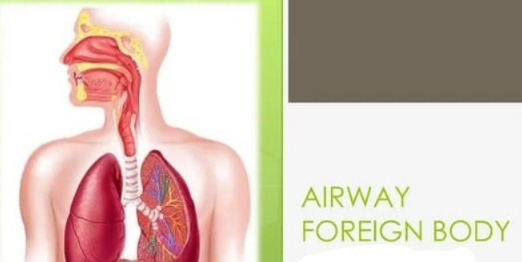
introduction
- A foreign body that gets stuck in the airway is called a medical emergency and requires immediate attention.
- Foreign bodies that get stuck in the airway in many places.
- Children put small objects in their mouths and breathe deeply, which can cause the foreign object to travel down the trachea or esophagus. In the mouth, the loadeg occurs.
- More cases of this chocking are seen in children younger than four years and those children put the following items in their mouths.
Example:= nuts, seeds, carrot, toy parts, grapes, hot dogs, pebbles, buttons and coins.
symptoms
- chocking or gagging when the object is first inhaled.
- coughing.
- strider.
- Whistling sound (This whistling sound is when children exhale when they exhale.)
If the following
- If symptoms are seen, check for airway obstruction.
- stridor,
- wheezing,
- cough,
- pneumonia,
- voice change.
forien bodies in the esophagus
introduction
- When any food is swallowed, it passes through the mouth and then goes to the throat and then the throat reaches the esophagus and then reaches the stomach.
- Many times the object considered is too large and gets stuck in the esophagus.
- One of the main objects included is a coin.
- In children under four years of age, any food or object that is stuck in the esophagus Too much gets stuck.
symptoms
- an initial shocking episode.
- drooling.
- Vomiting when ever trying to eat.
- Inability to eat.
assessment
- Assess the patient’s airway and respiratory function.
- Check the patient’s vital signs.
- Open the mouth and examine the oropharynx in a bright light.
- Perform laryngoscopy and examine the pharynx.
- Palp the throat and check the position of the trachea. To do.
- Examine the chest and listen to lung sounds.
- Do a cardio vascular examination.
- Carefully examine the abdomen.
treatment
- Treatment varies depending on the location of the foreign body.
- If an object is in the airway,
- Laryngoscopy and bronchoscopy are performed to remove the object.
- In this procedure, a rigid scope is inserted into the airway and then air is injected into the airway. The object is visualized and located, and then the foreign object is removed using forceps.
- If an object is lodged in the esophagus, esophagoscopy is performed and the stuck or swallowed item is removed using some special instruments.
- When the foreign body is removed, the child Recovers immediately.
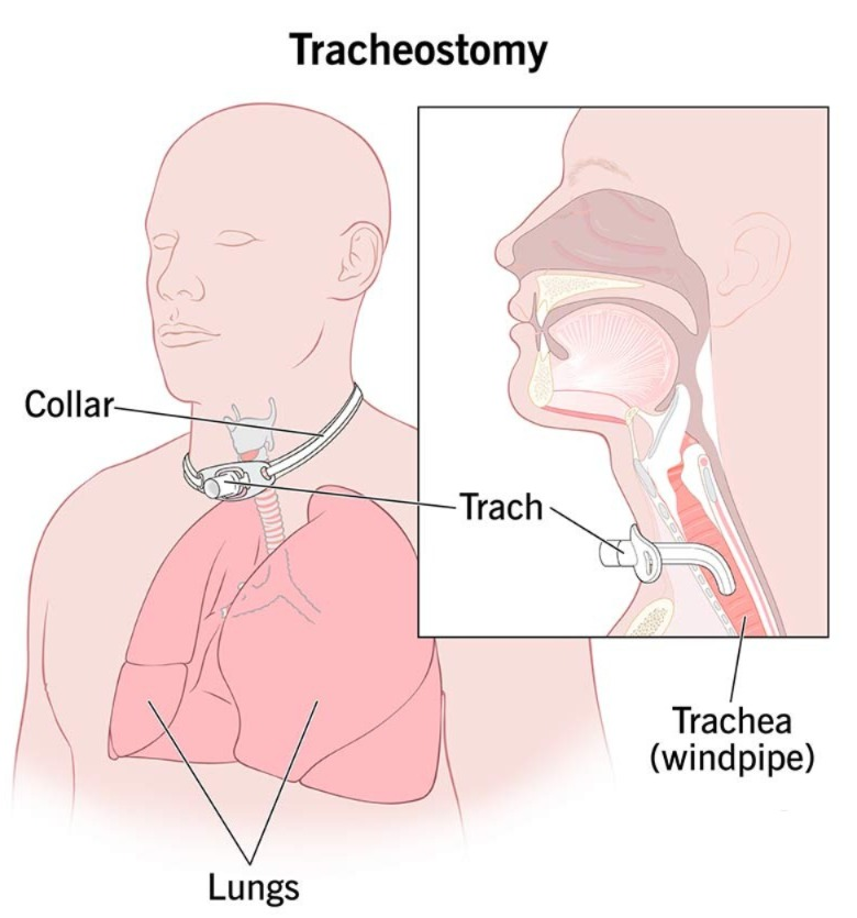
tracheostomy
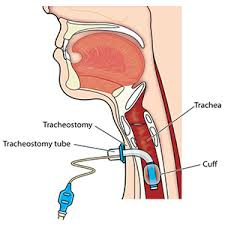
introduction
- A tracheostomy is a surgically created hole, usually between the third and fourth cartilages, and this opening is called a stoma.
- A tracheostomy is performed when oral and nasal intubation are not possible. To relieve airway obstruction that is insufficient.
tracheostomy indications
- 1) to relieve the upper Airway obstruction:=
- There is a foreign body.
- There is trauma.
- Acute infection.
- Acute epiglottitis Diphtheria etc.
- Glottic oedema.
- Bilateral abductor paralysis of the vocal cord.
- Having a tumor in the larynx.
- congenital atresia.
- 2. To improve respiratory functions .
- Fulminating
- Bronchopneumonia.
- Chronic bronchitis.
- Emphysema.
- Chest injury or flail chest.
- 3) respiratory paralysis
- Unconscious head injury.
- bulbur poliomyelitis.
- tetanus.
type of tracheostomy
1) a temporary tracheostomy. 2) a permanent tracheostomy .
1) a temporary tracheostomy
- This tracheostomy is done when the patient is on respiratory support for a long time and is unable to breathe on his/her own Unable to take in air.
- A tracheostomy tube is inserted to keep respiration patent.
2) a permanent tracheostomy
- This tracheostomy is done when the trachea has been removed from the skin and sutured to the neck.
- And this stoma is kept open.
- This procedure is elective and the patient is properly prepared for it.
tracheostomy tubes
- A tracheostomy tube is a type of indwelling tube that maintains the patency of a tracheostomy.
- This tracheostomy tube is made of metal or plastic.
- This tube may or may not be cuffed and may be fenestrated which helps the patient speak.
parts of tracheostomy tube
- Tracheostomy tube consists of three parts:=
- the outer cannula
- This is an outer tube that keeps the tracheostomy open.
- a neck plate.
- inner cannula.
- obturatore.
tracheostomy ( procedure):=
- Lay the patient in a supine position.
- At the same time, keep his neck and head extended.
- Place any pillow or sand bag inside the solder so that the neck is properly visible.
- Local anesthesia or general anesthesia is used in this procedure.
- Using a knife, a horizontal cut is made in the neck one centimeter above the sternal notch.
- This incision is extended to the mastoid muscle of the sternum.
- The skin is then separated and the trachea is exposed.
- Dissection of the facial plantar fascia, followed by anterior Retract the jugular vein, retract the strap muscles and then divide the thyroid isthmus.
- Place the cricoid hook over the second tracheal ring.
- Incise the third and fourth tracheal rings. To insert and place the tracheostomy tube.
- Before introducing the tracheostomy tube, the trachea should be properly suctioned to remove any secretions or blood, if any.
- Then, using an obturator, introduce a tracheostomy tube of the appropriate size.
- When choosing a tube, choose the smallest possible tube. The general rule is that the tube should cover a third to a fourth of the trachea.
- The cuff of the tube should be two to five ml of air is taken and inflated and placed in place using a neck tie.
- Then this in-situ is sutured using a skin suturer.
- Then a dressing is applied.
- And this The dressing should not be too tight, but should be loose enough to fit a finger.
post operative tracheostomy care
- Suction in a way that does not cause repeated trauma.
- Humidify the air and oxygen that we breathe.
- Provide him with a Fowler position to avoid breathing in this way.
- Ensure adequate fluid intake.
- Wash your mouth frequently. Perform.
- Provide mucolytic agents.
- Advise for cuffing and provide physiotherapy.
- Occasional bronchial lavage.
- Maintain aseptic technique while suctioning the tube.
- Give the patient prophylactic antibiotic medication.
- Deflate the cuff for five minutes every hour.
- Be careful not to let the tube touch the posterior tracheal wall.
routine tracheostomy care
- Irrigate any secretions around the stoma.
- Care for the tube daily using aseptic technique to prevent infection around the tube.
- Care for the tracheostomy tube once a day.
- A person on a ventilator needs more care.
- Dress the tracheostomy side and drain any secretions there.
- If there is irritation around the tube, rub it there.
Equipment
- Trachostomy care kit or tray,
- Sterile cotton tip applicator,
- Gauze piece,
- Sterile suction catheter Kit.
- Sterile normal saline.
- Hydrogen peroxide.
- Trachostomy ties.
- Seizures.
- Two sterile cups or clean disposable paper cups.
- A small blanket or towel Roll.
- Antibiotic ointment.
- Disposable bag.
procedure
- Proper hand washing for tracheostomy care.
- Explain the procedure according to the patient’s understanding.
- Make the patient lie down in a comfortable position, i.e. supine position.
- Then extend his neck.
- Then place a pillow or a small towel roll under his Keep the shoulder down so that the tracheostomy side can be properly visualized.
- Properly place the tracheostomy side on top of the Suctioning.
- Remove the dressing on the tracheostomy side and put it in a proper disposable bag.
- Remove the inner cannula and clean it with hydrogen peroxide, then clean it with normal saline, then check its patency and cleanliness by keeping it at eye level.
- If the tracheostomy tie has become wet, remove it and apply another tie.
- When we change the tie When there is a tracheostomy tube, place it in its place with one hand and check the skin inside the tie.
- Check the incision site.
- Dip the skin in hydrogen peroxide with a cotton tip and clean the tracheostomy site.
- Clean in a round shape.
- Clean from the inside to the outside and use one swab for one stitch only.
- Be careful not to let any liquid get inside the tracheostomy tube.
- Apply antibiotic ointment.
- Then apply a sterile tracheostomy dressing.
- While applying the tracheostomy dressing, check that the tracheostomy tube is properly supported.
- Wash your hands after tracheostomy.