ENGLISH-ENT (PART-1)
As per INC Syllabus
Ear :
a) Review of Anatomy and Physiology of Ear
◙ EAR..
Ears are located one on both sides of the head. It is an organ involved in hearing and maintaining body balance.
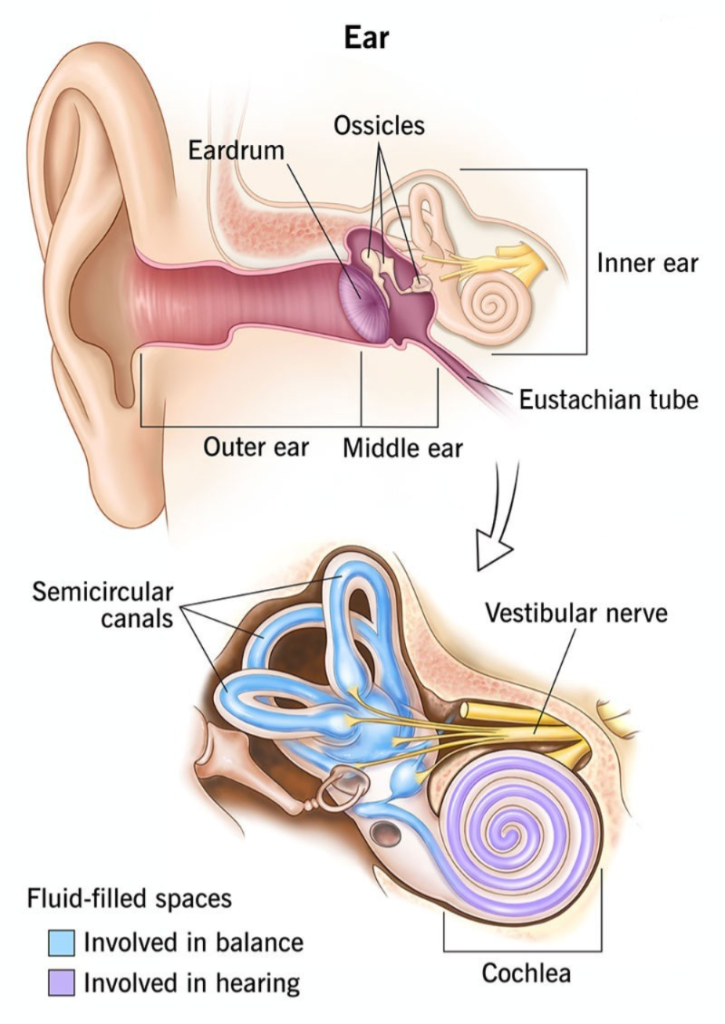
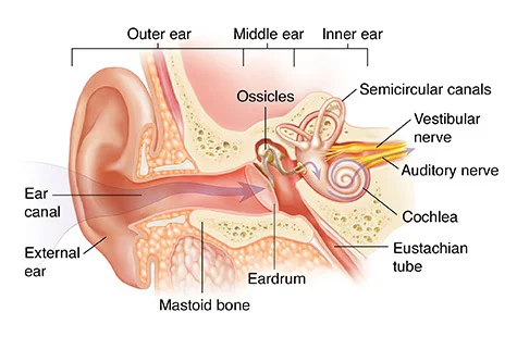
Structure-wise, the ear is anatomically divided into three parts
- External or outer ear
- Middle Ear
- Inner Ear
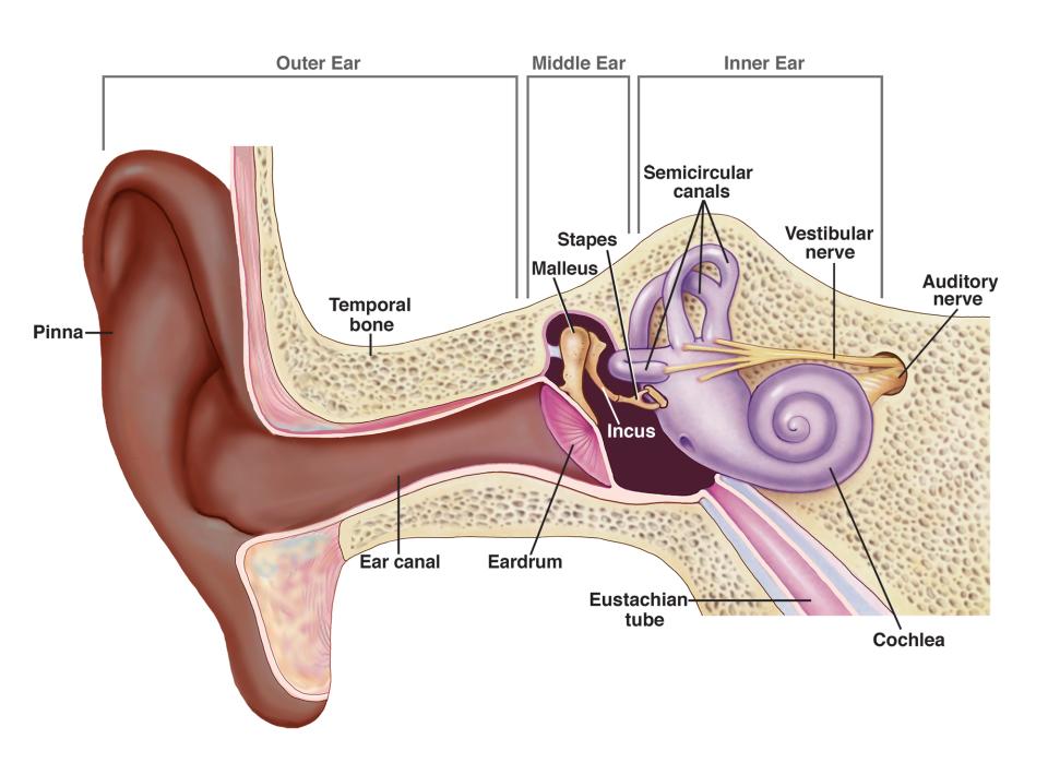
1. External or outer ear
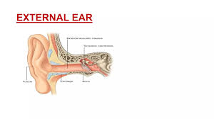
The external ear collects sound waves and transfers them to the inside. The following parts of the external ear are included.
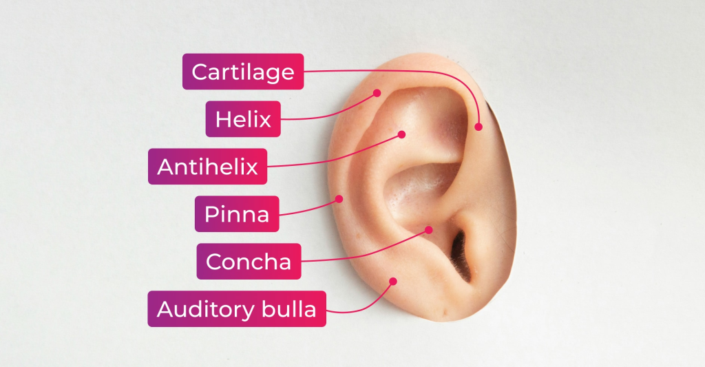
♦ Auricle or pinna (Oricles or pinna)
External acoustic meatus i.e. auditory canal
Oricles (Oricles or pinna)
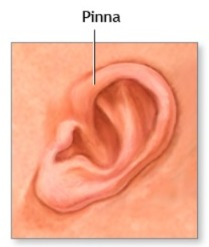
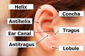
- It is a flap-like part on both sides of the head, made of elastic cartilage and covered with skin on the outside. It collects sound waves coming from the outside environment and transmits them to the inner hole. Its uppermost part is called the helix and the soft part hanging below is called the lobule.
External Acostic Meatus
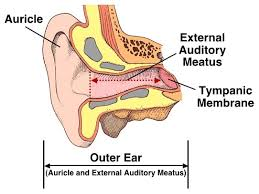
- Also known as the auditory canal, it is a curved tube in the shape of an S. Its length It is a canal that is about 2.5 cm long and is elongated on the inside and the glands in its wall secrete a wax-like viscous fluid, also known as cerumen. Some hairs are also present in this canal which, combined with the viscous fluid, prevent dust and foreign bodies from reaching the inner wall, i.e. the tympanic membrane.
Tympanic Membrane
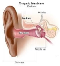
- It is a thin membrane that separates the external ear and the middle ear. Separates and is oval in shape. This membrane is made up of epithelium tissue and connective tissue, which contains fibroblast cells.
2. Middle ear
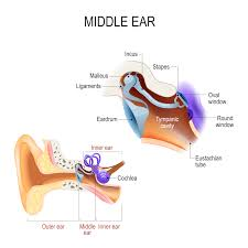
- It is a small air-filled cavity located in the temporal bone. The lining of this cavity is the epithelium. It is made of tissue and is separated from the external ear by the tympanic membrane, or ear drum, and the inner ear is separated from the middle ear by the oval and round windows.
- Its anterior wall has an opening called the Eustachian tube. This tube connects the middle ear to the structures of the nasal passages and maintains pressure within the tube cavity, which causes the tympanic membrane to expand during yawning and swallowing. The rupture of the membrane stops.
♦ Auditory ossicles (Auditory ossicles)

- The bones in the middle ear are called auditory ossicles, which include the malleus, incus, and stapes bones. Their number is one in each ear, meaning there are a total of 6 auditory ossicles in the body.
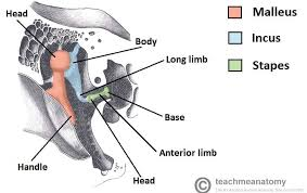
▲Malleus
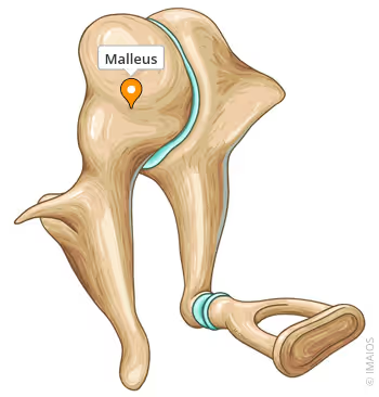
- It is hammer-shaped and its handle is attached to the wall of the tympanic membrane. The head of the malleus is connected to the incus bone.
▲Incus (incus)
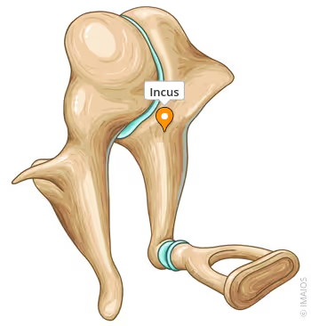
- It is the middle bone which is in the shape of an anvil. It is connected to the malleus bone and its head is connected to the stapes bone. There are fibrous tissues in the joint.
▲Stapes
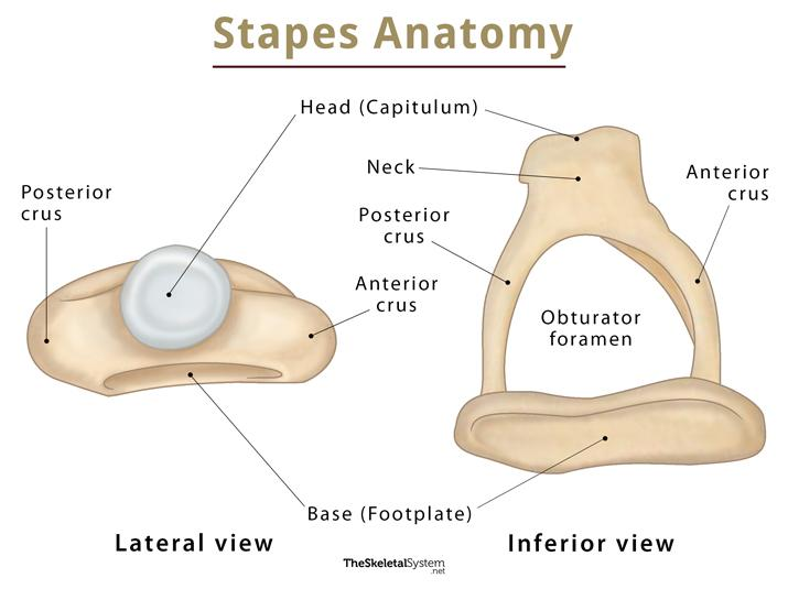
- It is stirrup-shaped It is a bone whose head connects to the incus bone and its foot plate is attached to the oval window, at the bottom of which there is a round window.
- Thus, the structure from the tympanic membrane to the oval and round windows is called the middle ear.
3. Inner Ear (Inner Ear)
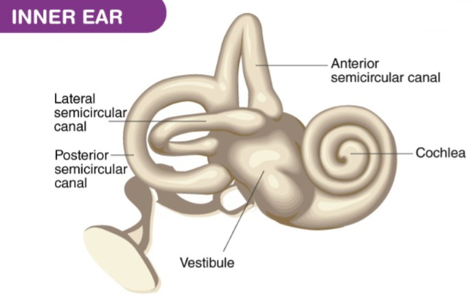
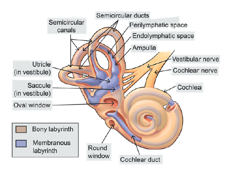
him Also known as a labyrinth. It has a complex structure and is involved in hearing and balance.
It has two parts: the bony labyrinth and the membranous labyrinth.
♦ Bony Labyrinth
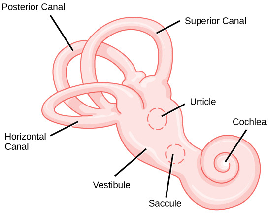
- It is a cavity located in the petrous portion of the temporal bone, whose wall is made of a layer of periosteum, inside which is a fluid called perilymph. It has three parts. A vestibule, a cochlea, and three semicircular canals.
♦ Vestibule
- It is the middle oval-shaped portion of the bony labyrinth, which contains the utricle and saccular structures, and its lateral walls contain the oval and round windows.
♦ Cochlea (choclea)
- This part of the bony labyrinth is associated with hearing. It is a ball-like structure called the Snail Cells Also known as.
♦Semi-circular canal (Semi-circular canal)
- The structure of this canal is associated with balance. The three tubes of the semicircular canal are arranged at right angles to each other.
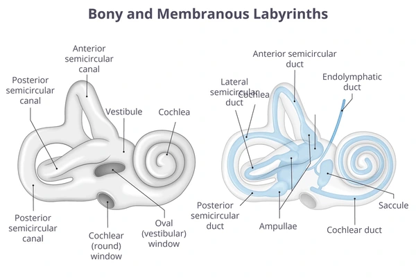
◙ Membranous Labyrinth
- Inside the bony labyrinth is a membranous tube called the membranous labyrinth, which contains a fluid called endolymph. The property of this fluid is csf Like.
- Vestibule which The utricle and the saccule are located.
- Vestibule and semicircular canal…
The vestibule and semicircular canals in the inner ear work to maintain the body’s balance.
In which the semicircular canal, saccule, and utricle help to maintain the body’s dynamic equilibrium. The semicircular canal is a tube-like structure that lies behind and above the vestibule and these canals are arranged at an angle to each other. The semicircular canal and cochlea, which have a bony labyrinth as their outer wall and a perilymph fluid inside, have another tube called the membranous labyrinth inside which contains endolymph.
The utricle is a membranous sac that is part of the vestibule and all its ducts open into a dilated portion called the ampulla. The saccule is part of the vestibule and communicates with the utricle and cochlea.
The walls of the utricle, saccule and ampulla contain small hair-like projections of specialized epithelial cells, which contain sensory nerve endings, which form the vestibular part and through which the vestibulocochlear nerve passes.
3. Cochlea
The cochlea, also known as the snail, is a coiled structure that resembles a snail. Inside the cochlea is a membrane called the cochlear duct, which is mainly involved in the process of hearing. The following parts are seen in the cross section of the cochlea.
♦Scala vestibuli
♦Scala Media or Cochlear Duct
♦Scala Tympany
The cochlear duct is a triangular-shaped tube. The bony part of the cochlea is divided into two parts, upper and lower, with the upper part called the scala vestibuli and the lower part called the scala tympani. In the middle part lies the cochlear duct and its roof is the basilar membrane. The organ of Corti, also known as the hearing organ, is located on the basilar membrane.
◙ Physiology of Hearing
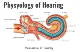
- The physiology of hearing is the act of hearing. The wavelength of the waves for hearing is 20 to 20,000 Hz. The human ear has a frequency capacity between 500 and 5,000 Hz. Sound waves The frequency of vibration is known as pitch. As the vibration increases, its pitch increases.
- Every sound produces sound waves and they hit the outside of the auricle and from there enter through the external auditory canal. These sound waves vibrate the tympanic membrane, i.e. the ear drum, which is the junction between the external ear and the middle ear.
- The malleus bone is connected to this tympanic membrane. These waves go from the malleus bone to the incus and from the incus to the stapes, and this stapes bone further connects to the oval window. It is connected to the oval window. These sound waves reach the perilymph fluid which goes to the cochlea and from there it goes to the endolymph and the round window vibrates and the vestibule goes to the cerebrum through the cochlear nerve and the sound is recognized.
◙ Balance and Ear (Balance of Ear)
♦Vestibule and Semi Circular canal (Vestibule & Semi circular canal)
- The vestibule and semicircular canals in the inner ear work to maintain the body’s balance.
- The semicircular canals sacule and utricle help to maintain the body’s dynamic equilibrium. The semicircular canals are tube-like structures that lie behind and above the vestibule and are arranged at an angle to each other. The semicircular canals and cochlea, whose outer walls are made up of the bony labyrinth and which contain perilymph fluid, have another tube inside them called Membranous labyrinth is called which contains endolymph fluid.