ENGLISH MODELS AND BONES GNM FIRST
MODELS
- HEART
Heart is an important organ of the circulatory system.
The heart is an organ made up of blood and muscles. It weighs approximately 310 grams in males and approximately 250 grams in females.
Location of the heart..
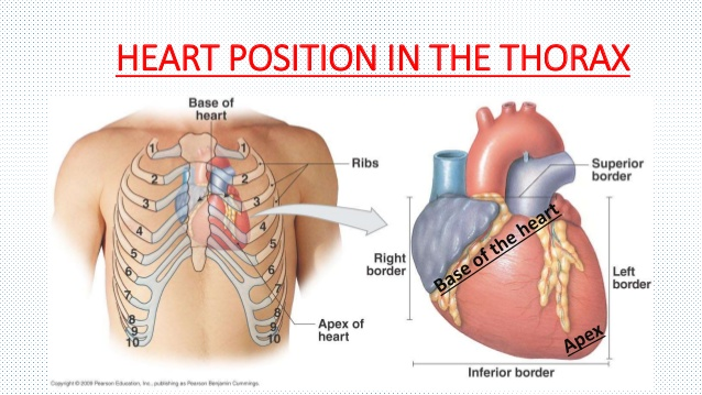
The heart lies above the diaphragm in the mediastinum space between the two lungs in the thoracic cavity.
The heart is a rough cone shape. In it, its upper broad part is known as the base and the lower angled part is known as the projection.
The heart is the size of a man’s closed fist. It measures 12 cm in length, 9 cm in width and 6 cm in thickness.
The heart is arranged slightly to the left between the two lungs in the thoracic cavity.
Organs around the heart.
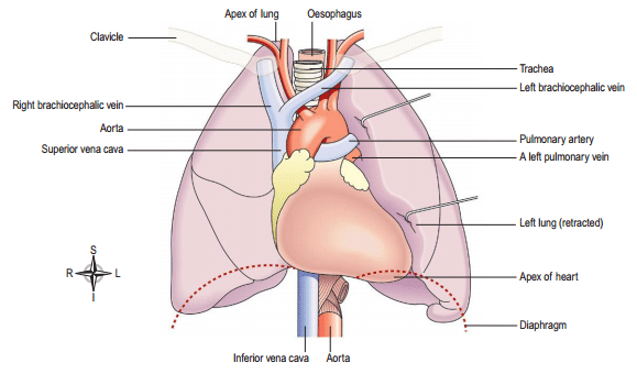
The heart is located in the thoracic cavity. It has one lung on both its left and right sides.
Underneath lies the diaphragm and central tendon.
On the upper side of the heart are the vena cava and the aorta and the pulmonary artery and pulmonary vein.
Posterior to the heart are the esophagus, trachea, bronchi and bronchioles as well as the descending aorta and thoracic vertebrae.
Anterior to the heart are the sternum bone and the ribs and intercostal muscles
Layers of the Heart..
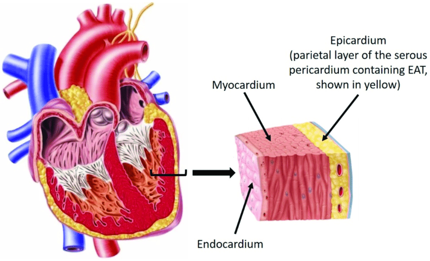
Heart is an organ made up of semi-muscles. Its wall is made up of three types of tissue layers.
The outermost layer of the heart wall is called epicardium or pericardium.
Epicardium or Pericardium.
It is thin and transparent and covers the heart from the outside. It is made up of fibrous connective tissue. In which there is a layer of fibrous tissue on the outermost side and a serous membrane on the inner side of the fibrous tissue which is found in a double layer. The outer layer of the serous membrane is known as the parietal layer and the inner layer as the visceral pericardium layer.
Myocardium..
Myocardium is the middle layer of the heart. It lies below the pericardium. It is made up of a special type of cardiac muscle tissue. The pumping action of the heart is seen due to the contraction of these muscles.
endocardium..
It is the innermost layer of the heart wall. It is in contact with layer blood. This layer is made up of epithelium tissue and connective tissue. This layer is smooth and shiny which is important for easy blood flow inside the heart.
Chambers of the Heart.
Heart is mainly divided into two parts. Right side and left side.
Between this right and left side lies the septum of the heart. It is again divided into four chambers above the valve and below the valve by the valves located between the right side and the left side of the heart.
Both chambers above the valve are called atrium or auricle or atrium. Both chambers below the valve are called ventricles. Thus the heart is divided into a total of four chambers.
Valve of the heart..
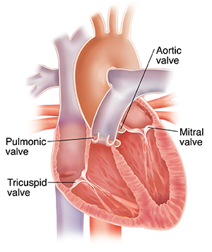
The flaps of tissue inside the heart are called valves. There are two types of valves in the heart.
Atrioventricular valve.
Semilunar valve.
Atrioventricular valve…
These valves are located between the atrium and the ventricle in the heart.
The valve between the right atrium and ventricle is called the tricuspid valve. This valve is made up of three tissue flaps.
The valve between the atrium and ventricle on the left side of the heart is called the bicuspid valve or the mitral valve.This valve is made up of two tissue flaps.
Semilunar valve…
These valves are C-shaped or crescent-shaped, so they are called semilunar valves.
These valves are present in the aorta and pulmonary artery. The valve in the aorta is called the aortic valve and the valve in the pulmonary artery is called the pulmonary valve. Which are located at the opening of its vessels. This valve also opens in one direction.
Great Vessels Associated with Heart..or Openings of the Heart..
Large blood vessels are mainly connected to the heart. Through which the blood comes from the body to the heart and the heart is pumped and the blood goes out to the body. The vessels and their openings are as follows.
Superior Wanakewa..
This brings deoxygenated blood from the upper part of the thoracic cavity, head and neck to the right atrium of the heart. It is in the number of one.
Inferior Wanakewa..
These blood vessels bring deoxygenated blood from the inferior part of the body and the lower part of the thoracic cavity and open into the right atrium of the heart. It is in the number of one.
Pulmonary artery..
It carries deoxygenated blood from the right ventricle of the heart out of the heart to the lungs. Its number is one.
Pulmonary vein..
Two pulmonary veins from both lungs bring the oxygenated blood to the left atrium of the heart. Its number is four.
Aorta..
The left ventricle of the heart takes oxygenated blood and circulates it throughout the body. Its number is one.
Thus a total of eight blood vessels and their openings are directly connected to the heart. Which is connected with the blood circulation in the heart.
Blood supply of the heart.
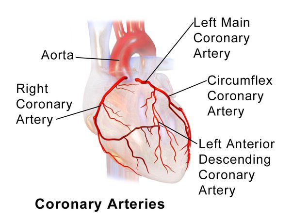
Oxygenated blood supply to the heart to function As soon as the aorta exits the left ventricle of the heart, it branches off into the right and left coronary arteries, which supply oxygenated blood to the heart. These coronary arteries are branches of the ascending aorta.
- Functions of the Heart…
Heart provides oxygenated blood supply to all organs and tissues of the body.
Heart is an important organ of the cardiovascular system. It functions as a vital organ without which the human body cannot survive.
The heart circulates the blood towards the lungs so that the blood can be oxygenated and purified.
Circulations like pulmonary circulation and systemic circulation are regulated by the heart.
The heart also regulates the heart rate according to the needs of the body and according to the body temperature.
The heart also regulates body temperature as it circulates blood to every part of the body.
As the heart pumps blood to the body’s excretory organs, the blood can be filtered and waste products removed from the blood.
Gross Structure Of Kidney
There are 2 kidneys in the human body. They are located in the right and left abdominal cavities on either side of the vertebral column on the posterior side of the body.
Kidney is a bean shaped organ. It lies from the level of the 12th thoracic vertebra to the level of the 3rd lumbar vertebra.
Kidney is 11 cm long by 5 to 6 cm wide. Its weight is approximately 150 grams. The right kidney is positioned slightly lower than the left kidney because the liver occupies a larger portion on the right side.
Veins around the kidney.
The kidney is an organ located in the abdominal cavity. One is located on both the right and left sides. Abdominal cavity organs like liver, small intestine, adrenal glands, stomach, spleen, pancreas etc. are located around both kidneys.
Structure of the Kidney..
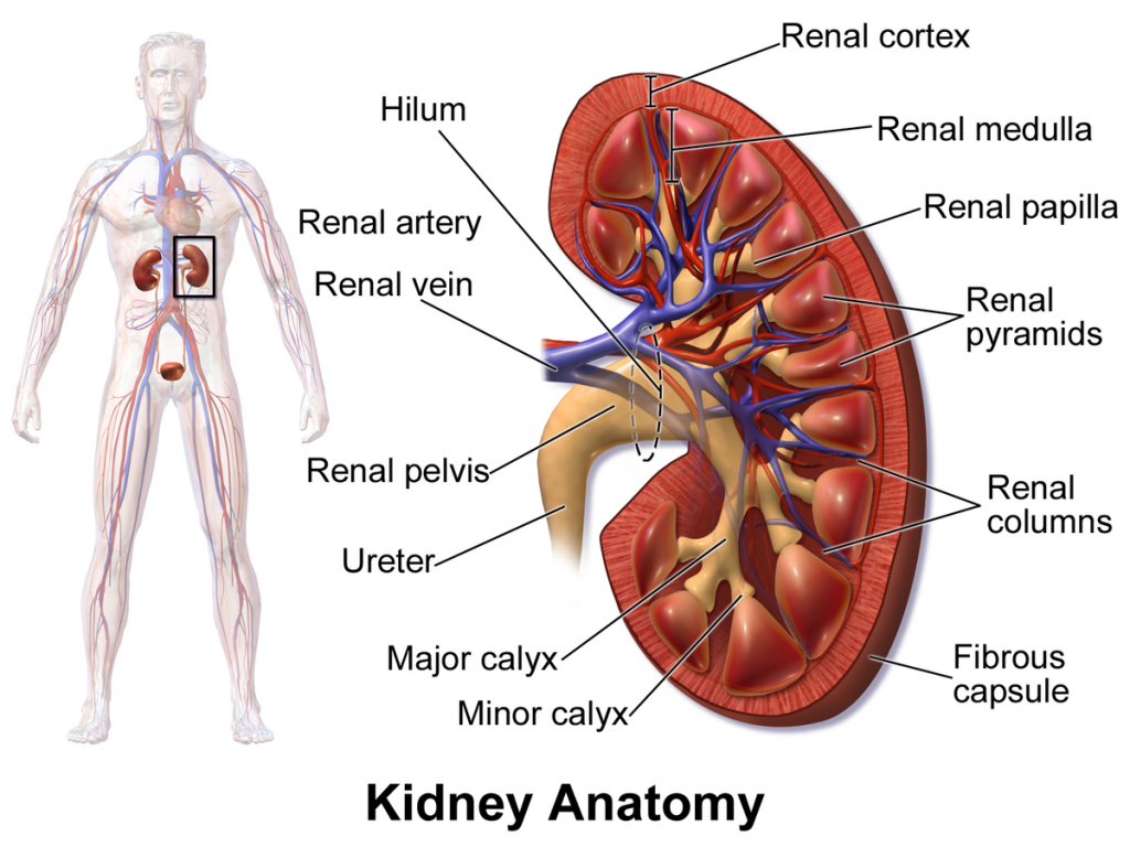
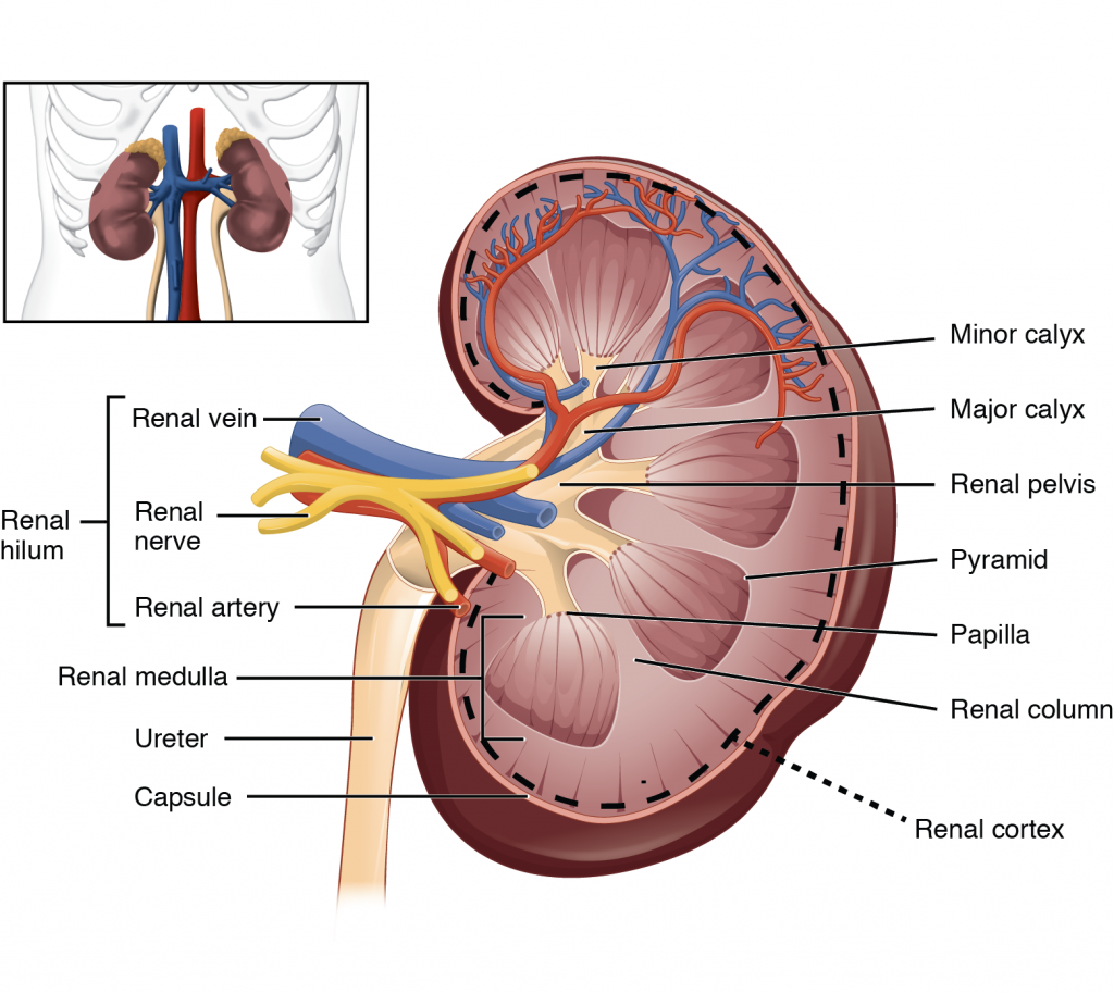
Kidney is a shapeless organ. A groove in the middle is called hilum or renal hilum. Through which the structures of renal artery, renal vein, nerves, lymph vessels and ureter enter and exit.
When the kidney is viewed in a longitudinal section, it is seen to be distributed into three kidney structures.
- Fibrous capsule.
It is part of the fibrous tissue that surrounds the kidney. This membrane is arranged around the kidney. Which acts as a layer to protect and maintain the shape of the kidney.
- Cortex.
It is redis brown in color made up of tissue. Which is located under the kidney capsule.
- Medulla.
In the kidney, the inner part from the cortex is called the medulla. It also has redish brown color. The triangular pyramid-like structures are called renal pyramids. The base part of this renal pyramid is towards the cortex and the pointed part of the pyramid i.e. the part of the renal papilla is arranged inwards towards the hilum.
The renal papilla forms an anterior cup-like structure called the calyx. The part with larger space is called major calyx and the part with smaller space is called minor calyx. The minor calyx opens into the major calyx. Beyond this calyx is the wide funnel-shaped portion called the renal pelvis.
The urine filtered by the kidney falls into the wide part of the calyx, the funnel shape, i.e. the renal pelvis. Urine collects here and then exits the kidney through a narrow structure called the ureter from the renal pelvis anteriorly to the urinary bladder.
Functions of the Kidney..
Kidneys are mainly responsible for urine formation.
Kidneys filter the blood and remove the waste products through urine.
The function of the kidney is to maintain the normal balance of electrolytes.
It works to maintain blood pH.
The body functions to remove waste products accumulated at the end of metabolism from the body.
Kidneys secrete a hormone called erythropoietin which plays a very important role in the production of RBCs.
Kidneys secrete a hormone called renin which plays a very important role in maintaining blood pressure.
Kidneys are responsible for maintaining water balance in the body.
- Lung
Lung is an important organ of respiratory system. They are located on either side of the mediastinum space in the thoracic cavity, totaling 2 in number.
The lungs take in oxygen from the atmosphere and expel carbon dioxide from the body.
Lungs are located in the thoracic cavity in number of 2. They are conical in shape.
The lungs are separated from the heart and the thoracic cavity by the mediastinum space.
The lungs are made up of spongy tissue within which many air field cavities are located. Its color is brown or grey.
The weight of the right lung is approximately 625 grams and the weight of the left lung is approximately 575 grams. The right lung is heavier in weight and larger in structure than the left lung.
Lungs are divided into lobes. The right lung has 3 lobes, namely the superior lobe, middle lobe and inferior lobe, while the left lung has 2 lobes, the superior lobe and the inferior lobe. These lobes are separated by a fissure. There are two fissures in the right lung. A fissure is seen in the left lung.
Lungs are classified into the following parts
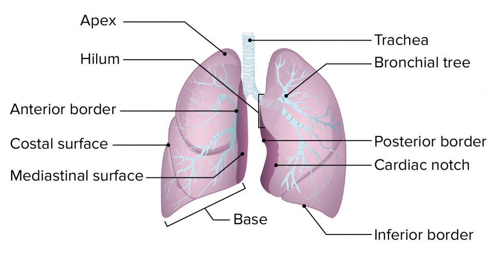
- Apex..
The upper triangular and round part of lunge is called Apex.Which is seen up to the level of the clavicle bone.
- Bayes.
The lower broad part of the lung is called the base. The base portion is attached to the diaphragm at the bottom. This part is of concave shape.
- Anterior border..
It is thin. It is shorter than the posterior border. It has a cardiac notch. In which the heart part is arranged.
- Posterior border..
It is thick. It is found from the 7th cervical vertebra to the 10th thoracic vertebra.
- Inferior border..
It is located at the bottom of the lung. It separates the costal surface and the medial surface. The costal surface is large and convex. It is in contact with the costal pleura. It is attached to the ribs and intercostal muscles by costal cartilage.
- Medial surface
It is concave. There is a groove in the middle of it which is called hilum. The hilum lies at the level of the fifth, sixth and seventh thoracic vertebrae. Through this hilum, bronchi, pulmonary blood vessels, lymphatic vessels and nerves enter and exit each lobe of the lung.
pleura..
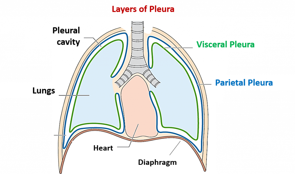
The pleura is the serous membrane surrounding both lungs. Which is found in double layer. The outer layer is known as parietal pleura and the inner layer is known as visceral pleura.
Between the parietal pleura and the visceral pleura lies a cavity called the pleural cavity. There is serous fluid which is also called pleural fluid.
Due to the pleural fluid in the pleural cavity, the two layers do not rub against each other and due to this, the lungs get enough space for expansion.
Difference between Right Lung and Left Lung..
Right lung has two fissures to separate the lobes while left lung has only one fissure to separate the lobes.
Right lung has three lobes superior, middle and inferior while left lung has two lobes superior and inferior.
The right lung has a straight anterior border while the left lung has an interrupted anterior border because of the cardiac notch on the side of the left lung where the heart is located.
Right lung is heavier and larger in weight. Its weight is approximately 625 grams. While the left lung is lighter and smaller in weight. Its approximate weight is 575 grams.
The right lung is short and wide while the left lung is long and narrow.
Function of Lung..
It is an important organ for respiration.
Through inspiration, oxygen reaches the lungs and mixes with blood to deliver oxygen to the whole body.
The waste product of the body, carbon dioxide, is removed from the body through the act of exhalation.
Oxygenated blood is delivered to the heart through the pulmonary circulation.
The body excretes excess water through the act of respiration.
Other gaseous wastes produced in the body are removed from the body through the act of exhalation.
- BRAIN.
The normal weight of brain is 1200 to 1400 grams
It is located in the cranium cvt
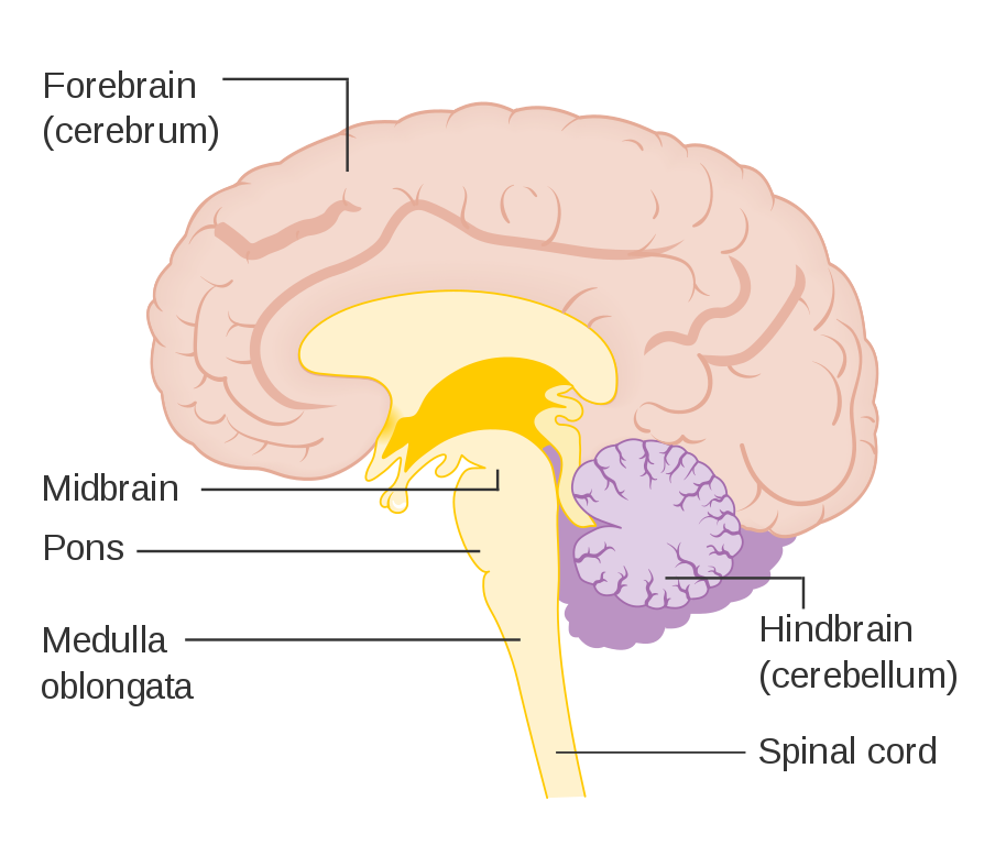
Its parts are as follows
Cerebrum
Mead brain
Pons Veroli
Medulla oblongata
The cerebellum
Blood supply to the brain is from anastomoses of several arteries of the circle of Wills.
CEREBRUM.
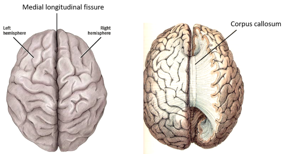
The cerebrum is composed of the right and left cerebral hemispheres.
The two cerebral hemispheres are connected by the corpus callosum. Each hemisphere has a cavity called a lateral ventricle.
Gray matter is located in the structure of the superficial part of the cerebrum called the cerebral cortex. White matter is located in the deep part of the cerebrum.
The cerebral cortex consists of elevated areas called gyri or convolutions and these are separated by notches called fissures or fissures.
The deepest of these fissures is the longitudinal fissure that separates the right and left hemispheres.
The hemispheres of the cerebrum are divided into different lobes which are as follows.
•Frontal lobe
•Parietal lobe
•Temporal lobe
•Occipital lobe..
- An important fissure on the side of the cerebrum;
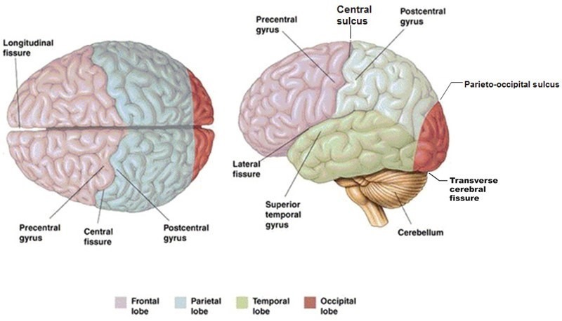
•Longitudinal sulcus or fissure which is the deepest and divides the cerebrum into two hemispheres.
•Central sulcus which lies between the frontal and parietal lobes.
- Lateral sulcus which is a deep groove and separates the temporal lobe from the frontal and parietal lobes.
- Parieto-occipital sulcus which separates the occipital lobe from the two parietal lobes….
•Nerve fiber tracks are located inside the cerebrum viz
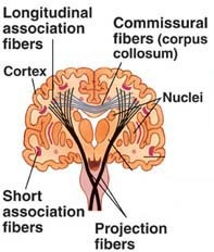
•Association fibers
•Commissural fibers
•Projection fibers
These fibers are interconnected and connect one area to another area and help in the transmission of impulses.
FUNCTIONS OF THE CEREBRUM.
- The control of intelligence, memory, reasoning, thinking, speaking, reading, writing, etc. is seen in the cerebrum.
2.No control is seen for the perception of sensory perception like pain, temperature, touch, sight, hearing, taste, smell etc.
- Control of skeletal muscle contraction is seen from this area.
- Functional areas of the Cerebrum.
There are several areas in the hemispheres of the cerebrum which are as follows.
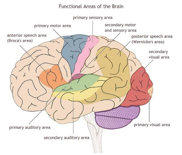
- Motor area Which is mainly located in the anterior part of the cerebral hemisphere. In which the following areas are found.
1.Primary motor area which is located in the frontal lobe and controls the voluntary contraction of muscles.
- Motor speech area also called Broca’s area which is located in the frontal lobe.
•Sensory Areas of Cerebrum
•Primary somatosensory area or general area located in the parietal lobe behind the central sulcus is the sensory area for touch pain and temperature.
•Primary visual area located in the occipital lobe that interprets vision.
•Primary auditory area located in the temporal lobe and associated with hearing.
•Primary gestatory area located behind the central sulcus in the parietal cortex which is connected to the testes.
- Primary olfactory area which is located in the temporal lobe. Area connected with smell is..
OTHER AREAS OF CEREBRUM.
There are some special areas in the cerebrum which are connected with the transmission of impulses and other important functions which are as follows.
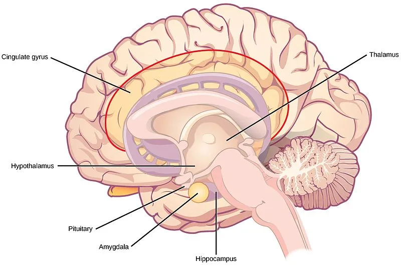
1.Basal ganglia..it is an area located in each cerebral hemisphere which is responsible for co-ordination of muscle tone, posture and voluntary muscle movement..
- Thalamus..It is located in the middle of the brain at the top of the brain stem. It acts like a relay station of sensory impulses coming to the sensory area of the cerebral cortex, which leads to the interpretation of proper sensation and some movement control.
- Hypothalamus…Hypothalamus is made up of a group of nerve cells located below the thalamus whose functions are as follows..
- Hypothalamus controls the following functions.
•Body temperature
- Hunger and thirst
•Emotional reaction
•Autonomic Nervous System
•Sexual behavior
•Biological clock or circadian rhythm…
- Secretion of certain hormones All of the above is done by the hypothalamus.
MENINGIES.
•Dura mater
The dura mater is the outermost layer of the brain and spinal cord. The dura mater is dense and tough and lies in a double layer. The outer layer forms the outer lining and the inner layer is attached to it except that the dura mater forms a partition in some places.
•Arachnoid matter
It is a delicate serous membrane that lies between the dura mater and the basal mater and contains collagen fibers and elastic fibers. The space between the skull bone and the dura mater is called the epi dural space
The space below the dura mater is called the sub-dural space.
The space between the arachnoid mater and the base mater is called the subarachnoid space where the cerebrospinal fluid is located.
- Base Matter It is the innermost layer. It is a thin and vascular membrane attached to the spinal cord which contains a large number of collagen fibers and fine elastic fibers and also contains many blood vessels which supply nutrition and oxygen to the spinal cord.
VENTRICLES OF THE BRAIN.
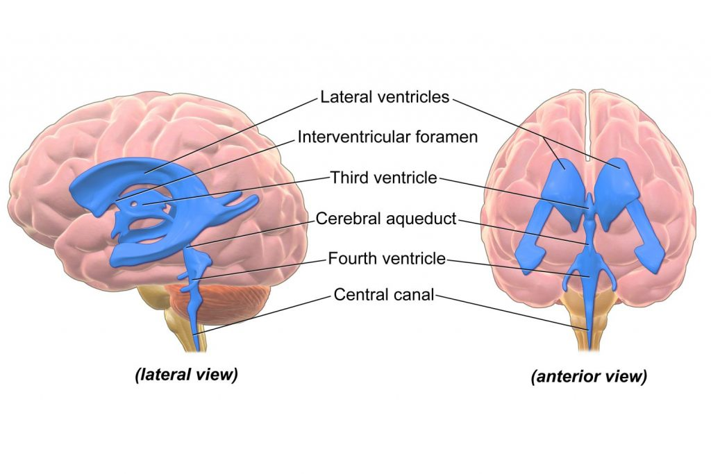
Cavities within the brain are called ventricles in which fluid is produced and circulates around the brain and spinal cord.
- Right and Left (Lateral Ventricles)
- Third ventricle
- Fourth ventricle
1.Lateral ventricle
These cvts lie beneath the corpus callosum of both cerebral hemispheres separated by the septum leucidum. Its lining is composed of epithelium tissue and its wall contains a network of capillaries called choroid plexuses from which cerebrospinal fluid is produced. It connects to the third ventricle through the interventricular foramen.
- Third ventricle The narrow cavity below the right and left lateral ventricles is called the third ventricle. It connects to the fourth ventricle through the cerebral aqueduct
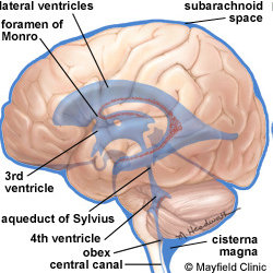
- Fourth ventricle It is diamond shaped, lies below the third ventricle and is continuous with the central canal of the spinal cord. The foramen (Lashka and Magendi) on its roof are connected to the subarachnoid space.
BRAIN STEM.
The brain stem consists of the following parts
- Mid brain
2.Pons Veroli
- Medulla oblongata
•Mid brain
It forms the upper part of the brain stem. On its side, the cerebral aqueduct connects the third and fourth ventricles.
At the top of the midbrain are the important centers of vision and hearing.
In the lower part of the midbrain passes the motor pathway that goes through the pons veroli and medulla oblongata to the spinal cord.
The midbrain contains the control centers for balance and eye movement.
•Pons Veroli
It forms the middle part of the brain stem.
It also has the same ascending and descending nerve pathways as the midbrain, and many nerve tracks are connected to the cerebellum and cerebellar cortex.
It helps in the transmission of impulses.
•Medulla oblongata
It forms the lowest part of the brain stem and connects the pons veroli to the spinal cord
It is approximately 2.5 cm long and has the following centers.
1.Respiratory center
- Cardio Vascular Center
3.Vazzo Motor Centre
- Reflex center for vomiting, gagging and swallowing.
Medulla oblongata has some special effects which are as follows
- Descending motor pathways from the medulla cross and pass through the spinal cord, providing impulses primarily to the skeletal muscles.
- Similar to the motor pathway, the sensory pathway also crosses the medulla to the brain.
- Cardio vascular center located in medulla which controls heart rate and force Sympathetic stimulation increases heart rate and force while parasympathetic stimulation decreases heart rate and force.
- The medulla houses the respiratory center which controls the rate and depth of respiration in which the intercostal muscles and dia pharm receive nerve impulses for inspiration and expiration.
- The medulla houses the vasomotor center which controls the diameter of the blood vessels causing vasoconstriction and vasodilatation.
- The reflex center in the medulla controls vomiting, gagging and hiccups, this is also a protective response.
CEREBELLUM.
It is the second largest area of the brain behind the medulla and pons veroli.
It is separated from the cerebrum by a transverse fissure where the inner layer of dura mater is inserted to form the tentorium cerebelli.
•The cerebellum performs the following functions
- It regulates posture and postural activity
- It plays an important role in coordination of muscles
- Plays an important role in maintaining body balance.
IMPORTANT BONES
- SKULL:
Skull is found in the upper part of the body above the vertebral column.
In which there are 2 parts as follows.
- cranium
- face
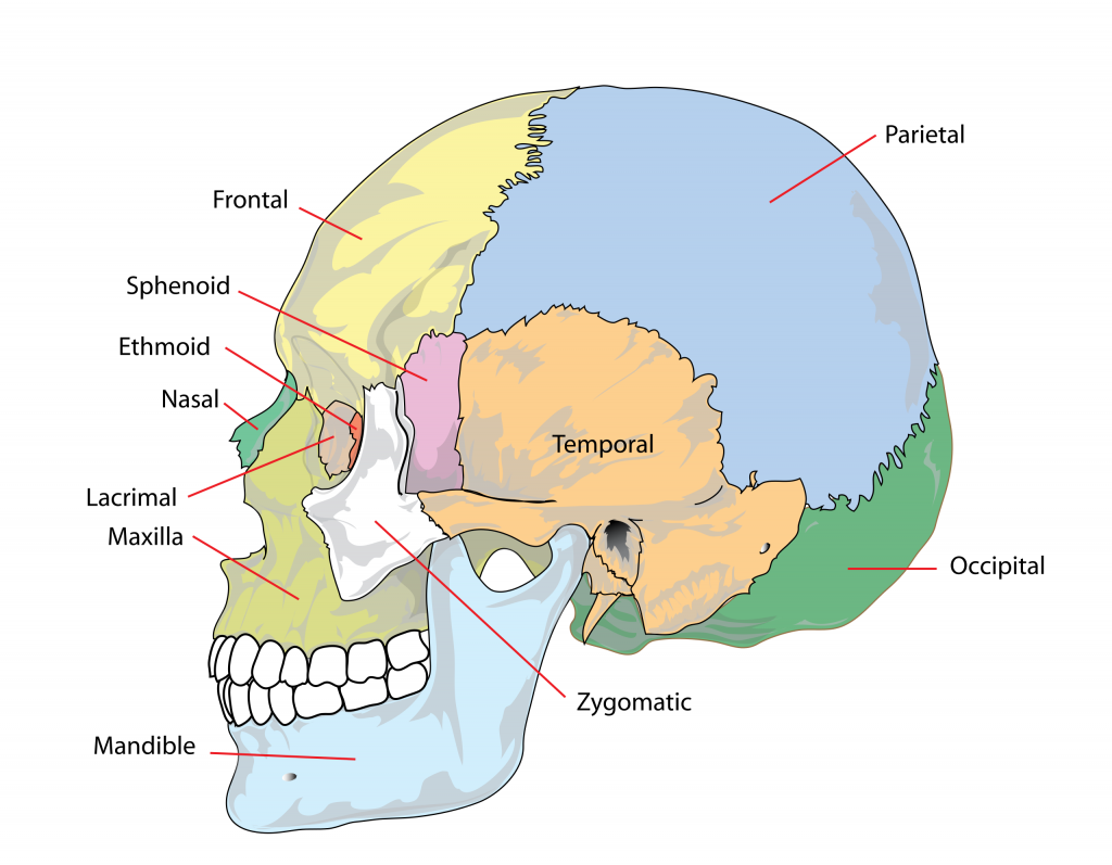
- Cranium:
The upper part of the skull bone is known as cranium.
8 irregular cranial bones together form the cranial cavity.
The human brain is located in the cranial cavity.
The lower part of the cranium is known as the base and the upper part as the vault. Its shape is like a dome shape.
Cranial bones join each other to form immovable joints known as sutures.
Sutures of the cranium bone.
The cranium is made up of irregular bones. These flat bones join to form an immovable joint. This joint is known as suture. Indications for cranium cavity are as follows.
Coronal suture
Suture between frontal bone and 2 parietal bones is called coronal suture.
Sagittal suture.
Suture between 2 parietal bones is called sagittal suture.
Lambdoidal suture.
2 The suture between the parietal bone and the occipital bone is called the lambdoidal suture.
Squamous suture.
The suture between the parietal bone and the temporal bone is called the squamous suture.
The above suture forms a depression like structure between the joints called fontanelles. These fontanelles are in the number of 2 in the infant skull.
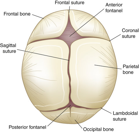
The Anterior Fontanelle is located near the junction where the coronal and sagittal sutures join at the front of the skull. It can also be known as bregma. It is big in size. By the time the child is 1.5 years old, it closes due to skeletal maturity (Growth). It is of diamond shape.
The Posterior Fontanelle is located near the junction where the sagittal and lambdoidal sutures join at the back of the skull. It can also be known as lemda. It is small in size. By the time the child is 6 to 8 weeks old, it closes due to skeletal maturity (Growth). It is of tri-angular shape.
The cranium in the skull is made up of many different flat and irregular bones. Cranium consists of 8 bones in total, which are as follows:
- Frontal bone 1
- Parietal bone 2
- Temporal bone 2
- Occipital bone 1
- Sphenoid bone 1
- Ethmoid bone 1
Frontal bone.
These bones are numbered 1 in the most anterior part of the cranium KV. It is also called fore head bone. It lies above the orbital cavity and nasal cavity. Above the orbital cvt there is a raised margin on both sides of the frontal bone, which is called the supra-orbital margin. A foramen is also located near this margin. It is called supra orbital foramen. The part between the two orbital margins is known as the glabella.
Parietal bone.
There are 2 of these bones in the cranial cavity and they form the roof of the cranial cavity. It is the bone attached to all the sutures of the cranium cavity.
Temporal bone.
It is in the number of 2. On either side of the cranium cvt is a bone near the ear. The temporal bone is connected to the zygomatic bone by the zygomatic process. It also forms the only movable joint of the skull with the mandible bone, the temporomandibular joint. The mastoid portion of the temporal bone is connected to the middle ear by an air cell.
Occipital bone.
It is the number 1 bone in the cranium cavity. It forms the back of the skull and part of the base. At the bottom of this bone is a large foramen, known as the foramen magnum. Through which the spinal cord emerging from the brain passes.
On either side of this foramen there are raised parts. Which is called condyle. The first vertebra of the vertebral column joins the atlas forming a hinge joint. The most prominent part of the back of the occipital bone is called the occipital protuberance.
Ethmoid bone.
It is a bone located anteriorly at the base of the cranium cvt. It is in the number of 1. It is a cubical shaped bone. The olfactory nerve passes through its path from the nose to the brain.
Sphenoid bone.
It is in the number of 1. It lies anterior to the occipital bone at the base of the cranium cvt. It is a butterfly shaped bone. There is a depression in the middle of it. It is known as hypophysial fossa or sella tarsica. Pituitary gland is located in this part. This bone is connected to all the bones of the cranium cavity.
Facial bones.
The bones of the face are called facial bones. It is 14 in total. These bones are found as follows.
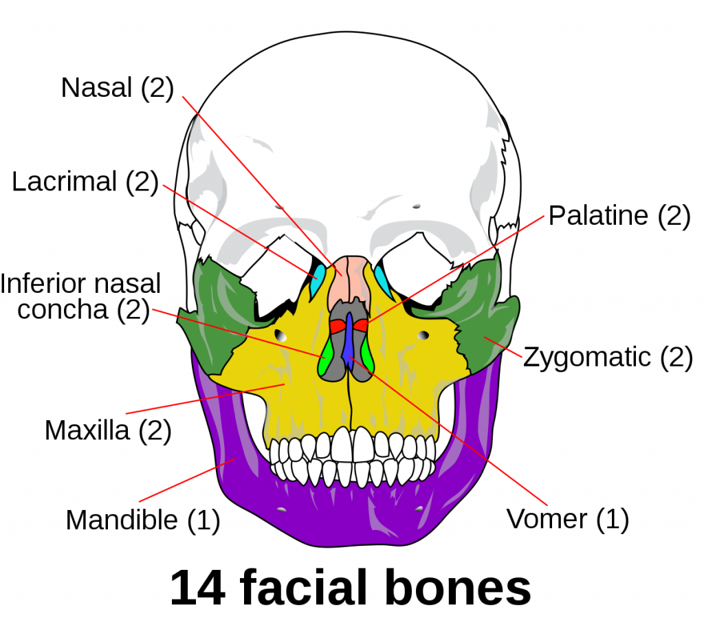
Maxilla bone.
It is also known as upper jo bone and is numbered 2. Upper teeth come in its margin. This bone forms the roof of the mouth and the floor of the nasal cavity and orbital cavity. A portion of this bone also forms the hard palate.
Zygomatic bone.
It is known as cheek bone. They are in number of 2. It forms the upper part of the cheek. This bone articulates anteriorly with the maxilla bone and with the temporal bone.
Mandible bone.
It is the bone at the bottom of the face. It is the only movable and strong bone of the skull. Which plays a very important role in chewing. It is in the number of 1.
The upper edge of this bone is known as the alveolar ridge. Teeth are arranged along this ridge.
The front part of this bone is called the body. A curved part is called an angle. From there the flattened part on the upper side is called the ramus. The next part is divided into two processes. In which coronoid process and condylar process are seen.
Muscles attach to the coronoid process while the temporal bone attaches to the condylar process to form the temporomandibular joint.
Nasal bone.
They are flat bones numbering 2. It forms the bridge of the nose. It forms the superior and lateral walls of the nasal cavity.
Lacrimal bones.
They are bones in number of 2. It forms the medial wall of the orbital cavity and carries eye secretions to the nose through the nasolacrimal duct. This bone is shaped like a finger nail.
Vomer bone.
It is the number 1 bone. The middle bone of the nasal cavity is attached anteriorly to the septum of cartilage. It separates the nasal cavity into 2 parts. On the upper side it is attached to the ethmoid bone.
Palatine bones.
They are bones in number of 2. It creates a hard palette. It has 2 L shape bones joined together to form part of the palette.
Inferior conchae.
They are in number of 2. It is a bar-shaped bone located in the lateral wall of the nasal cavity. It works by filtering and warming the incoming air.
Common Characteristics of Vertebrae.
All the vertebrae of the vertebral column have the following common characters
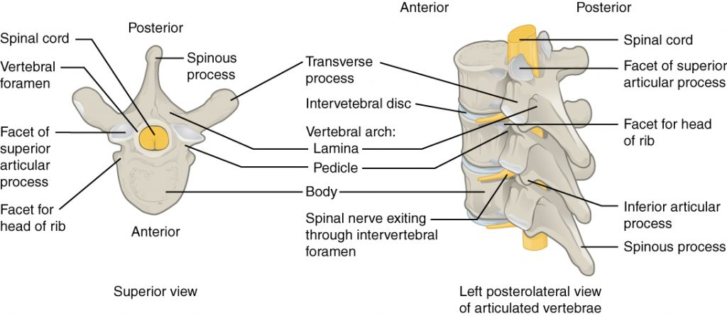
Body
At the front of the vertebra is a flattened body part. Its size varies according to the vertebra of each region. As such, the body segment is found to be smallest in the cervical vertebrae and increases in size towards the thoracic and lumbar vertebrae. The body is the largest and thickest in the lumbar vertebrae
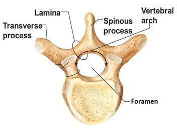
Process of Vertebrae
The two processes extending posteriorly from the body of the vertebra are called pedicles. This pedicle extends posteriorly and forms a portion of a V septum, the portion known as the lamina. From the junction of these laminae, a straight process called the spinous process emerges posteriorly.
At the junction of the vertebra pedicle and the lamina, a horizontal process called the transverse process arises on either side.
Between all these processes a foramen is formed, called the vertebral arch or neural arch. The spinal cord passes through this arch. This arch is the largest found in the cervical vertebra. The opening of this arch becomes smaller as it moves towards the thoracic and lumbar vertebrae.
In addition, the superior surface and inferior surface of each vertebra have articulating processes to join the vertebrae.
Sternum Bone Sternum Bone.
The sternum bone is the bone of the axial skeleton. It is the anterior medial bone in the thoracic cavity.
It is the bone immediately below the skin. Sternum bone is also known as breast bone. The sternum is a flat bone.
The ribs are attached to the sternum bone with the help of costal cartilage. Hence it is a very important bone for building the thoracic cage.
Below the sternum bone is divided into three parts.
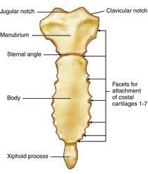
- Manubrium.
The uppermost part of the sternum bone is called the manubrium. It forms the upper broad part of the sternum. It has a rough triangular shape.
The notch in the middle of the uppermost part of the manubrium is called the suprasternal notch. The suprasternal notch is also known as the second jugular notch.
On the lower side of the suprasternal notch of the manubrium, there is a small groove called the clavicular notch. At this notch, the clavicular bone joins and forms the sternoclavicular joint.
The first pair of ribs are attached to the menorium by the costal cartilage.
The second pair of ribs are joined by costal cartilages near the angle between the manubrium and the body.
- Body…
The body forms the longest part between the sternum bone no. On its upper side is the manubrium and on its lower side is the xiphoid process. The part between these two is known as the sternum body.
There are many small grooves on both sides of the body. On either side of this groove, the third, fourth, fifth, sixth and seventh pairs of ribs are connected with the help of costal cartilage.
- Xiphoid process..
The lowest part of the sternum bone is called the xiphoid process. This part is attached to the muscles of the abdominal wall as well as the diaphragm.
Characteristics of Rib.

A rib is a flat bone. This bone has two extremities and a saft part.
At the posterior end of the rib towards the vertebral end is a flattened part, called the head of the rib. This head part is attached to the body of the thoracic vertebrae.
The narrow part after the head is called the neck. Then there is a raised part called tubercle. Part of this tubercle is attached to the facet of the transverse process of the thoracic vertebra.
The bend of the rib after the tubercle is known as angle of rib.
The middle part of the rib is called the clean part. The superior border of this part is smooth. Also its inferior border is bordered. It has a groove called the costal groove. The costal nerve and blood vessels pass through this groove.
The front part of the surface of the rib is called the anterior surface. Intercostal muscles are attached to it. When the posterior surface of the rib is in contact with the pleura of the lung.
Towards the end of the anterior side of the rib i.e. this end is also called the sternum end. Where the anterior side of the rib is attached to the sternum bone with the help of costal cartilage.
The space between two ribs is called intercostal space. Intercostal muscles are attached to this part. Due to the contraction and relaxation of these intercostal muscles, respiration occurs and the diameter of the thoracic cavity fluctuates.
Pelvis.
A pelvis is a basin-shaped cavity. Which is formed by two innominate bones, a sacrum and coccyx bone.
This creates a rounded area in the middle of the pelvis, called the pelvic brim. The part below the pelvis is called the true pelvis because of its importance in obstetrical terms. The upper part of the pelvic brim is called the false pelvis.
Innominate bone..
The innominate bone is the bone of the pelvic girdle. It is also known as hip bone.
They are two in number in the body. It is located one on both the right side and left side of the pelvic cavity. Both the innominate bones articulate with the sacrum bone behind and form the pelvic cavity.
The innominate bone is the larger bone. It is a flat and irregular type of bone.
Each innominate bone consists of three bones
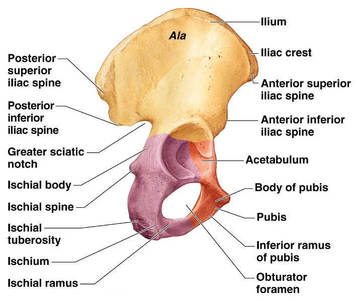
Ilium
Two ischiums
pubis.
Ilium
Innominate Bone The ma ilium bone is a flat bone on the upper side. At its uppermost point there is a ridge called the iliac crest. It falls below the iliac crest.
Anterior superior iliac spine (top of front)
Anterior inferior iliac spine (at the bottom of the front)
Posterior superior iliac spine (upper back)
Posterior inferior iliac spine (lower back)
The ilium bone forms the sacroiliac joint where it joins the sacrum bone at the back.
There is a big groove at the bottom of this joint. The notch is called the greater sciatic notch. From where the sciatic nerve and blood vessels pass to the lower extremities.
The gluteal muscles attach to the posterior surface of the ilium bone. And that makes up the gluteal region.
The anterior surface of the ilium bone is known as the iliac fossa. In this part there is a depressed part where the muscles are attached.
ischium
Below and behind the ilium bone is the ischium bone.
Between the ilium bone and the ischium bone, the posterior side is an inferior pointed part. This part is called ischial spine.
Below it lies a small notch called the laser sciatic notch.
This laser has a strong thick process below the sciatic notch. Which is called ischial tuberosity. Body no weight comes on this part while sitting in sitting position. This has a stronger structure than a weight beer.
pubis
The pubis bone forms the most anterior part of the innominate bone. Both the innominate bones of the pubis bone join anteriorly to form the symphysis pubis joint.
There is a large foramen at the bottom of this pubis bone. It is called obturator foramen. Through which nerves and blood vessels pass downwards towards the extremity.
The three bones ilium, ischium and pubis located in the hip bone form a cavity-like structure called the acetabulum cavity. The head of the femur bone joins this KV and forms the hip joint.
Femur Bone (Femur Bone).
The femur bone is the bone in the lower extremity. It is the longest and strongest bone among all the bones in the body.
This bone has two extremities and a smooth part. Which are as follows.
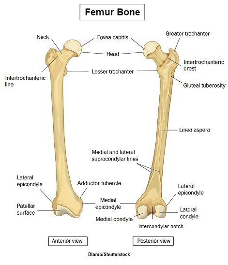
Upper extremity.
The uppermost one-third of the femur bone is called the upper extremity. It has the following structure.
head..
The frontmost part of the femur bone is the round bone part.The part which is called head of femur.
This round part joins with the acetabulum cavity of the innominate bone to form the hip joint.
The hip joint is a synovial joint so all the characteristics of a synovial joint can be seen here.
Neck..
The narrow part after the head is called the neck. Which is found in four to five centimeters long and round shape.
Greater trochanter and lesser trochanter..
Where the neck ends, two large rough and raised portions of bone are seen. Which is called trochanter.
The larger part that is raised on the outside is called the greater trochanter.While the small portion raised on the inner side is called the lesser trochanter. The line connecting these parts of the trochanter is called the intertrochanteric line. This part is where the muscles are attached.
After the upper extremity is completed, the middle part of the bone, i.e. the middle part of the bone, is known as the saft. Its structure is as follows.
Linea Aspera..
A bony ridge is raised on the posterior side of the face.It is called linea aspera. This part is where the muscles are attached. The smooth part of the femur bone is cylindrical and round in shape.
Lower extremity..
The lower one-third of the femur bone is called the lower activity. In which the lower side is seen as two rounded bony parts. Which is called condyle. The condyle on the medial side is the medial condyle and the lateral side is the lateral condyle. The part separating these two condyles is called the intercondylar notch.
The anterior surface of this condyle is a smooth surface called the patellar surface where the patella bone articulates.
A triangular surface is formed at the back of the lower extremity of the femur bone. It is called the popliteal surface. Popliteal vessels and nerves are seen in this part.
Tibia Bone.
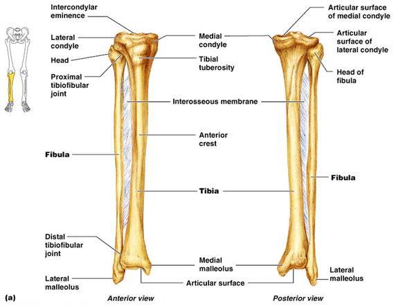
It is also known as shin bone. The tibia is a long bone in the lower extremity. It has two extremity and saft. This bone is a strong and weighty beer bone.
The upper extremity of the tibia bone has two condylar surfaces on the superior surface. The lateral and medial condyles of the femur bone join this surface to form the knee joint.
On the anterior surface of the tibia bone is a rough raised area called tuberosity of tibia.
The fibula bone joins on the lateral side of the tibia bone to form the proximal tibiofibular joint.
The smooth part of the tibia bone is irregularly triangular. There is a sharp crest on its anterior surface, known as crest of tibia. The part of this crest that lies immediately under the skin can be felt by touching with the hand.
A smooth elongated process is present on the medial side of the lower extremity of the tibia bone. It is called medial malleolus. This part joins with the tarsal bone to form the ankle joint.
The lower extremity joins the tibia and fibula to form the distal tibiofibular joint.