ENGLISH-microbiology unit-ii-Micro Organisms
UNIT-2-Micro Organisms(PART-1)
AS PER INC SYLLABUS
Micro Organisms
a) Classification, characteristics,
(Structure, size, method and rate of
reproduction)
b) Normal flora of the body.
c) Pathogenesis & common diseases.
d) Methods for study of microbes,
culture & isolation of microbes.
1.Microorganisms
Microorganisms are organisms that are so small that they can only be seen under a microscope (not with the naked eye). Thus, microorganisms are very microscopic, Unicellular living organisms. They are not only microscopic but each microorganism has its own distinct existence.
Microorganisms have existed on earth for many years and the reason for many processes is these microorganisms with their different characters.
Classification Of Micro organism :-
- 1.BACTERIA (Bacteria)
- 2.FUNGI (Fungi)
- 3.RECKETTSIAE (Rickettsia)
- 4.SPIROCHAETES (Spirochaetes)
- 5.PROTOZOA (Protozoa)
- 6.WARMS
- 7.VIRUS (Virus)
- 8.MYCOPLASMAS (Myco plasma)
- 9.CHLAMYDIAS (Chlamydias)
- 10. BACTEROIDS AND FUSOBACTERIA (Bacteroides and Fusobacteria)
- 11.Algae
- other parasites
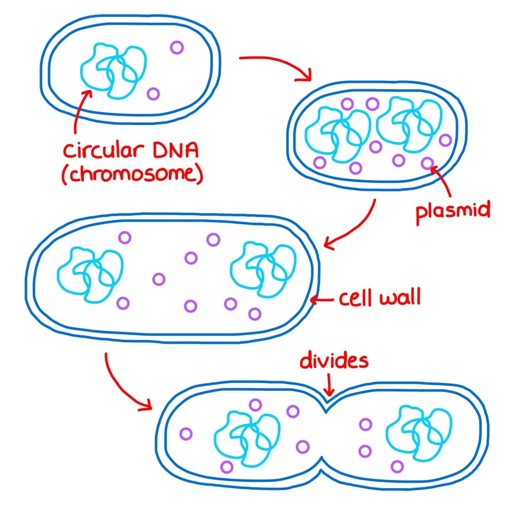
1.Bacteria
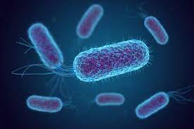
- Bacteria are unicellular (single-celled organisms).
- Their DNA is circular
- They do not contain any other types of organelles (small organelles)
- The unit Micron (μ) (micron) is used to measure it.
- 1 micron (μ) =1/1000 mm
- The normal size of bacteria is 1μ to 0.5 μ in diameter
- Its length>is usually 3 μ to 10 μ in length.
- Width 0.1 μ to 2 μ
- In some cases Size of 100 μ is seen
- Bacteria can be seen through a normal microscope.
♦ Classification of bacteria based on their shape:
Bacteria are classified based on their shape as follows.
- Cocci (cocci)-These are not round in shape
- Baccili (bacilli)-These are not cylindrical in shape
- Spiral (spiral)– These are twisted.
1.Cocci (cocci)–
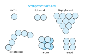
A.Micrococci (Micrococci)– These are single which are also known as coccus
B.Diplococci (Diplococci)– These are found in pairs
C.StrepteptoCocci (Streptococci)-These are found in chains
D.Styphylococci– These are found in groups
E.Tetrad (Tetrad)– These are found in groups of four.
F.Sarcina– These are found in groups of eight Is
2.Baccili (bacilli-rod)–

1.Cocobacillus– This is a single one which is also known as coccus
2.Diplobacili-These are found in pairs
3.Sreptobacili-These are found in chains
4.Palisades– A group of bacilli that form a fence of spikes to protect them from attack
3.Spiral– They are twisted

These include coma-shaped bacteria like Vibrio cholerae, Treponema pallidum, etc.
♦ Classification of bacteria based on their flagella
The tiny hair-like protein fibers found in motile bacteria and attached to the cell are called flagella.
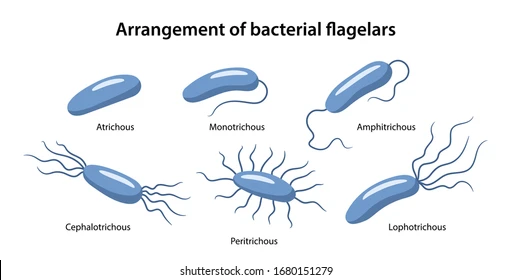
- Atrichous:- If the bacteria does not have a single flagella.
- Monotrichous:- Flagella are found at one end of the cell. E.g. Cholera Vibrio
- Amphitrichous:- Two flagella are found at opposite ends of the cell. E.g. : Alceligenes feacales.
- Lophotrichous:- Found in tufts of grass at one end of the cell. Eg. : Pseudomonas
- Cephalotrichous:- Found in tufts of grass at both ends of the cell
- Peritrichate:- Found in many flagella around the cell. Eg. : Typhoid bacilli
Morphology of Bacteria
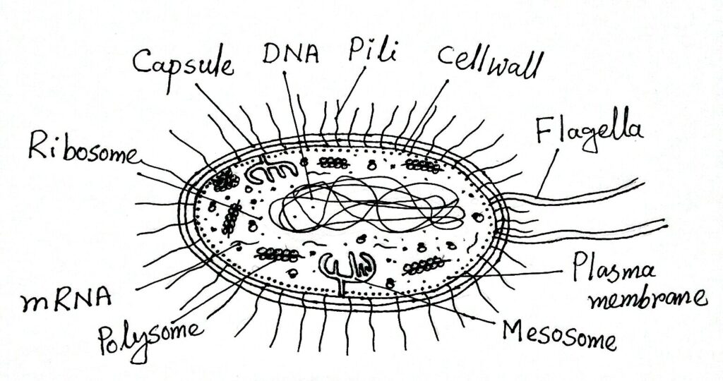
Fimbria/ Pilli (Fimbria/Pilli):
- These are hair-like structures of bacteria.
- Helps bacteria to stick to surfaces.
- Common pili (almost always called fimbriae) are commonly involved in the specific attachment of prokaryotes to natural surfaces.
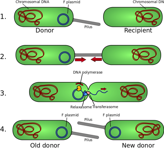
- Stabilize bacteria during transfer DNA during conjugation.
capsule (capsule)
- A thick layer of membrane surrounds most bacterial cells. This is known as the capsule. The distinction between the cell wall and the capsule is not always clear. When the capsule is in the form of loose secretion, it is called a slim layer. If it is very thin then it is called Micro – Capsule.
- It is made of polysaccharide, it inhibits the phagocytosis process.
- Contribution in virulence (ability to produce disease).
Cell wall
- Capsule’s It is a relatively stiff covering and gives shape to the bacterial cell. It varies in thickness, hardness and chemical structure, and is used to classify bacteria. Gram positive and Gram negative bacteria.
- Gram Positive bacteria have a relatively simple chemical structure than Gram Negative bacteria.
- Cell Wall is made up of Cellulose, Protein & Made of lipids.
Cell membrane (Cell membrane)
- Each cell has a barrier separating the inside and outside, known as the cell membrane.
- Cell membranes are very thin, flexible, and weak. And also known as Cytoplasmic membrane.
- Main function selective permeability regulation
- nutrients & Controls the path of waste products. (made of Phospholipid & Protein)
- The breakdown in this leads to cell death.
Cytoplasm
- It is a complex mixture of many types of micro molecules (Proteins, nucleicacid, Polysaccharides & Lipids) Organic or Inorganic solutes of colloid.
- They include Ribosomes, mesosomes, vacuoles and inclusions.
Rebosome, Mesosome, Inclusion (Ribosome, Mesosome Inclusion)
- Ribosome’s :- It is a center for the synthesis of Protein and RNA.
- Mesosomes :- It is a site of Respiratory enzymes.
- Inclusion :- They are sources of stored energy in bacterial cells.
- Vaculoes (Vacules ):- Fluid filled cavity
- Volutin Granules :- storage of inorganic components
Nucleus
- A tightly coiled structure within the cytoplasm that contains DNA (Deoxyribose nucleic acid). Bacteria do not have a developed nucleus like animals,
- It replicates by binary fission.
- Extrachromosome of DNA in bacteria called plasmid.
Bacterial endospore
- They are highly resistance to the environment
- Endospores are formed by a few bacteria as an intracellular structure .
- Many types of bacteria form a covering inside the parent cell under unfavorable conditions which is known as a spore. In this process the protoplasm inside the cell becomes concentrated (thickened) and forms a small sphere, which acts as a thick cell wall. Each cell usually produces an endospore which can survive even at very high temperatures.
Reproduction (Reproduction of Bacteria) :-
Binary Fission of Bacterial cell (Bacterial Cell Binary Fission)
Conjugation
2. Fungi (Fungi)
- It is found in filaments and is produced in a substance like stale bread. It is a non-pathogenic micro-organism. Its cell wall is hard. It takes its food from soluble nutrient substance through the cell wall by diffusion process.
- Ex.unicellular Mould, Yeats (Mold,Yeast)
- Multicellular- Mushroom
3.Rickettsiae
- This is a simple rod shape spiral organism First observed by Howard Taylor in 1909.
- Its size is smaller than bacteria. Length is 3 microns and diameter is 0.5 micron.
- It does not move much.
- Despite its small size, it passes many filters. And many do not pass through the filter.
- Whether it is a plant or animal micro-organism is not yet known.
- The diseases caused by this are not commonly seen in India,
- For example,Q- fever,
- Rocky mountain fever
- Trench fever (Trench Fever)
- It is a form between bacteria and viruses, because it has some properties of both bacteria and viruses.
4.Spirochetes
- Spirochetes have characteristics of both protozoa and bacteria Contains.
- 1- Venereal Treponeme
Ex. Treponeme Pellidum causes syphilis in humans
- 2-Non venereal Treponeme
ex.Lepto-spira- causing spirochetal jaundice
5.Protozoa (Protozoa)
- These are Animal Unicellular micro-organisms. Their structure is more complex than bacteria. They are of different types. And pathogens are
- i)Entamoeba – Dysentery, Hepatitis, Liver abscess
- (ii)Giardia – Diarrhoea
- (iii) Trichomonas hominis – Diarrhoea
- (iv) Trichomonas Vaginalis – Vaginitis
- (v) Trypanosoma – Sleeping sickness (Africa)
- (vi) Leishmania – Kala azar
- (vii) Plasmodium vivex – Beneign tertian malaria (Malaria)
- Plasmodium falciparum – Malignant Malaria
- Plasmodium malaria – Quartan malariamulurie.
- Plasmodium ovale-Beneign tertian malaria
- Belantidum (B.coli)-Desentry (dysentery)
Worm
- 1) Trematoda
They are mostly found in the liver and intestine and are responsible for Cirrhosis of liver, persistent diarrhoea, Gastro intestinal irritation, Enlarged liver, Eosinophil Urticaria, Haematuria (blood in urine).
- 2)Cestoda
They are found in the intestine and cause intestinal conditions.
Nematoda
They are found in the intestine, lymphatic vessels subcutaneous tissues. .Allergic reaction, Haemorrhage lesions in heart, Acute appendicitis, G. I. disturbances, ulcer, Rise of eosinaphil count and septic complication are responsible.
Virus
- Virus is a Latin word. Which means “Poison” or “Venom”
- Virus is an intracellular living organism.
- Can only be seen with an electron microscope.
- Diseases such as Polio, Mumps, Rabies, Herpes, Chicken pox, Dengue fever, Hepatitis B etc. are caused by different viruses. Not all Viruses are pathogenic. AIDS is also a disease caused by a virus.
History of virus
- In 1982, Russian biologist Iwanowski first discovered that the T. M. V. virus was responsible for tobacco mosaic disease in tobacco.
- In 1900, Walter Reed discovered the Yellow Fever Virus.
- In 1935, the electron microscope Virus study became possible. (Electron microscope can do 3,00,000 magnification.)
Characteristics of Virus
- Viruses have both living and non-living characteristics.
- Non-living characters of virus
- Viruses are not cellular organizations.
- They do not have protoplasm.
- They do not require respiration or food.
- They do not multiply by binary fission.
- They do not have independent existence.
- External stimuli respond to a They do not.
- Outside the cell, they behave like chemical molecules.
Living characteristics of Virus
- (i) Their reproduction takes place only in living cells
- (ii) They have the ability to grow in size and numbers.
- (iii) Living It mutates its genes through (dissection).
- (Iv) It adapts to its environment naturally.
- Therefore we can say that viruses come between Living and non – livings. But they are considered as living (in organisms) and are studied scientifically
- Viruses may be defined as, “Extremely small obligate intracellular living forms containing only one type of nucleic acid either DNA-RNA.”
- They are different in shape. The shape of a virus of the same type remains the same. But the shape of different types is not the same. They are found in Rod shaped, Cuboidal, Rhomboidal (multisided), needle shaped.
- Virus has a simpler structure than other micro-organisms,
- Virus can be grown artificially in any culture medium, for which living cells are the primary requirement. The following three methods are used for the cultivation of viruses.(i) Animal Inoculation:-This virus is introduced into the living water. (ii) Cultivation in chick embryo (iii) Cultivation in tissue culture:-
Fusobacteria (Fusobacteria )
- Long, thin, spindle shaped. With pointed end or cigar shaped They are found in Gums and tonsils of normal person. They are often associated with certain spirochetes in mixed infection causing fasospirochaetal disease
Bacteroides
- Anaerobic, Rod shaped, gm-ve Bacteria.
- Normally inhabitant in oral, respiratory, intestinal, urogenital cavities of human and animals,
- The infection of bacteroids (e.g. abdominal sepsis) are usually found in association with other organisms.
Non-Pathogenic Organism
- Bacteria with humans There is a very important relationship, some Non-Pathogenic bacteria destroy Pathogenic bacteria, in addition
- Some bacteria attach to the roots of plants and convert nitrogen (Nz) into useful chemicals. Which is used as a chemical fertilizer.
- Helps in making vinegar by making acetic acid from alcohol
- Some bacteria are helpful in making tobacco and rubber.
- Yeast processes flour, mando, and sugar and helps in raising bread.
- Produces a certain type of sodium in ghee, butter, and cheese.
- Yeast and actinomycetes produce antibiotics, which are useful in treating infections.
- Some bacteria help convert toxic gases from decaying or dead animal and plant matter into simple gases. This simple gas is useful as food for the growth and development of plants.
Growth factors of Bacteria
- Growth of bacteria can be seen in two ways
1. Increase in size
2.Increase total number of cells.
To see the growth of bacteria, it is shown in curved graph
- Lag phase :- Increase cell in size but it does not multiply
- Log phase :- Multiply at the exponential phase
- Maximal stationary phase:- Death & growth of bacteria are equal.
- Decline phase:- The progress of death cell of bacteria increases.
Factors affecting Bacterial Growth
- Like every other living organism, microbes also need a suitable environment and nutrients
- growth, maintenance & falls for multiplication
- Nutritional requirements for growth of bacteria :
- Protein or Protons, or nitrogen containing substance.-
- Energy food – Sugar
- Minerals – Sodium – Sugar, starch, beef etc..
- – Sodium chloride in small amount.
- Water in large amount.
- Blood, Glucose etc..
- Even minor changes in the environment affect bacterial growth. Spore forming type is the only one that can protect itself even in adverse conditions. By controlling the environment, we can increase or decrease bacterial growth.
(1) Moisture
- Every bacteria needs water as a nourishing food for growth. In fact, bacteria cannot get food in the absence of water, because all food elements need to be in a liquid state to pass through the bacterial cell wall. Every type of bacteria grows well in an aqueous medium, an environment completely devoid of moisture prevents their growth. Or destroys.
- In addition, cells cannot survive in low or high humidity.
2) Light (Light)
- Most bacteria are killed by direct contact with the ultraviolet rays in sunlight.
(3) Temperature :-
- Temperature is a very important factor affecting the growth of bacteria. Bacteria growth requires food, water, and an optimal temperature.
- Different bacteria have different optimal (favorable) temperatures. There are.
- 37° C is the optimal temperature for bacteria to grow in the human body.
- However, many bacteria are mesophilic ( meso = middle, phille = loving). The optimal temperature for it is 25 to 39* C.
- Most bacteria grow in this way.
- Except for psychrophilic (Psychro = cold) bacteria that grow better between 4 C to 10° C, some
- Thermophilic {Therma – Heat) are also found. Its growth is best between 55° C to 75 C.
- Temperatures above 75 C are fatal to bacteria. In fact, high temperatures are used to kill bacteria in various ways.
- Such as moist heat (steam), boiling water, pasteurization & autoclaving.
- Many species can survive at very low temperatures. Such as yeast, mould, viruses & Rickettsia, spirochetes (76* C can survive for years).
(4) Oxygen
- O2 also plays an important role in the life of bacteria. Many types of bacteria can only survive or grow in the presence of O2. They are called Aerobes (EX.Sarcina).
- On the contrary, Anaerobes can live or grow in the absence of 02 . Eg. Closteridium tetani-
- Apart from these, there are also bacteria. Which can live in the presence or absence of 02. They are known as facultative anaerobes . Eg. Salmonella typhi.
- Microaerophiles grow better in less oxygen than in air.
5) Hydrogen Ion Concentration: (Acidity and Alkalinity) PH Medium
- The acid or alkali concentration of the liquid in which bacteria grow affects its growth.
- This can be seen from the hydrogen ion concentration index is.
- PH – 0 (Zero) is the most acidic,
- PH – 14 is the least acidic concentration.
- PH – 7.) is neutral,
- PH <7 is acidic
- andPH >7 is alkaline
- Shows the condition.Most bacteria grow better between PH 5.0 and 8.5. There are some exceptions to this.
6) Osmotic pressure:-
- The life of bacteria also depends on more or less osmotic pressure. Bacteria that are immersed in a liquid whose osmotic pressure is too high or too low will collapse or become dormant due to the fluid escaping from the bacterial cell.
- Carbon dioxide is also necessary for the growth of bacteria.
Normal flora of the body (Normal flora of body)
- Normal flora of the body In humans, like other animals, a large number of micro-organisms are found inside and outside the body. Which cannot be completely removed from the body. Normal microbial flora plays an important role in the body.
- They become pathogens when the host’s immunity is down.
- They help prevent the colonization of pathogens.
- They strengthen the immune system of the person.
- Helps in the synthesis of Vitamin K and many Vit.B.
- Prevents or suppresses the entry of pathogens Is
Normal flora of the skin ((Normal flora of skin))
- Human skin is constantly exposed to organisms from the environment.
- It also gets contaminated by the person’s own secretions and excretions.
- Skin flora also depends on the body area and the clothes worn
- And the occupational environment also plays a role.
- The skin is commonly found to contain diptheroids, staphylococci, gram positive aerobic spore bearing bacilli, gram negative bacilli (e.colli) and pathogenic hemolytic strepto cocci.
Normal flora of the conjunctiva (Normal flora of the conjunctiva)
- Tear Due to the flushing action of the conjunctiva, it has a relatively low flora, although staphylococci and streptococci are often seen.
- Normal flora of Nose, Nasopharynx and Sinuses:-
Staphylococci, Streptococci, Haemophilus micro-organisms are found.
- Normal flora of mouth and URT (Upper Respiratory Tract)
Pigmented and non pigmented micro cocci, gm +ve spore bacilli, proteus, lactobacilli, Anaerobic micro cocci, streptococci, vibrios, fusiform bacilli, neisseria fungi, candida etc. are found.
Normal flora of Intestinal tract (Normal flora of Intestinal tract)
- bacilli, enterococci, staphylococci More flora can accumulate there depending on the pH of the stomach, Stomach of condition like carcinoma pyloric obstruction Gram+ve cocci and bacilli are found in the latter part of the duodenum. Mostly anaerobes, lactobacilli and mycoplasma candida are found
- Normal flora of the Genito-urinary tract :- Mycobacterium Smegmatis, Gram + Ve, Gram – Ve bacteria, Lacto bacillus, E.Coli, Yeast, neisseria o Spirochaetes are found.