ENGLISH microbiology unit-ii-Micro Organisms
Micro Organisms
a) Classification, characteristics,
(Structure, size, method and rate of
reproduction)
b) Normal flora of the body.
c) Pathogenesis & common diseases.
d) Methods for study of microbes,
culture & isolation of microbes.
- Microorganisms
Micro Organisms An organism that is so small that it can only be seen under a microscope (not with the naked eye). Thus, microorganisms are very microscopic, unicellular living organisms. They are not only microscopic but each microorganism has its own distinct existence.
Microorganisms have existed on earth for many years and are responsible for many processes.
Classification Of Microorganism :-
1.BACTERIA
2.FUNGI (Fungi)
3.RECKETTSIAE (Rickettsiae)
- SPIROCHAETES (Spirochites)
- PROTOZOA
6.WORMS (HELMINTHES)
- VIRUS
8.MYCOPLASMAS (Myco plasma)
- CHLAMYDIAS (Chlamydias)
- BACTEROIDS AND FUSOBACTERIA
11.Algaie
other parasites
- Bacteria
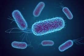
Bacteria are unicellular (one celled organism).
Its DNA is circular
It does not contain any kind of orgnelles (small-organ parts).
Micron (μ) unit is used to measure it.
1 micron (μ) = 1/1000 mm
The normal size of bacteria is 1μ to 0.5μ in diameter
Its length is usually 3 μ to 10 μ in length.
Width 0.1 μ to 2 μ
Size up to 100 μ is seen in some cases
Bacteria can be seen through an ordinary microscope.
Classification of bacteria based on their shape:-
Bacteria are classified as follows based on their shape.
Cocci (cocci-golanoo)-These are round in shape
Baccili (bacilli-dandanu)- these are cylindrical in shape
Spiral (Spiral)- These vaka-chunka are curved.
1.Cocci-
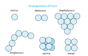
A Micrococci – These are singles also known as coccus.
B.Diplococci (Diplococci)- These are found in pairs
C.StreteptoCocci (Streptococci)-This is found in the form of cysts
D.Styphylococci (Staphylo cocci)- These are found in Jumkha
E.Tetrad (Tetrad)- is the number of four with mother.
F.Sarcina (Sarcina)- This number of eight (eight) is found with mother
2.Baccili (bacilli)-
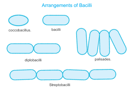
1.Cocobacillus- This is single also known as coccus
2.Diplobacili-These are found in pairs
3.Sreptobacili-These are found in the form of cysts
4.Palisades- An arrangement of palisades in the form of a palisaded fence to protect against attack.
3.Spiral (Spiral)- They are crooked
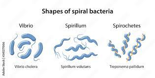
These include bacteria like coma-shaped Vibrio cholerae, Treponema pallidum no etc.
Classification of bacteria based on their flagella
The fibers found in motile bacteria are called flagella, which are composed of fine hair-like protein fibers and are attached around the cell.

Atrichous:- If the bacteria does not have any flagella.
Monotrichous:- Flagella is found at one end of the cell. Eg: Cholera Vibrio
Amphitrichous (Amphitrichus):- Two Flagella Cells are united at opposite ends. E.g. : Alceligenes feacales.
Lophotrichous:- At one end of the cell, it is found like in a tuft of grass. E.g. : Pseudomonas
Cephalotrichous (Cephalotrichoch) :– Seen at both ends of the cell like grass blades.
Peritrichate:- Many flagella are found around the cell. E.g. : Typhoid bacilli
Morphology of Bacteria
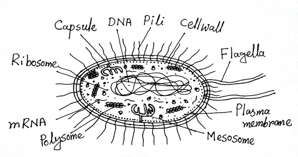
Fimbria/Pilli:
This bacteria has a hair like structure.
Helps bacteria stick to surfaces.
Common pili (almost always called fimbriae) are commonly involved in the specific attachment of prokaryotes to natural surfaces.
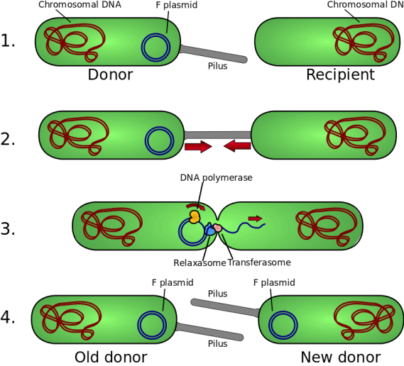
Sabilize bacteria during transfer DNA during conjugation.
capsule
There is a thick layer around the bacteria cell. Which is known as Capsule. Every time the difference between cell wall and capsule cannot be distinguished. When the capsule is in the form of loose secretion, it is called slim layer. If it is very thin then it is called Micro-Capsule.
It is composed of polysaccharides, it inhibits the phagocytosis process.
Contribution in virulence (ability to produce disease).
Cell wall
Immediately below the capsule is a relatively hard covering and gives shape to the bacterial cell. Depending on its thickness, hardness and chemical structure, bacteria are classified into gram positive and gram negative bacteria.
Gram Positive bacteria have a relatively simple chemical structure from Gram Negative bacteria.
Cell Wall is made up of Cellulose, Protein & Lipid.
Cell membrane
Each cell has a barrier that separates the inside and outside sides known as cell membranes.
Cell membranes are very thin, flexible and weak. And also known as Cytoplasmic membrane.
Main function selective permeability regulation
Controls the passage of nutrients & waste products. (made of Phospholipid & Protein)
This breakdown leads to cell death.
Cytoplasm
It is a complex mixture of many types of micro molecules (Proteins, nucleic acid, Polysaccharides & Lipids) Organic or Inorganic solutes of colloid.
It includes ribosomes, mesosomes, vacuoles and inclusions.
Rebosome, Mesosome, Inclusion
Ribosome’s :- It is the center for synthesis of Protein and RNA.
Mesosomes :- It is a site of respiratory enzymes.
Inclusion :- They are sources of stored energy in bacterial cells.
Vaculoes :- Fluid filled cavity
Volutin Granules :- Storage of inorganic components
Nucleus
The tightly coiled DNA (Deoxyribose nucleic acid) inside the cytoplasm. Bacteria do not have a developed nucleus like animals.
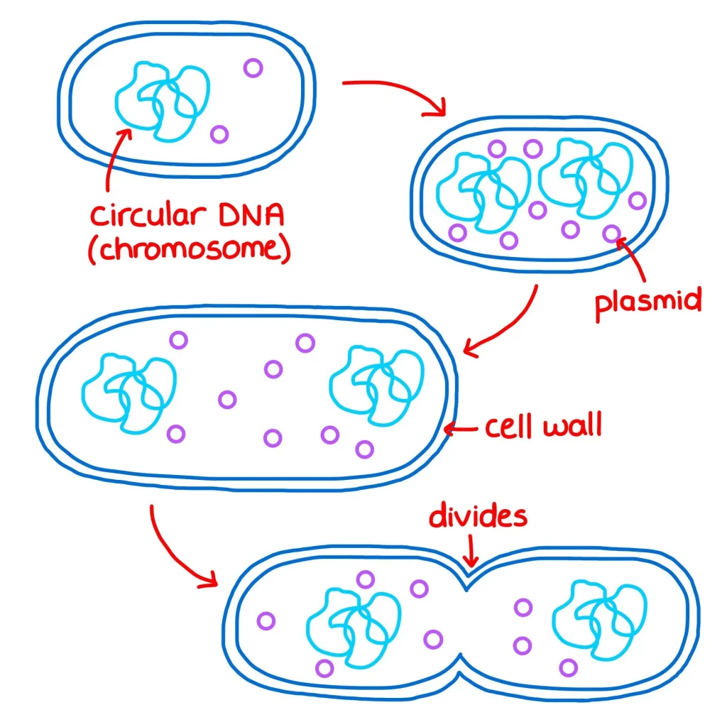
It replicates by binary fission.
Extrachromosome of DNA in bacteria called plasmid.
Bacterial endospore
They are highly resistant to the environment
Endospores are formed by a few bacteria as an intracellular structure.
Most of the bacteria under different conditions form a coating inside the parent cell which is called a spore. This process concentrates the protoplasm inside the cell to form a small sphere, which acts as a thick cell wall. An endospore is usually formed in each cell which sustains its existence even at very high temperatures.
Reproduction (Reproduction of bacteria) :-
Binary Fission of Bacterial cells
Conjugation
2. Fungi
It is found in tissues that are produced in a substance like stale bread. Non-pathogenic micro-organism. Its cell wall is hard. It takes its food from soluble nutrient substance through diffusion process through cell wall.
Ex.unicellular Mold, Yeats (mould,yeast)
Multicellular- Mushroom
- Rickettsiae
This is a simple rod shape spiral organism first observed by Howard Taylor in 1909.
Its size is smaller than bacteria. Length is 3 micron and diameter 0.5 micron.
They do not move much.
Although the size is small, many pass the filter. And many don’t pass through the filter.
It is not yet known whether it is plants or animal micro-organisms.
Diseases caused by this are not commonly found in India.
E.g. Q-fever,
Rocky mountain fever,
Trench fever
It is a form between bacteria and virus, because it has some properties of both bacteria and virus.
- Spirochetes
Spirochetes have characteristics of both protozoa and bacteria.
1- Venereal Treponeme
Ex. Treponeme Pellidum causes syphilis in humans
2-Non venereal Treponeme
ex.Lepto-spira- causing spirochetal jaundice
- Protozoa
These are animal unicellular micro-organisms. Its structure is more erratic than that of bacteria. They are of different types. And there are pathogens
i) Entamoeba – Dysentery, Hepatitis, Liver abscess
(ii) Giardia – Diarrhoea
(iii) Trichomonas hominis – Diarrhoea
(iv) Trichomonas Vaginalis – Vaginitis
(v) Trypanosoma – Sleeping sickness (Africa)
(vi) Leishmania – Kala azar
(vii) Plasmodium vivex – Beneign tertian malaria (Malaria)
Plasmodium falciparum – Malignant Malaria
Plasmodium malaria – Quartan malariamulurie.
Plasmodium ovale-Beneign tertian malaria
Belantidum (B.coli)-Desentry (dysentery)
Worm
1) Trematoda
They are mostly found in liver and intestine and are responsible for cirrhosis of liver, persistent diarrhoea, gastro intestinal irritation, enlarged liver, eosinophil urticaria, haematuria (blood in urine).
2) Cestoda
They are found in the intestine and cause intestinal conditions.
Nematoda
They are in intestine, lymphatic vessels subcutaneous tissues. Allergic reaction, Haemorrhage lesions in heart, Acute appendicitis, G. I. disturbances, ulcer, Rise of eosinaphil count and septic complication are responsible for.
- Virus
Virus is a Latin word. Which means “Poison” or “Venom”.
Virus is an intracellular living organism.
It can be seen only with an electron microscope.
Diseases like Polio, Mumps, Rabies, Herpes, Chicken pox, Dengue fever, Hepatitis B etc. are diseases caused by different viruses. Not all viruses are pathogenic. AIDS is also a disease caused by a virus.
History of viruses
In 1982 Russian biologist Iwanowski first discovered that the T. M. V. virus was responsible for tobacco mosaic disease in tobacco.
In 1900, Walter Reed discovered the yellow fever virus.
In 1935, electron microscope became possible to study virus. (Electron microscope can do 3,00,000 magnification.)
Characteristics of Viruses
Viruses have both living and non-living characteristics.
Non-living characters of virus
(i) Viruses do not have cellular organization.
(ii) They do not contain protoplasm.
(iii) It does not require respiration function or food.
(vi) A binary fission does not multiply.
It does not have independent existence.
(v) External stimuli do not produce a response.
(vi) Outside the cell it behaves like chemical molecules.
Living characteristics of Viruses
(i) Its reproduction takes place only in living cells
(ii) It has the capacity to grow in size and numbers.
(iii) Mutation of own genes through living.
(iv) It naturally adapts to its environment.
So we can say that viruses fall between living and non-living. But they are scientifically studied in Lliving
Viruses may be defined as, “Extremely small obligate intracellular living forms containing only one type of nucleic acid either DNA-RNA.”
They are different in shape. The shape of the same type of virus remains the same, but the shape of different types is not the same. They are found Rod shaped, Cuboidal, Rhomboidal (multisided), needle shaped.
Viruses have a simpler structure than other micro-organisms.
Living cells are the primary requirement for the virus to grow artificially in any culture medium. The following three methods are used for the cultivation of the virus. (i) Animal Inoculation :- The virus is introduced into the running water. (ii) Cultivation in chick embryo (iii) Cultivation in tissue culture :-
Fusobacteria
Long, thin, spindle shaped. With pointed end or cigar shaped They are found in Gums and tonsils of normal person. They are often associated with certain spirochetes in mixed infection causing fasospirochaetal disease
Bacteroides
Anaerobic, Rod shaped, gm-ve Bacteria.
Normally inhabitant in oral, respiratory, intestinal, urogenital cavities of human and animals,
The infection of bacteroids (e.g. abdominal sepsis) are usually found in association with other organisms.
Non-Pathogenic Organism
Bacteria have a very important relationship with human beings, some non-pathogenic bacteria destroy pathogenic bacteria, besides
(1) Some bacteria attach to plant roots and convert nitrogen (Nz) into useful chemicals. Which is used as chemical fertilizer.
(2) It helps to make vinegar from alcohol by making acetic acid
(3) Some bacteria help in the production of tobacco and rubber.
(4) Yeast is processed over flour, flour, and sugar to help the bread rise.
5) Ghee, butter and cheese produce certain types of sodium.
(6) Yeast and actinomytius produce antibiotic substances, which are useful in the treatment of infection.
(7) Certain bacteria help in converting poisonous gases from decayed or dead animal and vegetable matter into simple gases. This simple gas becomes useful as food for plant growth.
- Growth factors of bacteria
Growth of bacteria is seen in two ways.
- Increase in size
2.Increase total number of cells.
To see the growth of bacteria, it is shown in a curved graph.
Growth of bacteria is seen in two ways.
- Increase in size
2.Increase total number of cells.
To see the growth of bacteria, it is shown in a curved graph.
Lag phase :- Increase cell in size but it does not multiply
Log phase :- Multiply at the exponential phase
Maximal stationary phase:- Death & growth of bacteria are equal. Both are seen equally.
Decline phase :- Death cell progress of bacteria increases.
Factors affecting Bacterial Growth
Like every other organism, microbes also need a suitable environment and nutrients
falls for growth, maintenance & multiplication
Nutritional requirements for growth of bacteria:
Protein or Protons, or nitrogen containing substance.-
Energy food – Sugar
Minerals – Sodium – Sugar, starch, beef etc..
- Sodium chloride in small amount.
Water in large amount.
Blood, Glucose etc..
Even minor changes in the environment affect bacterial growth. Spore forming type is the only protection even in adverse conditions. can get By controlling the environment we can increase or stop bacterial growth
(1) Moisture
Like nourishing food, every bacteria needs water for growth. In fact, bacteria cannot get food in the absence of water, because every food element needs to be in a liquid state to pass through the wall of the bacteria. All types of bacteria grow well in liquid medium (Aqueous medium), an environment without complete moisture prevents its growth. or destroys.
Apart from this, cells cannot live in low or high humidity.
2) Light
Most bacteria are destroyed by direct exposure to ultraviolet rays in sunlight.
(3) Temperature :-
Temperature is a very important factor affecting the growth of bacteria. Optimal temperature with food, water is necessary for bacteria growth.
Different bacteria have different optimal temperatures.
37°C is the optimal temperature for bacteria growing in the human body.
However, many bacteria are mesophilic (meso = middle, phille = loving). The optimum temperature for it is 25 to 39* C.
Most bacteria grow this way.
Whereas psychrophilic (psychro = cold) bacteria grow better between 4 C to 10° C, some
Thermophilic (Therma – Heat) is also found. Its growth is best between 55°C to 75°C.
Temperature above 75 C is fatal for bacteria. In fact high temperatures are created to kill bacteria in different ways.
Like moist heat (steam), boiling water, pasteurization & autoclaving.
Many species can survive even at very low temperatures. Like yeast, mould, viruses & Rickettsia, spirochetes (76* C can survive for years).
(4) Oxygen
O2 also plays an important role in the life of bacteria. Many types of bacteria can only survive, or grow, in the presence of O2. They are called Aerobes (EX.Sarcina).
Conversely Anaerobes can live or grow in the absence of 02. E.g. Closteridium tetani-
Apart from this there are also bacteria. which can survive in the presence or absence of 02. They are known as facultative anaerobes. E.g. Salmonella typhi.
Microaerophils grow more in less oxygen than is present in air.
5) Hydrogen Ion Concentration: (Acidity and Alkalinity) PH medium
The acid or alkaline concentration of the liquid in which the bacteria grow affects the growth.
This is seen from the hydrogen ion concentration index.
PH – 0 (Zero) is the most acidic,
PH – 14 shows the lowest acidic concentration.
PH – 7.) A nutral (neutral),
pH < 7 is acidic
and alkaline at pH >7
Most bacteria grow best between pH 5.0 to 8.5. There are some exceptions to this too.
6) Osmotic pressure :-
The life of bacteria also depends on high or low osmotic pressure. If the bacteria are immersed in a liquid whose osmotic pressure is very high or very low, the bacterial cell collapses or becomes dormant due to the leakage of liquid.
Carbon Dioxide is also necessary for the growth of bacteria.
Normal flora of the body
Normal flora of the body In humans, like other animals, a large number of micro-organisms are found inside or outside the body. Which cannot be completely removed from the body. Normal microbial flora plays an important role for the body.
(1) They become pathogens when the immunity of the host is down.
(2) They help prevent colonization of pathogens.
(3) It strengthens the immune system of the person.
(4) Helps in synthesis of Vitamin K and many Vit.B.
(5) Prevents or suppresses the entry of pathogens
Normal flora of the skin
Human skin is constantly attacked by organisms from the environment.
It also gets contaminated by the person’s own secretion and excretion.
Skin flora also depends on body area and clothes worn.
And occupational environment also plays a part.
Diptheroids, staphylococci, gram positive aerobic spore bearing bacilli, gram negative bacilli (e.coli) and pathogenic hemolytic streptococci are usually found on the skin.
Normal flora of the conjunctiva
Because of the flushing action of tears, the conjunctiva contains relatively little flora, although staphylococci and streptococci are often seen.
Normal flora of Nose, Nasopharynx and Sinuses:-
Micro-organisms of Staphylococci, Streptococci Haemophilus species are found.
Normal flora of mouth and URT (Upper Respiratory Tract)
Pigmented and non pigmented micro cocci, gm +ve spore bacilli, proteus, lactobacilli, Anaerobic micro cocci, streptococci, vibrios, fusiform bacilli, neisseria fungi, candida etc. are found.
Normal flora of intestinal tract
bacilli, entrococci, staphylococci Due to the pH of Stomach, more flora can accumulate there, Stomach of conditions like carcinoma pyloric obstruction etc. gram+ve cocci and bacilli are seen. In the latter part of the duodenum, gram is seen to increase. Mostly anaerobes, lactobacilli and mycoplasma candida are found.
Normal flora of the Genito-urinary tract :- Mycobacterium Smegmatis, Gram + Ve, Gram – Ve bacteria, Lacto bacillus, E.Coli, Yeast, neisseria or Spirochaetes are found.
Growth media (Methods of cultivation of micro-organisms)
Examination of micro-organism shows only the morphology of bacteria.
But to know the bacteriological characteristics, it is necessary to know the culture of organisms.
Many factors are responsible for the growth of microbes
Like moisture, Nutrients, Absence of toxin substance, Osmosis, PH Value, 02 etc.
Certain culture media are used to cultivate bacteria. Micro-organisms grow very fast in this media.
Required for antigen production and vaccine preparation.
Importance of Culture Media
The same way Nutrition and Environment are necessary for the growth of bacteria in the natural environment, just like Nutrition and Environment Artificial bacteria in the lab. Mother is given.
In which water, agar, peptone, casein hydrosylate, meat extract, yeast extract etc. are used as culture media.
Type of growth media
1) Liquid media :-
Which is done in test tube, flask or bottle. It is also known as “Broth”. Use for large number of bacteria
(2) Solid media :- This is made by adding Agar-Agar in liquid. Which is made from red algae. Concentration 1.5 to 2.5 % (3) Semi-solid media:- In this liquid Agar- Agar is added and made to know micro-aerophilic bacteria or bacterial motility. (4) Basal media :- Water, agar, peptone are used.Ex staphylococcus
(5) Enriched media :- Blood is used as media. When the blood is heated to law temperature, the color of hemoglobin in it becomes dark brown, which is used as media, which is known as chocolate media.
(6) Cell culture media :- This is used for cultivation of Virus and Rickettsia. Tissue cell culture technique is used.
(7) Chick embryo media :-This is also used for culture of virus and Rickettsia. Eggs are kept in the incubator for 10-12 days after which the fertile egg is used as media.
8) MacConkey Agar :- This is commonly used for Enterobacteriaceae. It contains agar, peptone, sodium chloride, bile salt, lactose and neutral red.
It is a selective and indicator medium:
Selective as bile salt does not inhibit the growth of Enterobactericeae but inhibits the growth of many other bacteria.
Indicator medium as the colonies of bacteria that ferment lactose take a pink color due to production of acid. Acid turns the indicator neutral red to pink. These bacteria are called “‘lactose fermented, e.g. Escherichia coli. Colorless colony indicates that lactose is not fermented. i.e. the bacterium is non-lactose fermenter, e.g Salmonella, Shigella and Vibrio. 9) Transport media :-Clinical specimens must be transported to the laboratory immediately after collection to prevent overgrowth of contaminating organisms or commensals. This can be achieved by using transport media. Such media prevent drying (desiccation) of specimen, maintain the pathogen to commensal ratio and inhibit overgrowth of unwanted bacteria. Cary Blair medium and Venkatraman Ramakrishnan medium is used to transport feces from suspected cholera
10) Anaerobic Media :- is required to grow anaerobic bacteria, which need reduced oxidation-reduction potential and extra nutrients, Such media may be reduced by physical or chemical means.Boiling the medium serves to expel any dissolved oxygen, Addition of 1% glucose, 0.1% thioglycollate, 0.1% ascorbic acid, 0.05% cysteine or red hot iron filings can reduce a r medium. Robertson Cooked Meat (RCM) medium that is commonly used to grow Clostridium species contains a 2.5 cm column of bullock heart meat and 15 mL of nutrient broth. Thioglycollate broth 02 contains sodium thioglycollate, glucose, cystine, yeast extract and casein hydrolysate.The organism to be cultured is kept in an anaerobic jar so that the required organism could grow in oxygen free environment