ENGLISH – GUJARAT NURSING COUNCIL General Nursing & Midwifery (First Year) BIO-SCIENCES-2024 paper no 7
GNC BIO SCIENCE
Date: 22/04/2024
Q-1.a. Explain the gross structure of Heart. -Explain the gross structure of heart. 03 marks.
Heart..
Heart is an important organ of the circulatory system. Heart beats continuously during human life. Due to its pulsation, the blood circulates continuously in the blood vessels.
The heart is an organ made up of blood and muscles. It weighs approximately 310 grams in males and approximately 250 grams in females. The heart beats approximately one lakh times during the day.
Structure of the Heart..
Heart is an organ made up of semi-muscles. Its wall is made up of three types of tissue layers.
The outermost layer of the heart wall is called epicardium or pericardium.
Epicardium or Pericardium.
It is thin and transparent and covers the heart from the outside. It is made up of fibrous connective tissue. In which there is a layer of fibrous tissue on the outermost side and a serous membrane on the inner side of the fibrous tissue which is found in a double layer. The outer layer of the serous membrane is known as the parietal layer and the inner layer as the visceral pericardium layer.
The space between the parietal and visceral pericardial layers is called the pericardial space. This space contains fluid called serous fluid or pericardial fluid. Which prevents friction between the two layers.
This layer of outer pericardium protects the heart from the outside and this layer is also seen around the vessels coming out of the heart.
Myocardium..
Myocardium is the middle layer of the heart. It lies below the pericardium. It is made up of a special type of cardiac muscle tissue. The pumping action of the heart is seen due to the contraction of these muscles.
This myocardium layer is thin at the base and thick at the apex. Also, the layer of the wall of the left ventricle is thicker than that of the wall of the right ventricle.
The contraction of these muscles has an involuntary action that results in the pumping action of the heart and is controlled by the autonomic nervous system and the conducting system in the heart.
endocardium..
It is the innermost layer of the heart wall. It is in contact with layer blood. This layer is made up of epithelium tissue and connective tissue. This layer is smooth and shiny which is important for easy blood flow inside the heart. This layer also covers the valves inside the heart and this layer is continuous in the inner wall of the blood vessels leaving the heart.
B. Write down the functions of Heart. -04 marks.
Functions of the Heart..
Heart provides oxygenated blood supply to all organs and tissues of the body.
Heart is an important organ of the cardiovascular system. It functions as a vital organ without which the human body cannot survive.
The heart circulates the blood towards the lungs so that the blood can be oxygenated and purified.
Circulations like pulmonary circulation and systemic circulation are regulated by the heart.
The heart also regulates the heart rate according to the needs of the body and according to the body temperature.
The heart also regulates body temperature as it circulates blood to every part of the body.
As the heart pumps blood to the body’s excretory organs, the blood can be filtered and waste products removed from the blood.
c. Explain the pulmonary circulation. 05 marks.
Pulmonary Circulation..
Pulmonary circulation started from the right ventricle and the blood goes to the lungs and from there returns to the left atrium, so the circulation from the right ventricle to the left atrium is called pulmonary circulation.
In the pulmonary circulation, deoxygenated blood in the right ventricle exits the right ventricle through the pulmonary artery. As it exits, the pulmonary artery divides into a right and a left pulmonary artery and both enter the lung. In which two branches in the left lung and three branches in the right lung enter the pulmonary artery which is according to each lobe of the lung.
In the lungs, gas exchange takes place between the blood and the tissue of the lungs, and two pulmonary veins take oxygenated blood from both sides of the lungs and enter the left atrium of the heart by taking oxygenated blood from each lobe.
Pulmonary circulation converts deoxygenated blood in the heart to oxygenated blood via the lungs. This blood enters the left ventricle and supplies oxygenated blood to the whole body through the systemic circulation.
The circulation from right ventricle to left atrium is called pulmonary circulation.
OR
A. List out organs of urinary system. 03 marks.
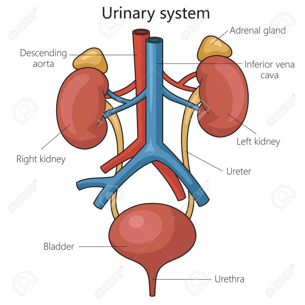
Urinary system is one type of excretory system of the body. It removes the waste products of the body out of the body. Any body substances that are not used in the body (metabolic waste) are removed from the body. It includes the following organs.
2 kidneys := which does the formation of urine. They are right and left 2.
2 ureters:= which carry urine from kidney to urinary bladder. They are right and left 2.
1urinary bladder := This is an organ in which urine is collected. It is in the number of 1.
1 urethra:= through which the urine collected in the urinary bladder is excreted outside the body.
B. Describe gross structure of Kidney. – Explain the gross structure of kidney. 04 marks.
Gross Structure Of Kidney.
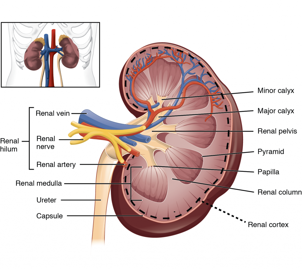
There are 2 kidneys in the human body. They are located in the right and left abdominal cavities on either side of the vertebral column on the posterior side of the body.
Kidney is a bean shaped organ. It lies from the level of the 12th thoracic vertebra to the level of the 3rd lumbar vertebra.
Kidney is 11 cm long by 5 to 6 cm wide. Its weight is approximately 150 grams. The right kidney is positioned slightly lower than the left kidney because the liver occupies a larger portion on the right side.
Veins around the kidney.
The kidney is an organ located in the abdominal cavity. One is located on both the right and left sides. Abdominal cavity organs like liver, small intestine, adrenal glands, stomach, spleen, pancreas etc. are located around both kidneys.
Structure of the Kidney..
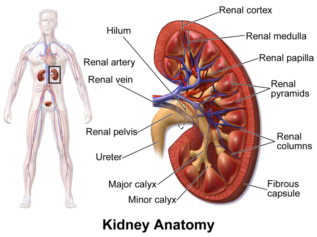
Kidney is a shapeless organ. A groove in the middle is called hilum or renal hilum. Through which the structures of renal artery, renal vein, nerves, lymph vessels and ureter enter and exit.
The inner border or hilum of the kidney is found on the side of the vertebral column.Its outer border is convex. The kidney is a hanging organ on both sides of the abdominal cavity. To hold it in position, it is surrounded by a network of fatty tissue and fibroelastic connective tissue called the renal fascia. With the help of this kidney can maintain its position and it also gets protection.
When the kidney is viewed in a longitudinal section, it is seen to be distributed into three kidney structures.
- Fibrous capsule.
It is part of the fibrous tissue that surrounds the kidney. This membrane is arranged around the kidney. Which acts as a layer to maintain the shape of the kidney and to protect it.
- Cortex.
It is redis brown in color made up of tissue. Which is located under the kidney capsule.
- Medulla.
In the kidney, the inner part from the cortex is called the medulla. It also has redish brown color. The triangular shaped pyramidal structure is called renal pyramid. The base part of this renal pyramid is towards the cortex and the pointed part of the pyramid i.e. the part of the renal papilla is arranged inwards towards the hilum.
The renal papilla forms a cup-like structure anteriorly called the calyx. The part with large space is called major calyx and the part with small space is called minor calyx. The minor calyx opens into the major calyx. Beyond this calyx is the wide funnel-shaped portion called the renal pelvis.
The urine filtered by the kidney falls into the wide part of the calyx, the funnel shape, i.e. the renal pelvis. Urine collects here and then passes anteriorly from the renal pelvis through a narrow structure called the ureter that exits the kidney and reaches the urinary bladder.
Urine filtered by the kidney passes from the minor calyx to the major calyx and from the major calyx to the renal pelvis. It then reaches the urinary bladder through the ureter. This action is not controlled by any kind of nervous system. In the wall of the renal pelvis there are special muscles and pacemaker cells due to the contraction of which this urine flows forward.
C. Write down functions of Kidney. 05 marks.
Functions of the Kidney..
Kidney is mainly responsible for urine formation.
Kidneys filter the blood and remove the waste products through urine.
The function of the kidney is to maintain the normal balance of electrolytes.
It works to maintain blood pH.
The body functions to remove waste products accumulated at the end of metabolism from the body.
Kidneys secrete a hormone called erythropoietin which plays a very important role in the production of RBCs.
Kidneys secrete a hormone called renin which plays a very important role in maintaining blood pressure.
Kidneys are responsible for maintaining water balance in the body.
Q-2 a. Describe the structural & functional unit of body. . 08 marks.
Cell is the smallest microscopic structural and functional unit of the human body. The cells in it are important for the function of every organ in the body. Every organ can perform normal function only with the function of this cell. There are many different types of cells in the body. Here we study about the basic cell of the body and its structure.
The components in Selma are as follows.
Cell membrane
Nucleus
Cytoplasm
Protoplasm
Mitochondria
Golgi apperatus
Ribosomes
Endoplasmic Reticulum (Smooth Endoplasmic Reticulum and Rough Endoplasmic Reticulum)
Write the structure and functions of cell.
Cell is the basic functional and structural unit of the human body. It is the main working tax unit.
A cell is known as a mass of protoplasm. Inside the cell are organelles that are covered by a plasma membrane.
A zygote is formed by the fusion of ovum and sperm in the human body.The growth and cell division of this zygote leads to the formation of the human body.
The fluid part inside the cell is known as cytoplasm. It contains many organelles. The structure of a cell is as follows.
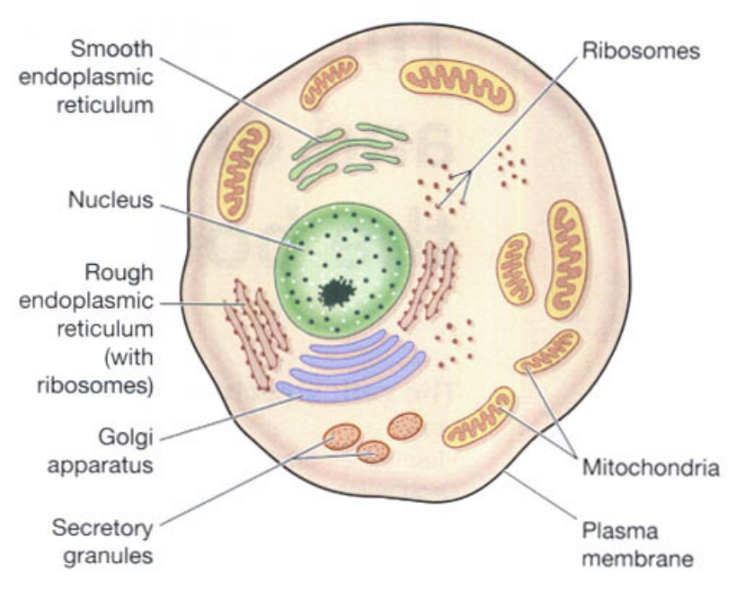
Plasma membrane.
The membrane surrounding the cell is called the plasma membrane. This membrane has selective permeability (movement of only selected substances). Due to which some substance can come inside the cell and some substance can go out of the cell. Thus the cell maintains the structure of its cytoplasm through this membrane.
The plasma membrane is a double layer membrane composed of phospholipids. It works to provide protection to the organelles of the cell and to maintain the shape of the cell.
the nucleus.
Nucleus is located in the center of the cell. It contains a liquid called protoplasm. The nucleus is surrounded by a nuclear membrane. This membrane also has selective permeability.The nucleus membrane partially separates the cytoplasm and protoplasm. Nucleus controls all the activities inside the cell and only with its help the cell can stay alive.
Inside the nucleus there are proteins like tension which are called Chromatin. This chromatin turns into chromosomes during cell division and performs the function of cell division.
These chromosomes inside the nucleus are responsible for the hereditary traits of an individual. Chromosomes are found in 23 pairs in the cells of the human body. 22 pairs of these chromosomes are called ordinary chromosomes while 1 pair is called sex chromosomes.
Mitochondria.
Mitochondria is a rod shape structure. Which is located in the cytoplasm inside the cell. There is a double membrane around it, the structure of the membrane is similar to the plasma membrane. The outer layer of this membrane is a smooth layer and the inner layer has many folds. This series of folds is called cristae.
Within these cristae are enzymes that release ATP.This is why mitochondria are called the power house of the cell.
ribosomes.
They are tiny granules in the cytoplasm. They are made up of proteins and RNA. It performs the function of protein synthesis from amino acids. Some ribosomes lie free in the cytoplasm and some are attached to the surface of the endoplasmic reticulum.
endoplasmic reticulum.
It is a series of interconnecting membrane or channel like structures. which connects one structure of cytoplasm with another structure.There are two types of it.
- Smooth endoplasmic reticulum..
Its surface is smooth. It functions in steroid hormone and lipid synthesis. It also helps to detoxify certain drugs.
- Rough endoplasmic reticulum..
Its surface is rough. Ribosomes are located on its surface.These ribosomes perform the function of protein synthesis.
Endoplasmic reticulum also helps transport substances from one place to another in the cytoplasm of the cell.
Golgi apparatus.
The Golgi apparatus is a bag-like structure with four to eight folds. These folds overlap each other. The end portion of this structure forms a pouch-like structure called a cisterna. Proteins synthesized by ribosomes are collected and stored in the form of secretory vesicles at the ends of these cisternae. When needed, these secretory vesicles release proteins into the cytoplasm. The Golgi apparatus is a structure located near the nucleus.
lysosomes.
Lysosomes are a type of secretory vesicles that are secreted through the membrane of the Golgi apparatus. These lysosomes contain the content of certain enzymes that break down certain large molecules in the cytoplasm of the cell. It works to protect cells from foreign material and microorganisms. These lysosomes also work to remove the waste material accumulated inside the cell.
There is also a sun-shaped centrosome in the cytoplasm of the cell, which plays an important role in cell division.
Apart from this, the cytoplasm of the cell also contains a network of microfilaments and microtubules which work to maintain the shape of the cell and to protect and support the structure of the cell.
Functions of the Cell.
A cell has the property of movement with the help of which the cell can move from one place to another. This function of the cell is called motile function or mobility.
The cell uses oxygen and performs cellular activity, finally the carbon dioxide accumulated in the cell goes out of the cell, thus the cell performs the function of respiration as it performs gas exchange.
The cell obtains the nutrient material from the blood and performs cell activity i.e. cell nutritional function.
The waste product deposited inside the cell exits the cell through the plasma membrane thereby performing the cell excretory function.
The cell transports the nutrient materials within itself from one cell to another and from one place within the cell to another, thus the cell also performs the function of circulation and transportation.
A cell performs the function of reproduction as it has the property of multiplying and dividing.
Due to the enzymes inside the cell, it works to destroy micro-organisms and pathogens, thus it performs the action of phagocytosis.
Cells have the ability to receive nerve impulses and pass them on, thus performing functions of irritability and conductivity. This cell function is mainly found in nerve cells.
B. Write down functions of Neuron. . 04 marks.
The smallest basic structural and functional unit of the nervous system is the neuron.
Brain has a large number (100 billion) of neurons.
Each neuron does the following:
A neuron has properties of conductivity and excitability, through which it responds to stimuli from the external environment, including mechanical, electrical and chemical stimuli.
Sensory impulses from the sense organs, muscles, joints and visceral organs are carried through the spinal cord to the central nervous system. In which the following Na arias are found.
Somatic cutaneous conveys impulses of sensations like pain, temperature, touch, vibration etc.
Special senses which convey impulses like taste, smell etc.
Proprioceptors transmit impulses from special senses such as vision, hearing, balance, and cranial nerves through the cranial nerve.
Motor neurons convey impulses from the central nervous system to effector organs, muscles or glands.
Sensory neuron Somatic nerves provide impulses to control the contraction of skeletal muscles.
Autonomic Nervous (sympathetic and parasympathetic) which causes contraction of smooth muscles, cardiac muscles, glands through cranial and spinal nerves.
Mix is with the sensory and motor nerves in the spinal cord while in the other part of the body it is distributed by connective tissue. It is called mixed nerve.
OR
a. Explain the gross structure & functions of Liver. . 08 marks.
Liver…
Among all the glands in the body, the liver is the largest gland.which lies below the diaphragm in the right quadrant of the abdominal cavity. It weighs approximately 1.4 kilograms inside an adult. It is located below the ribs of the chest. Ribs protect it. A part of it is also located in the region of the left abdominal cavity.
The upper surface of the liver is smooth. This portion is attached to the diaphragm and has an irregular surface and margin on the back and underside of the liver.
Organs around the liver.
Organs such as the diaphragm, anterior abdominal wall muscles, stomach, duodenum, kidney, inferior vena ceva, gallbladder etc. are arranged around the liver.
Liver is mainly divided into two lobes, right lobe and left lobe.
The right lobe is larger than the left lobe and both lobes are separated by the falciform ligament.
The quadrate lobe is found on the posterior side of the liver and the quadrate lobe is found on its inferior side.
Thus four lobes of liver can be seen anatomically.
Portal fissure…
The groove on the posterior surface of the liver is called the portal fissure. From this fissure some structures enter the liver and some structures exit the liver viz.
The portal vein carries deoxygenated and nutritious blood from the intestine and enters the liver through this fissure.
The hepatic artery carries oxygenated blood and enters the liver through this fissure.
Nerve fibers of the sympathetic and parasympathetic nerves enter the liver through this fissure.
The left and right hepatic ducts exit this fissure to carry bile from the liver to the gall bladder.
The hepatic vein exits this fissure to carry deoxygenated blood from the liver.
Structure of the Liver…
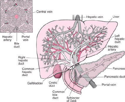
The liver is an organ located in the right quadrant of the abdominal cavity. It is mainly composed of 2 lobes.These lobes are made up of many lobules. These lobules are made up of special type of epithelium cells and cells called hepatocytes. These cells are found in a hexagonal pattern in the liver.
After entering the hepatic artery and portal vein within the liver, a capillary network of arteries and veins is formed. This network of capillaries is called sinusoids. Kupffer cells are found in the wall of these sinusoids, which Kupffer cells act to protect the liver from bacteria, foreign material or toxins and perform a protective function in the liver.
Hepatocytes cells within the liver secrete bile.This bile enters the bile canaliculi. These bile canaliculi carry the bile into small bile ducts. These small bile ducts join to form the right and left hepatic ducts. which drains bile from the liver through the common hepatic duct. The common hepatic duct joins with the cystic duct from the anterior gallbladder to form the common bile duct. Bile is drained into the small interstitium and plays an important role in the digestion of fat.
Blood supply of the liver.
The liver is supplied with blood by the hepatic artery and the portal vein carries nutritious blood and enters the liver.
The hepatic vein drains deoxygenated blood from the liver and joins the inferior vena cava.
Functions of the Liver..
Liver is a very important organ of our body. It is associated with many important functions.These functions are as follows.
The liver is responsible for the metabolism of carbohydrates.
The liver functions to maintain normal carbohydrate ie blood glucose levels. When blood glucose levels fall, glucose is produced from glycogen through the action of glycogenolysis and blood glucose levels are maintained.
When the amount of glucose in the blood increases, glucose is converted into glycogen by the action of glycogenesis.
The liver converts fat into fatty acids so that it can be used in the body, this is called desaturation of fat, thus it helps in lipid metabolism.
The liver helps in protein metabolism so that amino acids are synthesized in the body.
The liver converts ammonia and makes urea so that this waste product can be excreted through urine.
The death of red blood cells releases bilirubin, which is modified by the liver to help remove excess bilirubin from the body.
Liver helps to detoxify the toxic substances, alcohol, drugs etc. introduced in the body and remove them from the body.
The liver synthesizes bile salts, which are essential for emulsification of fats and hence absorption of lipids and cholesterol.
Helps to maintain the level of vitamin D in the body.
Since Kupffer cells are located inside the liver, they perform a protective function by protecting the liver from harmful substances and performing phagocytosis.
The liver acts as a storage of vitamins and minerals and releases these vitamins and minerals into the body when needed.
Liver is responsible for heat production in the body.
b. Describe Menstrual cycle. 04 marks.
Menstruation cycle occurs after puberty phase in females.In which changes are seen in the function of ovaries and uterus.
A menstrual cycle occurs every 26 to 30 days. This is seen due to changes in the level of hormones in the blood.
The onset of the menstruation cycle is known as menarche.
Females find this cycle continuous after the age of puberty. Which stops temporarily during pregnancy and stops completely after the period of menopause.
The onset of menstruation is due to the degeneration of the corpus luteum layer in the uterus and bleeding occurs through the vaginal cavity.
Menstruation cycle has the following phases.
- Menstrual phase..
This phase occurs every 28 days and lasts for about four days. When fertilization of the egg does not take place in the female, the hormones estrogen and progesterone that support the uterine wall decrease and the hormone oxytocin increases. So the stimulation of contraction of the uterus increases and degeneration of the corpus luteum layer of the wall of the uterus starts and blood drains from the uterus through vaginal discharge. This phase lasts from 1 to 4 days.
This menstrual flow contains endometrial glands, endometrial cells, blood and unfertilized ovum. Approximately 100 to 200 ml of blood is shed during the 3 to 5 days of this phase which is called the menstrual phase.
- Proliferative phase..
The menstrual phase ends on the 5th day. After that, the proliferative phase starts from day 6 and lasts for 14 days.
In this phase follicle stimulating hormone stimulates the ovarian follicles and hence increases estrogen production. This estrogen stimulates the proliferation of the endometrium.
The endometrium of the uterus begins to develop from the sixth day.Its cells multiply and due to this increase in mucus secreting glands and blood capillaries. Thus the endometrium of the uterus becomes bulky and vascular.
At the end of this phase, the inner wall of the uterus is ready for implantation of the fertilized egg. This phase ends with ovulation. Towards the end of this phase, there is a decrease in estrogen levels.
- Secretary phase.
After the proliferation phase is completed, the secretory phase begins. The secretory phase is observed from the 15th day of the menstrual cycle to the 28th day.
As progesterone hormone is important in this phase, this phase is also called progesterone phase.
When the mature egg is released by the ovary due to ovulation, the amount of estrogen and progesterone hormones decreases, but the corpus luteum of the uterine wall maintains the pregnancy by secreting progesterone.
As this mature ovum is not fertilized by a sperm, the corpus luteum decreases progesterone and due to the decrease in progesterone hormone, there is an increase in the amount of oxytocin hormone and the uterine muscles begin to contract.
The next cycle begins at the end of this phase due to the corpus luteum not receiving a fertilized ovum and increased uterine contractions.
Q-3 Write short answer (any two) 6X2=12
a) Describe the mechanism of hearing.
Physiology of hearing is the act of hearing. The wavelength for hearing is 20 to 20,000 Hz. The human ear is capable of frequencies between 500 and 5,000 hz.
The frequency at which the sound waves vibrate is known as the pitch, as the vibration increases, the pitch increases.
Every sound produces sound waves which strike the outer part of the auricle and from there enter through the external auditory canal, these sound waves vibrate the tympanic membrane i.e. the ear drum which is the junction between the external ear and the middle ear.
It is attached to the tympanic membrane by the malleus bone, to the incus with the malleus bone, and to the incus to the stapes, and the stapes bone is attached to the oval window in front.
These sound waves from the oval window reach the fluid of the perilymph that goes to the cochlea and from there to the endolymph and the round window vibrates and travels to the cerebrum through the vestibule cochlear nerve and the sound is recognized.
B. Write-down functions of Pituitary gland
The pituitary gland acts as the body’s master gland. Due to which many other glands help to maintain normal function.
The growth hormone of the pituitary gland maintains the normal growth of the body.
The pituitary gland regulates steroid hormones in the body.
The prolactin hormone of the pituitary gland plays an important role in milk production.
Hormones of the pituitary gland play an important role in maintaining fertility.
Hormones of the pituitary gland play an important role in maintaining normal delivery, breast development and breast feeding.
Hormones from the pituitary gland play an important role in maintaining body water balance and blood pressure.
C. Describe the mechanism of respiration
Respiration is gas exchange between two surfaces. In which air from the atmosphere enters the lungs. Gas exchange between lung tissue and blood is called external respiration. The gas exchange that takes place between each cell tissue of the body and the blood is called internal respiration.
During the act of respiration, oxygen enters the lungs through inhalation and carbon dioxide leaves the body through exhalation.
Normally, the act of respiration is observed 16 to 18 times in a minute.
The following mechanisms are found in the cycle of resuspension.
Inspiration
expiration
pos.
Inspiration
Inhalation of atmospheric air into the lungs is called inspiration.
When the brain receives nerve impulses for contraction of the diaphragm and intercostal muscles, the contraction of the diaphragm and intercostal muscles increases the size of the thoracic cavity. The air pressure inside the thoracic cavity decreases so that air from the outside atmosphere can enter the lungs through the action of inspiration. This action is called inspiration.
Contraction impulses to the diaphragm cause the diaphragm to flatten downwards and contraction of the intercostal muscles causes the ribs and intercostal muscles to move upwards and outwards. So the size of the thoracic cavity increases and negative pressure is created inside the cavity. As the air pressure in the outside environment is higher and the air pressure in the thoracic cavity is lower, the action of inspiration can take place. The act of inspiration is an active process.
Expiration
The process of expelling air from the lungs into the atmosphere is called expiration. The action of expiration is a passive process that begins after the action of inspiration is completed.
During exhalation, the contracted diaphragm and intercostal muscles relax. So the diaphragm returns to its original position and the ribs move downwards and inwards, reducing the size of the thoracic cavity and exhaling. In which the air from the lungs is thrown out into the atmosphere.
In the act of exhalation, the air pressure in the lungs is greater than the atmospheric pressure so that the act of exhalation takes place.
Pause.
This is the relaxed stage of the lung. In which no action of inspiration or expiration takes place. This period is called pause period.
Q .4 Write short notes. Write a short summary. (Any three) 12
A. Role of nurse in prevention of infection.
As a nurse, we must know how to protect ourselves and patients from exposure to pathogens.
It is very important for the nurse to understand and follow the infection control policy of the organization such as use of personal protective equipment, environmental sanitation, etc.
Standard Precautions :-
To break the chain of infection, standard precautions should be taken to prevent transmission from body fluids containing pathogens.
- Hand Hygiene :-
This is the number one weapon in preventing cross-infection. Most of the spread of micro-organisms occurs through the hands, so hand washing should be done before and after each procedure. It is very important to wash hands after touching every contaminated object
- Aseptic technique
In which to use contact precautions and aseptic technique during the procedure. Use only sterile items in invasive procedures
- Environmental infection control measures
The equipment used in the hospital and the surrounding environment should be kept clean and the floor of the hospital should be cleaned with anti-septic solution. Bio-medical waste generated during procedures or work should be properly disinfected and disposed of.
- Droplet pre-caution Mask should always be worn while working in the hospital and infected patients should also wear it, precautions should be taken while cuffing, sneezing etc.
- P.P.E
Cross-infection can be prevented from personal protective equipment to the nurse and the patient, whose cap, mask, gown are used. which should be worn during the care of highly infectious patients.
- Health Education :–
Provides health education to patients and staff working under them to prevent the spread of infection in the hospital
B. Factors affecting on microbial growth
1) Moisture
Like nourishing food, every bacteria needs water for growth. In fact, bacteria cannot get food in the absence of water, because every food element needs to be in a liquid state to pass through the bacterial wall. All types of bacteria grow well in liquid medium (Aqueous medium), an environment without complete moisture prevents its growth. or destroys.
In addition, cells cannot live in low or high humidity
2) Light
Most bacteria are destroyed by direct exposure to ultraviolet rays in sunlight.
3) Temperature :-
Temperature is a very important factor affecting the growth of bacteria. Optimal temperature with food, water is necessary for bacteria growth.
Different bacteria have different optimal temperatures.
37°C is the optimal temperature for bacteria growing in the human body.
However, many bacteria are mesophilic (meso = middle, phille = loving). The optimum temperature for it is 25 to 39* C.
Most bacteria grow this way.
Whereas psychrophilic (psychro = cold) bacteria grow better between 4°C to 10°C, some
Thermophilic (Therma – Heat) is also found. Its growth is best between 55°C to 75°C.
Temperature above 75 C is fatal for bacteria. In fact high temperatures are created to kill bacteria in different ways.
Like moist heat (steam), boiling water, pasteurization & autoclaving.
Many species can survive even at very low temperatures. Like yeast, mould, viruses & Rickettsia, spirochetes (76* C can survive for years).
(4) Oxygen
O2 also plays an important role in the life of bacteria. Many types of bacteria can only survive, or grow, in the presence of O2. They are called Aerobes (EX.Sarcina).
Conversely Anaerobes can live or grow in the absence of 02. E.g. Closteridium tetani-
Apart from this there are also bacteria. which can survive in the presence or absence of 02. They are known as facultative anaerobes. E.g. Salmonella typhi.
Microaerophils grow more in less oxygen than is present in air.
(5)Hydrogen Ion Concentration: (Acidity and Alkalinity) PH medium
The acid or alkaline concentration of the liquid in which the bacteria grow affects the growth.
This is seen from the hydrogen ion concentration index.
PH – 0 (Zero) is the most acidic,
PH – 14 shows the lowest acidic concentration.
PH – 7.) A nutral (neutral),
pH < 7 is acidic
and alkaline at pH >7
Most bacteria grow best between pH 5.0 to 8.5. There are some exceptions to this too.
6) Osmotic pressure :-
The life of bacteria also depends on high or low osmotic pressure. If the bacteria are immersed in a liquid whose osmotic pressure is very high or very low, the bacterial cell collapses or becomes dormant due to leakage of liquid.
Carbon Dioxide is also necessary for the growth of bacteria.
C. Role of nurse in bio-medical waste management Role of nurse in bio-medical waste management
Nurses have a very important role in the management of bio medical waste
Disinfect the biomedical waste generated in the ward as early as possible so that it does not become a source of infection.
To reduce as much as possible the transportation and storage of waste
According to the medical waste policy, it should be put in separate bags or containers
To discount disposable items so that they are not reused
Infectious plastic waste that can be recycled should be done only after disposal.
Sharp instruments should be kept in a puncture proof white container
Sharp instruments should be treated for dish effect before transport
A chemical like sodium hydrochloride should be used
If there is any mistake in segregation of bio medical waste, it should be corrected by the nursing staff
Bio medical waste should be surveyed in case of infection
Should be clean and used in dusting
Careful handling should be done to prevent
Bio medical waste should be segregated according to the color coding of bio medical waste
Education should be provided for buy medical waste
Proper records and reports of bio medical waste should be maintained
D. Precautions to take while collecting blood sample for culture test
The following precautions should be taken while taking samples for culture test:
Hand hygiene: Wash or sanitize your hands thoroughly before and after taking the sample.
Clean instruments: Use only sanitized and sterilized instruments.
Personal Protective Equipment (PPE): Use PPE such as gloves, masks, gowns, and eye protection.
Sample Containers: Use only sterilized containers for sample collection and seal them properly.
Timing of sample: Take sample on time, especially before starting antibiotic therapy.
Labeling: Label sample containers appropriately, including patient name, date, time, and sample type.
Storage and Transport: Store and transport samples at appropriate temperature to avoid spoilage.
Disinfectant Practice: Keep surrounding environment clean during and after sampling.
Patient preparation: Explain the procedure and its importance to the patient so that they cooperate.
Documentation: Record all necessary information and send it to the laboratory with the sample.
These precautions can help ensure quality sample collection and prevent possible cross-contamination.
Q-5 Define following (any six)Nise’s definition Lakhs (any six) 12
a) Microbiology-
“Micro” means minute and “bio” means life and “logy” means study. Thus, microbiology means the study of microscopic organisms that cannot be seen by the naked eye.
b) Sterilization-
Sterilization is the process in which all microorganisms, such as bacteria, viruses, fungi and their spores are completely destroyed. Sterilization is particularly used in the medical, pharmaceutical, laboratory and food processing sectors.
Some of the main methods of sterilization include:
Heat Sterilization:
Autoclaving: In this method sterilization is done using pressure and high temperature.
Dry Heat Sterilization: In this method sterilization is done using moisture without high temperature.
Chemical Sterilization:
Ethylene oxide: This is a gas used to sterilize medical devices and equipment.
Hydrogen Peroxide: This method is used for sterilizing equipment and analysis materials.
Filtration: A method used to remove microbes from liquids and gases.
Radiation:
Gamma Radiation: A method used to sterilize food and medical devices.
UV Light: A method used to sterilize soil and air.
The purpose of sterilization is to prevent infection and prevent the spread of infection, so as to ensure health and safety.
c) Cross infection –
Cross infection is a situation in which an infectious agent (such as bacteria, virus, or fungus) is spread from one person to another. This usually occurs in health care facilities where infections are spread through contact with patients, medical instruments, or healthcare staff.
Cross-infection can occur as a result of poor hygiene, inadequate disinfection practices, and inappropriate infection control policies.
d ) Respirnion-respiration
Respiration is gas exchange between two surfaces. In which air from the atmosphere enters the lungs. Gas exchange between lung tissue and blood is called external respiration. The gas exchange that takes place between each cell tissue of the body and the blood is called internal respiration.
During the act of respiration, oxygen enters the lungs through inhalation and carbon dioxide leaves the body through exhalation.
Normally, the act of respiration is observed 16 to 18 times in a minute.
e) Tidal volume-
The volume of air that moves into the lungs during normal inspiration and the volume of air that leaves the lungs during normal expiration during one respiration is called tidal volume. Normally it is around 500 ml.
f) Immunity
The resistance shown by the host against the condition caused by the micro organism and its products (toxin) is called Immunity.
or
Immunity means when any antigen or microorganism enters our body and our body protects against it by resisting it is called immunity.
g) Mycology-
Mycology is a branch of science that deals with the study of fungi. These include life cycle of fungi, classification, genetics, environmental importance, medicinal properties and fungal diseases. Mycology plays an important role in agriculture, medicine and environmental science.
h) Pandemic
Pandemic means any disease that spreads from one state to another state and from one country to another country and is seen all over the world is called a pandemic.
D. T. Swine flu covid 19 etc
Q-6(A) Fill in the blanks.05
- The left lung has …….. lobes.
The left lung has………lobes. 02 lobes.
2…….blood group is known as universal donor.
…….. blood group is known as universal donor. blood group O.
3.Tears are produced by ………..gland.
Ashu………gland Dara is produced. Lacrimal Gland.
4………..gives the color to the eye.
……….gives eye color. Melanin inside the iris
5.10th cranial nerve is……
Cranial nerve number 10 is… Vegas Nerve
B) (Tine or False) To know true and false.05
- Spinal cord is a part of peripheral nervous system. wrong
- The first cervical vertebra is known as Atlas. correct
- Dermis is an outermost layer of the skin. The outermost layer of the skin is the dermis. wrong
4.Fertilization of ovum is occurred in fallopian tube. correct
5.Fumigation method is used to disinfect the instrument.
C) March the following – Jodka Jodka.
A) Adrenal cortes Adrenal cortex. (A) Melanocyte stimulating hormone
Melanocyte Stimulating Hon
B) Adrenal medulla (B) Glucocorticoids
(C) Thymus glund- Thymus gland (C) Melatonin- Melatonin
(D) Pineal gland- Pineal gland (D) Thymosin- Thymosin
(E) Pituitary gland Pituitary end (E) Epinephrine- Epinephrine
A – B
B – E
C – D
D – C
E – A
BEST WISHES FROM MY NURSING APP..