ENGLISH – GNM FY BIOSCIENCE Anatomy 2023 PAPER 4
GNC BIO SCIENCE PAPER 2023 Que. 1 (a) List out the organs of the digestive system
List the organs of digestive system. 03
Organs of the Digestive System…
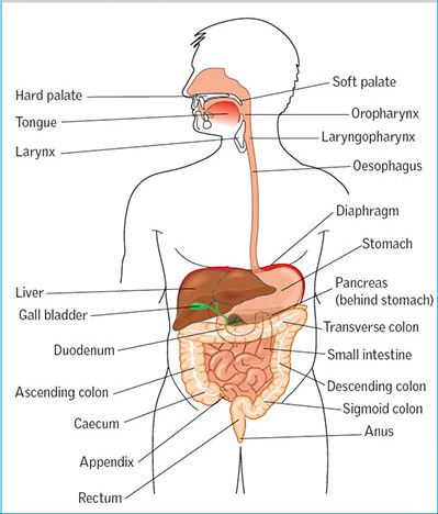
Digestive tract also called alimentary tract is a hollow tube which starts from mouth and extends to anus and its organs are as follows.
mouth
Fairings
Esophagus
Stomach
The small intestine consists of the duodenum, jejunum, and ileum.
Large intestine which includes cecum, ascending colon, transverse colon, descending colon, sigmoid colon, rectum and anal canal.
Accessory organs of the digestive track…
These organs do not come in the main track of the digestive tract but are located in the side which pours its secretion into the alimentary canal and helps in the process of digestion so it is called accessory organ.
3 pairs of salivary glands
Liver and biliary tract
Goal Bladder
Pancreas.
(b) Discuss about the digestive process in the stomach. 04
Food taken by mouth reaches the stomach through the esophagus. Mechanical digestion of food begins due to the charming movement of the stomach due to the contraction of the muscular layer of the stomach. Here all the food is converted from large molecules to small molecules by mechanical digestion.
After this mechanical digestion has gone on for some time, hydrochloric acid is secreted with the food through the glands in the inner wall of the stomach.
Chemical digestion begins when the chemicals present in this juice of the stomach mix with the food.
In addition to hydrochloric acid, this gastric juice contains enzymes, mainly pepsin and renin. This pepsin digests large molecules of protein into smaller molecules. Digestion of protein starts here.
Renin in the gastric juice digests the protein casein in milk and converts it into paracasein. Thus, digestion of protein mainly takes place in the stomach.
Digestion of carbohydrates and fats does not take place in the stomach, only mechanical digestion takes place here. After the partial digestion of this food in the stomach, it goes to the small intestine where it is completely digested and absorbed.
As the food in the stomach mixes with gastric juice, the harmful bacteria and viruses in the food are destroyed due to the acidic property of HCL in it.
Que. 1 (c) Describe the structure of the cell
Write in detail about the structure of cell 05
Cell is the basic functional and structural unit of the human body. It is the main working tax unit.
A cell is known as a mass of protoplasm. Inside the cell are organelles that are covered by a plasma membrane.
A zygote is formed by the fusion of ovum and sperm in the human body. The growth and cell division of this zygote leads to the formation of the human body.
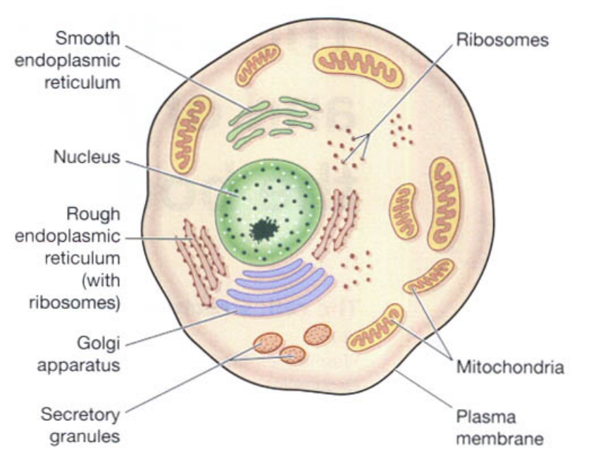
The fluid part inside the cell is known as cytoplasm. It contains many organelles. The structure of a cell is as follows.
plasma membrane..
The membrane surrounding the cell is called the plasma membrane. This membrane has selective permeability. Due to which some substance can come inside the cell and some substance can go out of the cell. Thus the cell maintains the structure of its cytoplasm through this membrane.
The plasma membrane is a double layer membrane composed of phospholipids. It works to provide protection to the organelles of the cell and to maintain the shape of the cell.
Nucleus..
Nucleus is located in the center of the cell. It contains a liquid called protoplasm. The nucleus is surrounded by the nuclear membrane. This membrane also has selective permeability. The nucleus membrane partially separates the cytoplasm and protoplasm. Nucleus controls all the activities inside the cell and only with its help the cell can stay alive.
Inside the nucleus are stringy proteins called chromatin. This chromatin turns into chromosomes during cell division and performs the function of cell division.
These chromosomes inside the nucleus are responsible for the hereditary traits of an individual. Chromosomes are found in 23 pairs in the cells of the human body. 22 pairs of these chromosomes are called ordinary chromosomes while 1 pair is called sex chromosomes.
Mitochondria…
Mitochondria are rod-shaped structures. Which is located in the cytoplasm inside the cell. There is a double membrane around it, the structure of the membrane is similar to the plasma membrane. The outer layer of this membrane is a smooth layer and the inner layer has many folds. This series of folds is called cristae.
Within these cristae are enzymes that release ATP. This is why mitochondria are called the power house of the cell.
Ribosomes..
They are tiny granules in the cytoplasm. They are made up of proteins and RNA. It performs the function of protein synthesis from amino acids. Some ribosomes lie free in the cytoplasm and some are attached to the surface of the endoplasmic reticulum.
Endoplasmic reticulum…
It is a series of interconnecting membrane or channel like structures. which connects one structure of cytoplasm with another structure. There are two types of it.
Smooth endoplasmic reticulum..
Its surface is smooth. It functions in steroid hormone and lipid synthesis. It also helps to detoxify certain drugs.
Rough endoplasmic reticulum..
Its surface is rough. Ribosomes are located on its surface. These ribosomes perform the function of protein synthesis.
Endoplasmic reticulum also helps transport substances from one place to another in the cytoplasm of the cell.
Golgi apparatus…
The Golgi apparatus is a bag-like structure with four to eight folds. These folds overlap each other. The end portion of this structure forms a pouch-like structure called a cisterna. Proteins synthesized by ribosomes are collected and stored in the form of secretory vesicles at the ends of these cisternae. When needed, these secretory vesicles release proteins into the cytoplasm. The Golgi apparatus is a structure located near the nucleus.
Lysosomes…
Lysosomes are a type of secretory vesicles that are secreted through the membrane of the Golgi apparatus. These lysosomes contain the content of certain enzymes that break down certain large molecules in the cytoplasm of the cell. It works to protect cells from foreign material and microorganisms. These lysosomes also work to remove the waste material accumulated inside the cell.
A sun-shaped centrosome is also present in the cytoplasm of the cell, which plays an important role in cell division.
Apart from this, the cyto plasm of the cell also contains a network of microfilaments and microtubules which function to maintain the shape of the cell and to protect and support the structure of the cell.
Or
Que 1(a) draw the diagram of the heart with label
Draw a labeled diagram of a heart. 03

Que 1(b) write the functions of the heart
Functions of the Heart … 04
Heart provides oxygenated blood supply to all organs and tissues of the body.
Heart is an important organ of the cardiovascular system. It functions as a vital organ without which the human body cannot survive.
The heart circulates the blood towards the lungs so that the blood can be oxygenated and purified.
Circulations like pulmonary circulation and systemic circulation are regulated by the heart.
The heart also regulates the heart rate according to the needs of the body and according to the body temperature.
The heart also regulates body temperature as it circulates blood to every part of the body.
As the heart pumps blood to the body’s excretory organs, the blood can be filtered and waste products removed from the blood.
QUE. 1(c) explain pulmonary circulation 05
Pulmonary circulation started from the right ventricle and the blood goes to the lungs and from there returns to the left atrium so the circulation from the right ventricle to the left atrium is called pulmonary circulation.
In the pulmonary circulation, deoxygenated blood in the right ventricle exits the right ventricle through the pulmonary artery. As it exits, the pulmonary artery divides into a right and a left pulmonary artery and both enter the lung. In which two branches in the left lung and three branches in the right lung enter the pulmonary artery which is according to each lobe of the lung.
In the lungs, gas exchange takes place between the blood and the tissue of the lungs, and two pulmonary veins take oxygenated blood from both sides of the lungs and enter the left atrium of the heart by taking oxygenated blood from each lobe.
Pulmonary circulation converts deoxygenated blood in the heart to oxygenated blood via the lungs. This blood enters the left ventricle and supplies oxygenated blood to the whole body through the systemic circulation.
The circulation from the right ventricle to the left atrium is called the pulmonary circulation.
Que 2 (a) List out the reproductive organs and describe about uterus
List the organs of reproductive track and write about uterus 08
Reproduction means the ability of any individual to give birth to a new organism is called reproduction.
In which a male and a female of different sexes start a new life by joining their germ cells. That is called reproduction.
The fusion of male germ cell i.e. spermatozoa and female germ cell i.e. ovum forms a new cell i.e. zygote. From this zygote, the process of birth of a new life is gradually done by its cell division. This entire function is called reproduction.
The organs of the reproductive tract are as follows.
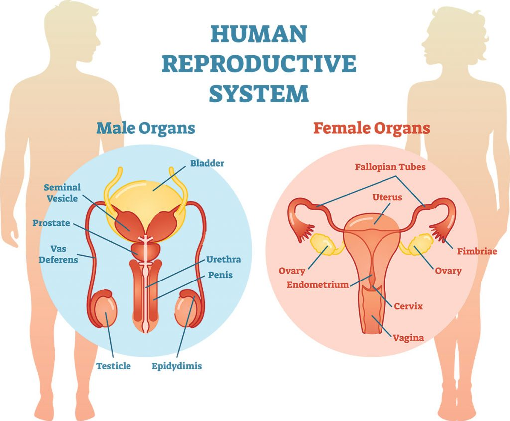
Organs of the female reproductive tract.
The organs of the female reproductive tract are divided into external and internal.
External Organs
Mons pubis
Labia majora
Labia minora
High man
The clitoris
Bartholien’s Gland
Vestibular glands
Perineum.
Internal Organs.
Overy 02
Fallopian Tubes 02
Uterus 01
Vaginal and vaginal CVD
Organs of the Male Reproductive System.
The organs of the male reproductive tract are also divided into external and internal.
External Organs.
Pennies
scrotum.
Internal Organs.
Testis. 02
The epididymis. 02
Vasdifferentus duct 02
Spermatic cord 02
Seminal vesicles. 02
Ejaculatory duct. 02
prostate gland. 01
Uterus..
The uterus is a hollow muscular organ located in the pelvic cavity. It is a pear shaped organ. It lies behind the urinary bladder in the pelvic cavity and in front of the rectum. It is 7.5 cm long, 5 cm wide and 3 cm thick.
Its weight is approximately 30 to 40 grams. Its weight increases during pregnancy and is estimated to be 900 to 1000 grams.
In women, the uterus is arranged in the normal anatomical position of anteversion i.e. forward and anteflexion i.e. leaning forward.
Uterus is divided into three main parts according to its anatomical parts.
fundus..
The uppermost dome-shaped part of the uterus is called the fundus. One fallopian tube emerges from both sides of this part.
the body.
The most central part of the uterus is called the body. It is a narrow canal like structure.
Cervix..
The lowest, narrow, round portion of the uterus is called the cervix. The opening on the inside of this part is called the internal os. The opening to the outside of the vaginal cavity is called the external os.
Structure of the uterus.
The wall of the uterus is divided into three types of layers
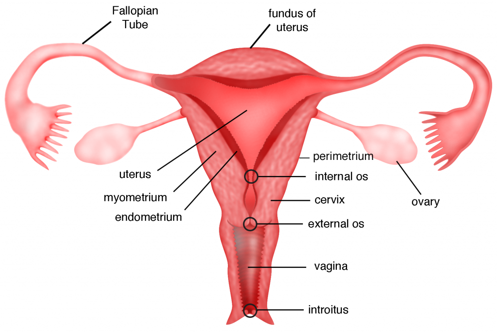
Perimetrium layer…
It forms the outermost wall of the uterus. It is the serous layer. It is attached to the peritoneum layer on the fundus side of the uterus and also to the urinary bladder. There forms a vesicouterine pouch. This layer forms the rectouterine pouch below the body of the uterus and the cervix where it joins the rectum. It is also called Pouch of Douglas. Areolar connective tissue and epithelium tissue are present in its wall.
Myometrium layer.
It forms the middle layer of the uterus. Muscles are located in this layer. These muscles are smooth muscles. The process of child birth is seen due to the contraction of these muscles during child birth.
Endometrium layer.
This is the innermost layer of the uterus.
Its inner wall is known as mucus membrane. It is composed of epithelium tissue and secretes mucus. During pregnancy this inner layer is known as decidua.
Ligaments of the uterus.
The uterus lies in the pelvic cavity. The following ligaments are present to hold and support it in its normal position.
Broad ligament
Round ligament
Utero sacral ligament
Transverse cervical ligament etc.
Blood supply of uterus.
Blood supply to the uterus is through branches of the uterine artery and the internal iliac artery. Venus return also occurs through its own Venus branches.
Nerve supply to the uterus is by sympathetic and parasympathetic nerves.
Functions of the Uterus..
Uterus helps in fertilization of ovum and sperm.
After fertilization, it helps the zygote to implant in the inner wall of the uterus and maintain the pregnancy.
In pregnancy, the content inside the uterus increases, the size of the uterus also increases so that the pregnancy can continue.
It functions to provide nutrition to the fetus in the uterus during pregnancy.
The muscles of the uterus contract to help the baby come out during delivery.
The endometrium, the inner wall of the uterus, breaks down during the menstrual cycle. As this cycle continues every 26 to 30 days, the chance of infection is reduced due to influx of wbc.
Que 2(b) Discuss the menstrual cycle
Describe menstrual cycle 04
Menstruation cycle occurs after puberty phase in females. In which changes are seen in the function of ovaries and uterus.
A menstrual cycle occurs every 26 to 30 days. This is seen due to changes in blood hormone levels.
The onset of the menstruation cycle is known as menarche.
Females find this cycle continuous after the age of puberty. Which stops temporarily during pregnancy and stops completely after the period of menopause.
The onset of menstruation is due to the degeneration of the corpus luteum layer in the uterus and bleeding exits through the vaginal cavity.
Menstruation cycle has the following phases.
Menstrual phase..
This phase occurs every 28 days and lasts for about four days. When fertilization of the egg does not take place in the female, the hormones estrogen and progesterone that support the uterine wall decrease and the hormone oxytocin increases. So the stimulation of contraction of the uterus increases and degeneration of the corpus luteum layer of the wall of the uterus starts and blood drains from the uterus through vaginal discharge. This phase lasts from 1 to 4 days.
This menstrual flow contains endometrial glands, endometrial cells, blood and unfertilized ovum. Approximately 100 to 200 ml of blood is shed during the 3 to 5 days of this phase which is called the menstrual phase.
Proliferative phase..
The menstrual phase ends on the 5th day. After that the proliferative phase starts from day 6 and lasts till day 14.
In this phase follicle stimulating hormone stimulates the ovarian follicles and hence increases estrogen production. This estrogen stimulates the proliferation of the endometrium.
The endometrium of the uterus begins to develop from the sixth day. Its cells multiply and due to this increase in mucus secreting glands and blood capillaries. Thus the endometrium of the uterus becomes bulky and vascular.
At the end of this phase, the inner wall of the uterus is ready for implantation of the fertilized egg. This phase ends with ovulation and towards the end of this phase there is a decrease in estrogen levels.
Secretary phase.
After the proliferation phase is completed, the secretory phase begins. The secretory phase is observed from the 15th day of the menstrual cycle to the 28th day.
As progesterone hormone is important in this phase, this phase is also called progesterone phase.
When the mature egg is released by the ovary due to ovulation, the amount of estrogen and progesterone hormones decreases, but the corpus luteum of the uterine wall maintains the pregnancy by secreting progesterone.
As this mature ovum is not fertilized by a sperm, the corpus luteum decreases progesterone and due to the decrease in progesterone hormone, there is an increase in the amount of oxytocin hormone and the uterine muscles begin to contract.
The next cycle begins at the end of this phase due to the corpus luteum not receiving a fertilized ovum and increased uterine contractions.
Or
Que 2(a) Describe the structure and function of the skin
- Type in the structure of the skin and expand it
A skin is a covering that completely covers the body from the outside. It is also called integumentary system. It is the largest organ or part of our body.
The total surface area of the skin is about 2 meters square and its thickness is approximately 1 to 2 mm.
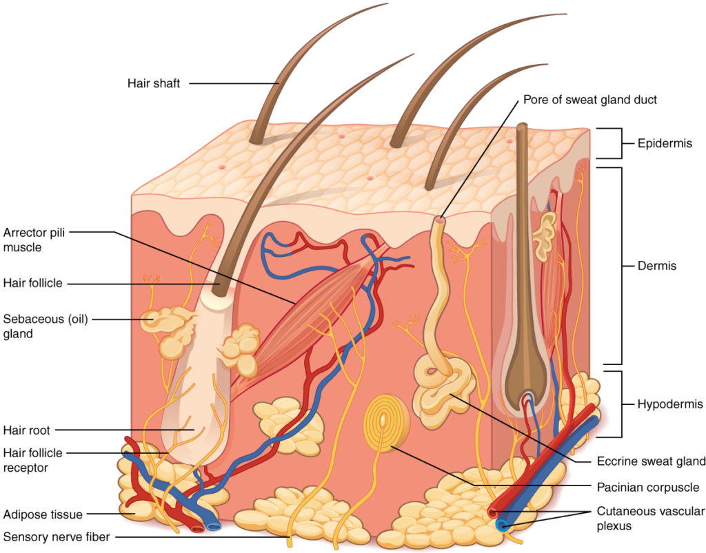
Structure of the skin..
The structure of the skin consists of layers of skin, glands, nails and hair.
Layers of the skin..
There are mainly three layers of skin. Epidermis, dermis, hypodermis
Epidermal…
It is the most superficial and outermost layer of the skin. Its structure consists of stratified columnar epithelium tissue. This layer does not contain blood vessels.
The epidermis is not evenly distributed throughout the body. Somewhere it is thicker like the soles of the feet and the palms of the hands. Somewhere its thickness is also less, like the part of the cornea of the eye.
In the epidermis layer, cells grow from the basement layer and up to the superficial layer. It takes about 35 to 40 days for the entire epidermis to be replaced.
The epidermal layer consists of the following layers.
A. Stratum corneum.
This is the outermost layer among all the layers. Dead cells are arranged in a line. These cells are flat and very thin.
Keratin is present in this layer. It is a protein. It works to protect the cells inside and prevents them from drying out.
It maintains the elasticity of the skin and helps to keep it soft.
This layer is constantly worn away due to external wear and tear.
B. Stratum lucidum.
This layer is also made up of dead and flattened cells. It is also called block layer because the cells in this layer do not contain water and nucleus.
This layer contains a protein called LEDN. It works to protect the skin from the ultraviolet rays coming from the sun.
Ç. Stratum granulosum..
Because these cells contain granules, it is called the granulosum layer.
This layer is 2 to 4 cell rows thick.
D. Stratum germinative.
This layer is the innermost layer of the epidermis.
This layer produces new cells from time to time and they rise towards the cell surface. Two types of cells are found here, precal cells and basal cells.
Dermish layer..
This layer of skin is below the epidermis. It contains connective tissue. This layer contains collagen fibers, elastic fibers and reticular fibers.
Due to which the elasticity of the skin is maintained. These fibers strengthen the skin and are very important in providing support.
Cells in the dermis include fat cells, fibroblasts and macrophages cells.
The dermis layer is composed of a papillary layer and a reticular layer. Both of which are connected to each other, cannot be separated.
The dermis layer consists of blood vessels, lymph vessels, hair follicles, sensory nerve endings,
Sweat gland, sebaceous gland etc. structures are present.
Hypodermis layer..
This layer is below the dermis layer. It is also called the subcutaneous layer.
This layer is made up of loose fibrous connective tissue. It is thicker than the dermis. It contains blood vessels, lymph vessels and nerves.
Functions of the Skin.
The skin forms a continuous covering around the outside of the body, thereby acting as the first line of protection.
Prevents micro-organisms from entering the body directly.
Prevents any external injury or any harmful elements from entering the body.
It plays a very important role in maintaining normal body temperature.
The skin provides the outer frame work to the body. It is the largest organ in the body. By covering all the organs on the outside, it works to give shape to the body.
It synthesizes vitamin D in the body. In which there is a chemical called Seven D Hydrocholesterol in the skin. which converts ultra violet rays from the sun into vitamin d3 and cholecalciferol.
Thus it acts to synthesize vitamin D.
The skin also functions to excrete waste products and harmful substances from the body. It also excretes some waste products from the body through the secretion of perspiration.
Some substances are also absorbed through the skin. This is also a no route to medication. In which the skin absorbs certain ointments and medicines applied to it and sends them into the systemic circulation.
Sensory nerve endings are located in the skin. Which helps in transmitting the impulses of touch, temperature and pain etc. to the brain. This gives us an interpretation of every stimulation.
The skin also serves to store certain nutrient materials such as fat.
Skin plays an important role in wood healing.
Que 2(b) Explain posterior pituitary gland. 04
The pituitary gland is known as the master gland of the body because the functions of other glands are regulated by the hormones of this gland.
The pituitary gland is located in the hypophysial fossa of the sphenoid bone of the cranium cvt. Pituitary gland is also known as hypophysis. The pituitary gland is strongly connected to the hypothalamus, so many functions are regulated due to this inter-connectivity.
The size of the pituitary gland is 1.2 to 1.5 cm long. It weighs approximately 0.5 grams. It is a peanut shaped gland.
The pituitary gland consists mainly of three lobes. Different hormones are secreted from each lobe which can be described as follows. Hormones secreted from each lobe are influenced by releasing and inhibiting hormones secreted from the hypothalamus. Releasing hormones secreted from the hypothalamus increase the secretion of the pituitary gland while inhibiting hormones decrease the secretion of the pituitary gland.
Posterior Pituitary Gland.
This is a lobe of the posterior pituitary gland.
It is known as neurohypophysis.
This lobe of the pituitary gland is connected to the hypothalamus by nerves and nerve fibers. Due to which it is known as neurohypophysis. The following hormones are secreted by this lobe.
A. oxytocin..
It is a hormone secreted by the posterior lobe. It is the hormone responsible for initiating normal labor pain during delivery and the contraction of the myometrium muscles in the uterus. By which the process of child birth can be done well.
Oxytocin hormone also acts to eject the milk from the breast when the baby sucks the mother’s breast milk.
This hormone is also responsible for smooth muscle contractions during sexual intercourse that allow sperm to travel from the vaginal cavity and uterus to the fallopian tubes.
B. Antidiuretic hormone..
This hormone is also known by another name of vasopressin. This hormone prevents large amount of urine coming out of the body which leads to absorption of water in the renal tubules due to which urine excretion is controlled.
It plays a very important role in maintaining water content in the body and maintaining fluid balance.
It causes the contraction of smooth muscles which is important for increasing blood pressure.
Que 3 Write short answer (any two). 6+6 =12
a. Explain the methods of steam sterilization. Explain methods of stream sterilization.
Autoclave
Heat under pressure is a practical and useful agent in sterilization.
In this method 15 lbs pressure and 121° C temp. 30 minutes is required for sterilization.
Pressure does not kill organisms, but steam under pressure can do the job.
Time is how much bulky material to sterilize. Depends on it. This is considered one of the best method of sterilization. Which destroys all bacteria and spores.
The equipment used for this process is called an autoclave machine, which is similar to a home pressure cooker.
An utoclave is a double walled metal instrument.
which has an airtight chamber. They usually have 2 gauges.
It reads the steam presser of the outer inner chamber.
It has a safety valve to prevent explosion due to high pressure, which releases steam as the pressure increases.
There is also an exhaust valve to release the steam from the inner chamber.
There is also a valve to send steam from the outer chamber to the inner chamber.
Some autoclaves also have thermometers fitted inside the chamber.
After keeping the object on the plate inside the inner chamber, after closing the lids tightly, the heating is turned on.
15 lbs/inch, 120°C 30 minutes autoclave advantages are. That can kill the organism including the spore.
And fixed temperature and pressure of steam can be maintained. This method can be used to sterilize culture media, rubber goods, linens, dressing, syringes and…Inruments and many other important consumables can be used in sterilization. It is one of the best, most practicable, dependable method of sterilization.
Tyndallization
It is also called Tyndallization as it was developed by British scientist John Tyndall.
In this process 100* C temp. 20 min. until required.
And this is done for 110 4 days.
In other words, repeated heating at day intervals (12 to 24 hrs) is used, it also destroys vegetative forms bacteria and spores.
In the third heating, almost all remaining organisms are destroyed, except for the preparation of culture media, this method is not used.
Arnold, a German bacteriologist developed a special apparatus called Arnold steam sterilizer, in which steam is used without pressure.
Que 4(a) short answer : cerebrum 04 marks
- The cerebrum is composed of right and left cerebral hemispheres.
- The two cerebral hemispheres are connected by the corpus callosum. Each hemisphere has a cavity called a lateral ventricle.
- Gray matter is located in the structure of the superficial part of the cerebrum called the cerebral cortex. White matter is located in the deep part of the cerebrum.
- Cerebral cortex consists of elevated areas called gyri or convolutions and these are separated by notches called fissures or fissures.
- The deepest of these fissures is the longitudinal fissure that separates the right and left hemispheres.
- The hemispheres of the cerebrum are divided into different lobes which are as follows.
- Frontal lobe
- Parietal lobe
- Temporal lobe
- Occipital lobe..
- An important fissure on the side of the cerebrum;
- Longitudinal sulcus or fissure which is the deepest and divides the cerebrum into two hemispheres.
- Central sulcus which lies between the frontal and parietal lobes.
- Lateral sulcus which is a deep groove and separates the temporal lobe from the frontal and parietal lobes.
- Parieto-occipital sulcus which separates the occipital lobe from the two parietal lobes….
- Nerve fiber tracks are located within the cerebrum viz
- Association fibers
- Commissural fibers
- Projection fibers
These fibers are interconnected and connect one area to another and help in the transmission of impulses.
FUNCTIONS OF THE CEREBRUM
Cerebrum controls intelligence, memory, reasoning, thinking, speaking, reading, writing etc.
No control is seen for the perception of sensory perception like pain, temperature, touch, sight, hearing, taste, smell etc.
Control for the contraction of skeletal muscles is found in these aria.
Que 4(c) innominate bone 04
The innominate bone is the bone of the pelvic girdle. It is also known as hip bone.
They are two in number in the body. It is located one on both the right side and left side of the pelvic cavity. Both innominate bones articulate with the sacrum bone behind and form the pelvic cavity.
The innominate bone is the larger bone. It is a flat and irregular type of bone.
Each innominate bone consists of three bones.
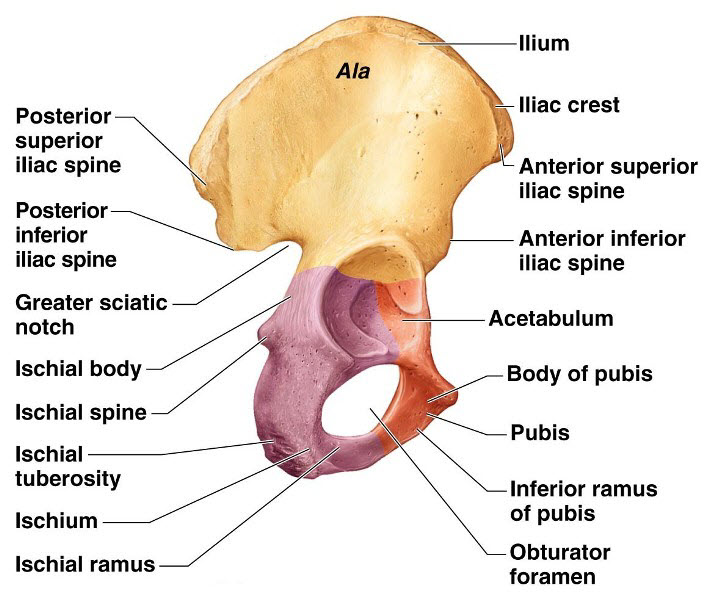
Ilium
ischium
pubis.
Ilium…
Innominate Bone The ilium bone is a flat bone on the upper side. At its uppermost point there is a ridge called the iliac crest. It falls below the iliac crest.
Anterior superior iliac spine (top of front)
Anterior inferior iliac spine (at the bottom of the front)
Posterior superior iliac spine (upper back)
Posterior inferior iliac spine (lower back)
The ilium bone forms the sacroiliac joint where it joins the sacrum bone at the back.
There is a big groove at the bottom of this joint. The notch is called the greater sciatic notch. From where the sciatic nerve and blood vessels pass to the lower extremities.
The gluteal muscles attach to the posterior surface of the ilium bone. And there make up the gluteal region.
The anterior surface of the ilium bone is known as the iliac fossa. In this part there is a depressed part where the muscles are attached.
ischium…
Below and behind the ilium bone lies the ischium bone.
Between the ilium bone and the ischium bone, the posterior side is an inferior pointed part. This part is called ischial spine.
Below it lies a small notch called the laser sciatic notch.
This laser has a strong thick process below the sciatic notch. Which is called ischial tuberosity. Body no weight comes on this part while sitting in sitting position. This has a stronger structure than a weight beer.
Pubis…
The pubis bone forms the most anterior part of the innominate bone. Both the innominate bones of the pubis bone join anteriorly to form the symphysis pubis joint.
There is a large foramen at the bottom of this pubis bone. It is called obturator foramen. Through which nerves and blood vessels pass downwards towards the extremity.
The three bones ilium, ischium and pubis located in the hip bone form a cavity-like structure called the acetabulum cavity. The head of the femur bone joins this KVT and the hip joint is formed there.
Que-4(d) kidney
Describe the gross structure of kidney 04
There are two kidneys in the human body. They are located one on both sides of the vertebral column on the poster side of the body on the right and left sides of the abdominal cavity.
Kidney is a shapeless organ. It lies from the level of the twelfth thoracic vertebra to the level of the third lumbar vertebra.
Kidney is 11 cm long by 5 to 6 cm wide. Its weight is approximately 150 grams. The right kidney is positioned slightly lower than the left kidney because the liver occupies a larger portion on the right side.
Veins around the kidney.
The kidney is an organ located in the abdominal cavity. One is located on both the right and left sides. Abdominal cavity organs like liver, small intestine, adrenal glands, stomach, spleen, pancreas etc. are located around both kidneys.
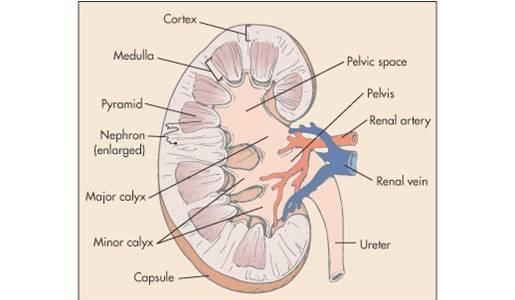
Structure of the Kidney..
Kidney is a shapeless organ. A groove in the middle is called hilum or renal hilum. Through which the structures of renal artery, renal vein, nerves, lymph vessels and ureter enter and exit.
The inner border or hilum of the kidney is found on the side of the vertebral column. Its outer border is convex. The kidney is a hanging organ on both sides of the abdominal cavity. To hold it in position, it is surrounded by a network of fatty tissue and fibroelastic connective tissue called the renal fascia. With the help of this kidney can maintain its position and it also gets protection.
When the kidney is viewed in a longitudinal section, it is seen to be distributed into three kidney structures.
Fibrous capsule.
It is part of the fibrous tissue that surrounds the kidney. This membrane is arranged around the kidney. Which acts as a layer to maintain the shape of the kidney and to protect it.
Cortex.
It is redis brown in color made up of tissue. Which is located under the kidney capsule.
Medulla.
In the kidney, the inner part from the cortex is called the medulla. It also has redish brown color. The triangular shaped pyramidal structure is called renal pyramid. The base part of this renal pyramid is towards the cortex and the pointed part of the pyramid i.e. the part of the renal papilla is arranged inwards towards the hilum.
The renal papilla forms a cup-like structure anteriorly called the calyx. The part with large space is called major calyx and the part with small space is called minor calyx. The minor calyx opens into the major calyx. Beyond this calyx is the wide funnel-shaped portion called the renal pelvis.
The urine filtered by the kidney falls into the wide part of the calyx, the funnel shape, i.e. the renal pelvis. Urine collects here and then passes anteriorly from the renal pelvis through a narrow structure called the ureter that exits the kidney and reaches the urinary bladder.
Urine filtered by the kidney passes from the minor calyx to the major calyx and from the major calyx to the renal pelvis. It then reaches the urinary bladder through the ureter. This action is not controlled by any kind of nervous system. In the wall of the renal pelvis there are special muscles and pacemaker cells due to the contraction of which this urine flows forward.
Write kidney not done
Kidney is mainly responsible for urine formation.
Kidneys filter the blood and remove the waste products through urine.
The function of the kidney is to maintain the normal balance of electrolytes.
It works to maintain blood pH.
The body functions to remove waste products accumulated at the end of metabolism from the body.
Kidneys secrete a hormone called erythropoietin which plays a very important role in the production of RBCs.
Kidneys secrete a hormone called renin which plays a very important role in maintaining blood pressure.
Kidneys are responsible for maintaining water balance in the body.
Kidney prevents the elements that are needed in the body from leaving the body.
Que 5 define following (any six) 12
(a) Pasteurization
Pasteurization is a method of sterilizing milk. In which only harmful bacteria are destroyed. But lactic acid and necessary organisms are not destroyed. Some changes occur in the protein and sugar in the milk
(b) DISINFECTION
The process of removing or killing pathogenic micro-organisms, which does not necessarily include non-pathogenic organisms, is called dis-infection.
(c) Immunity
Micro organism and
By its products (toxin).
Immunity is the resistance shown by the host against the condition
or
Immunity ie
When any antigen or microorganism enters our body and our
His body
It is called immunity by registering against it and giving protection
(d) Carrier
Someone who
Animals with specific diseases
It contains producing micro-organisms that can spread diseases or
carried
But it does not show any signs or symptoms of those diseases
Carrier says.
E.g. Typhoid
(e) Tidal volume Tidal volume..
Tidal volume is the volume of air entering the lungs during inspiration and the volume leaving the lungs during expiration which is called the titular volume during normal respiration of a person. Tidal volume in a normal adult is approximately 500 ml.
(f) Pandemic Pandemic
Pandemic ie
Any disease can spread from one state to another and from one country to another and
It is called a pandemic if it occurs all over the world
D. T. Swine flu
covid 19 etc
(g) Hypersensitivity
(h) Oogenesis.
The process of formation of female gametes i.e. female ovum cells is called oogenesis. This process is also called gametogenesis.
Que 6(a) multiple choice questions 05
The smallest bone of the ear is the stapes
A Melius
B Inks
C stapes
Secreted by cerebrospinal fluid..choroid plexus..
A choroid plexus
B spinal cord
C Cerebrum
The outer layer of the bone is called the periosteum.
A Myocardium
B periosteum
C Perimetrium
Total chambers of heart are .. 4 ..
A 2
B 4
C 6
Total cranial nerves are .. 12..
A 12
B 14
C 16
Que 6(b) fill in the blank 05
Bile is stored in _ Bile is stored in .. Gallbladder.. Anti diuretic hormone is secreted by__ Anti diuretic hormone is secreted by ..pituitary gland..
Normal pH value of blood is_______ The normal pH value of blood is ..7.35 to 7.45..
Tears are produced by______ Gland. It is produced by the lacrimal gland.
The largest part/organ of human body is _ The largest part of the human body is skin.
State Que 6(b) True False 05
Lifespan of platelet is 230 days. (false)
Tongue is an involuntary muscle. The tongue is an involuntary muscle. (false)
Alpha cell of pancreas secretes insulin. Insulin is secreted from the alpha cells of the pancreas. (false)
Sternum is the bone of thoracic cage. The sternum is a bone of the thoracic cage. (correct)
Spleen is a largest lymphatic organ. The spleen is the largest lymphatic organ. (correct)