ENGLISH – GNM FY BIO SCIENCE -2016 PAPER 5
Q-1 a. Write the gross structure and functions of the heart. 08
Write the gross structure of heart and its functions.
Heart is an important organ of the circulatory system. Heart beats continuously during human life. Due to its pulsation, the blood circulates continuously in the blood vessels.
The heart is an organ made up of blood and muscles. It weighs approximately 310 grams in males and approximately 250 grams in females. The heart beats approximately one lakh times during the day.
Location of the heart..
The heart lies above the diaphragm in the mediastinum space between the two lungs in the thoracic cavity.
The heart is a rough cone shape. In it, its upper broad part is known as the base and the lower angled part is known as the projection.
The heart is arranged slightly to the left between the two lungs in the thoracic cavity.
Structure of the Heart..
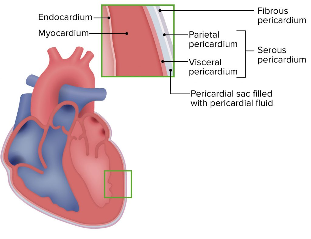
Heart is an organ made up of semi-muscles. Its wall is made up of three types of tissue layers.
The outermost layer of the heart wall is called epicardium or pericardium.
Epicardium or Pericardium.
It is thin and transparent and covers the heart from the outside. It is made up of fibrous connective tissue. In which there is a layer of fibrous tissue on the outermost side and a serous membrane on the inner side of the fibrous tissue which is found in a double layer. The outer layer of the serous membrane is known as the parietal layer and the inner layer as the visceral pericardium layer.
The space between the parietal and visceral pericardial layers is called the pericardial space. This space contains fluid called serous fluid or pericardial fluid. Which prevents friction between the two layers.
This layer of outer pericardium protects the heart from the outside and this layer is also seen around the vessels coming out of the heart.
Myocardium..
Myocardium is the middle layer of the heart. It lies below the pericardium. It is made up of a special type of cardiac muscle tissue. The pumping action of the heart is seen due to the contraction of these muscles.
This myocardium layer is thin at the base and thick at the apex. Also, the layer of the wall of the left ventricle is thicker than that of the wall of the right ventricle.
The contraction of these muscles has an involuntary action that results in the pumping action of the heart and is controlled by the autonomic nervous system and the conducting system in the heart.
endocardium..
It is the innermost layer of the heart wall. It is in contact with layer blood. This layer is made up of epithelium tissue and connective tissue. This layer is smooth and shiny which is important for the smooth flow of blood inside the heart. This layer also covers the valves inside the heart and this layer is continuous in the inner wall of the blood vessels leaving the heart.
Functions of the Heart
Heart provides oxygenated blood supply to all organs and tissues of the body.
Heart is an important organ of the cardiovascular system. It functions as a vital organ without which the human body cannot survive.
The heart circulates the blood towards the lungs so that the blood can be oxygenated and purified.
Circulations like pulmonary circulation and systemic circulation are regulated by the heart.
The heart also regulates the heart rate according to the needs of the body and according to the body temperature.
The heart also regulates body temperature as it circulates blood to every part of the body.
As the heart pumps blood to the body’s excretory organs, the blood can be filtered and waste products removed from the blood.
b. Explain pulmonary circulation. 04
Pulmonary circulation started from the right ventricle and the blood goes to the lungs and from there returns to the left atrium, so the circulation from the right ventricle to the left atrium is called pulmonary circulation.
In the pulmonary circulation, deoxygenated blood in the right ventricle exits the right ventricle through the pulmonary artery. As it exits, the pulmonary artery divides into a right and a left pulmonary artery and both enter the lung. In which two branches in the left lung and three branches in the right lung enter the pulmonary artery which is according to each lobe of the lung.
Gas exchange takes place between the blood in the lungs and the tissues of the lungs and two pulmonary veins carry oxygenated blood from each lobe and enter the left atrium of the heart from the lungs on both sides.
Pulmonary circulation converts deoxygenated blood in the heart to oxygenated blood via the lungs. This blood enters the left ventricle and supplies oxygenated blood to the whole body through the systemic circulation.
Circulation from right ventricle to left atrium is called pulmonary circulation.
Q-2 Answer the following questions (ANY THREE) 12
a. Explain the structure of long bone. Explain the structure of long bone.
Long bones are the long bones in the body. In which the length is more than the width. The structure of this long bone includes the following parts.
Epiphyses..
It is also called the upper and lower extremity of the bone. It is the proximal and distal part of a long bone. Its outer lining consists of compact bone and calcareous bone on the inside.
Epiphyseal cartilage separates the epiphyses from the diaphyses.
diaphysis.
It is called the middle part of the long bone. The middle medullary canal is located in the middle of this part. In which the bone marrow resides.
Metaphysis.
In a long bone, the metaphysis is located where the epiphyseal plate joins the diaphysis.
Articular cartilage.
At the end of the epiphysis there is a layer of this type of cartilage. Which are located between connecting with other bones. It prevents friction between the two bones and makes the movement pain-less.
periosteum..
The membrane on the outer wall of the bone is called the periosteum layer. It forms a covering around the outside of the bone. It is not located in the articular cartilage. This membrane is made up of fibrous tissue. It contains blood supply, nerve supply and lymphatic vessels.
Medullary canal.
It is a narrow canal between Long Bone. Bonmero is located in this part of the canal.
Endosteum..
This is the membrane surrounding the medullary canal which contains stem cells and osteoclast cells.
b. Explain the digestive activities in stomach. Explain the process of digestion in the stomach.
Food taken by mouth reaches the stomach through the esophagus. Mechanical digestion of food begins due to the charming movement of the stomach due to the contraction of the muscular layer of the stomach. Here all the food is converted from large molecules to small molecules by mechanical digestion.
After this mechanical digestion has gone on for some time, hydrochloric acid is secreted with the food through the glands in the inner wall of the stomach.
Chemical digestion begins when the chemicals present in this juice of the stomach mix with the food.
In addition to hydrochloric acid, this gastric juice contains enzymes, mainly pepsin and renin. This pepsin digests large molecules of protein into smaller molecules. Digestion of protein starts here.
Renin in the gastric juice digests the protein casein in milk and converts it into paracasein. Thus, digestion of protein mainly takes place in the stomach.
Digestion of carbohydrates and fats does not take place in the stomach, only mechanical digestion takes place here. After the partial digestion of this food in the stomach, it goes to the small intestine where it is completely digested and absorbed.
As the food in the stomach mixes with gastric juice, the harmful bacteria and viruses in the food are destroyed due to the acidic property of HCL in it.
c. List the functions of cerebrum
Cerebrum controls intelligence, memory, reasoning, thinking, speaking, reading, writing etc.
No control is seen for the perception of sensory perception like pain, temperature, touch, sight, hearing, taste, smell etc.
Control for the contraction of skeletal muscles is found in these aria.
Functions related to vision and speech.
Spatial areas in the cerebrum are associated with specific functions such as temperature regulation. No control for hunger and thirst and no action for satisfaction.
The cerebrum is also involved in the coordination of certain motor movements.
The words spoken by you are also interpreted by the cerebrum.
This aria also works for emotions as well as certain psychological functions.
d. Explain regulation of body temperature in human.
Regulation of heat in the human body means maintaining the balance of heat production and heat loss in the body. Generally, normal body temperature in humans is found to be 36.5 to 37 degrees Celsius. Only if this temperature is kept normal, every function in the body is kept regular and internal homeostasis can be maintained.
The structure that regulates body temperature in humans is the thermoregulatory center located in the hypothalamus. Nerve impulses of temperature are received by the hypothalamus through the thermoreceptors located in the body and the hypothalamus acts accordingly to know the normal body temperature.
Heat production occurs in the body due to metabolic activity, movement of skeletal muscles etc. When this heat is higher than normal in the body, the hypothalamus stimulates the sweat glands to lose excess heat from the body through the action of perspiration and helps to maintain a regular body temperature.
When the body heat is less than normal, sweat gland activity is not observed and the body maintains normal body temperature by preserving heat.
Thus hypothalamus plays an important role in maintaining normal body temperature.
Q-3. Write the shortnotes on (ANY THREE) of the following :- 12
a. Sterilization –
Microbes are almost everywhere. But we are not conscious of it. Their presence is excessive in hospitals where the patient leaves a large number through their urine, stool, sputum, secretion.
Apart from this, microbes are also present in blood, food, water, sewage, air and soil etc.
Since they are found in many ways, different methods have been developed to destroy them.
Which includes physical and chemical methods, which are used according to the knowledge and requirements of the operator.
A person’s personal comforte also depends on his knowledge of this matter and the method of microbe control.
Like, Environment, Equipments, Cleanings, Food & kitchen, Body care etc. There are three main reasons for destroying or inhibiting micro-organisms
(1) To prevent infection and transmission of disease.
(2) To prevent decomposition and spoilage of food.
(3) To prevent contamination of material used in culture.
There are several methods of sterilization which are as follows
Physical agents
(1) Heat:- Direct Flaming, Boiling
(2) Sunlight:- Sterilization is done by this natural method in water tank, rivers, lakes etc.
(3) Cold
(4) Drying and desiccation
(5) Radiation
(6) Filtration
(7) Sound Wave and Ultra Sonic Vibration )
Chemical agents
(1) Phenol and cresol compound
(2) Alcohol
(3) Halogen
(4) Dia
(5) Aldehyde
(6) Acid
(7) Alkali
8) Gas
9) Metallic salts
(10) Oxidizing agent
(11) Surface active agents
b. Thyroid gland –
The thyroid gland is an important gland of the endocrine system. It is located in the soft tissue of the neck. This gland is a butterfly shaped gland.
The weight of this gland is approximately 30 grams. Its length is 5 cm and width is 3 cm.
This gland is located from the level of the fifth cervical vertebra to the level of the first thoracic vertebra.
Thyroid gland has one lobe on both sides. Which is covered with fibrous tissue around it. The middle part connecting the two lobes is called the isthmus. The lobes of the thyroid gland have a pyramidal shape.
The tissue in the thyroid gland is made up of tiny structures called follicles. Each follicle is composed of simple cuboidal glandular epithelium tissue. which is connected with secretion.
Thyroid stimulating hormone is a hormone released from the pituitary gland that regulates the function of the thyroid gland.
The thyroid gland secretes the following hormones.
- Triiodothyronine T3… The main function of this hormone is to maintain normal physical growth and development in the body. This hormone also plays a useful role in maintaining heart rate and certain metabolic activities.
- Thyroxine T4… This hormone also performs the same function as the T3 hormone. That is, the body maintains metabolic activity and functions to maintain normal physical growth and development. This hormone increases the basal metabolic rate.
- Calcitonin..
This hormone is secreted by the parafollicular cells of the thyroid gland. Which affects the metabolism of calcium in the blood.
c. Physiology of respiration –
Respiration is gas exchange between two surfaces. In which air from the atmosphere enters the lungs. Gas exchange between lung tissue and blood is called external respiration. The gas exchange that takes place between each cell tissue of the body and the blood is called internal respiration.
During the act of respiration, oxygen enters the lungs through inhalation and carbon dioxide leaves the body through exhalation.
Normally, the act of respiration is observed 16 to 18 times in a minute.
The following steps are observed in the cycle of resuspension.
Inspiration
expiration
pos.
Inspiration…
Inhalation of atmospheric air into the lungs is called inspiration.
When the brain receives nerve impulses for contraction of the diaphragm and intercostal muscles, the contraction of the diaphragm and intercostal muscles increases the size of the thoracic cavity. The air pressure inside the thoracic cavity decreases so that air from the outside atmosphere can enter the lungs through the action of inspiration. This action is called inspiration.
Contraction impulses to the diaphragm cause the diaphragm to flatten downwards and contraction of the intercostal muscles causes the ribs and intercostal muscles to move upwards and outwards. So the size of the thoracic cavity increases and negative pressure is created inside the cavity. As the air pressure in the outside environment is higher and the air pressure in the thoracic cavity is lower, the action of inspiration can take place. The act of inspiration is an active process.
Expiration…
The process of expelling air from the lungs into the atmosphere is called expiration. The action of expiration is a passive process that begins after the action of inspiration is completed.
During exhalation, the contracted diaphragm and intercostal muscles relax. So the diaphragm returns to its original position and the ribs move downwards and inwards, reducing the size of the thoracic cavity and exhaling. In which the air from the lungs is thrown out into the atmosphere.
In the act of exhalation, the air pressure in the lungs is greater than the atmospheric pressure so that the act of exhalation takes place.
Pos…
This is the relaxed stage of the lung. In which no action of inspiration or expiration takes place. This period is called pause period.
d.Uterus-
Uterus..
The uterus is a hollow muscular organ located in the pelvic cavity. It is a pear shaped organ. It lies behind the urinary bladder in the pelvic cavity and in front of the rectum. It is 7.5 cm long, 5 cm wide and 3 cm thick.
Its weight is approximately 30 to 40 grams. Its weight increases during pregnancy and is estimated to be 900 to 1000 grams.
In women, the uterus is arranged in the normal anatomical position of anteversion i.e. forward and anteflexion i.e. leaning forward.
Uterus is divided into three main parts according to its anatomical parts.
fundus..
The uppermost dome-shaped part of the uterus is called the fundus. One fallopian tube emerges from both sides of this part.
the body.
The most central part of the uterus is called the body. It is a narrow canal like structure.
Cervix..
The lowest, narrow, round portion of the uterus is called the cervix. The opening on the inside of this part is called the internal os. The opening to the outside of the vaginal cavity is called the external os.
Structure of the uterus.
The wall of the uterus is divided into three types of layers. Which can be classified as follows from outside to inside.
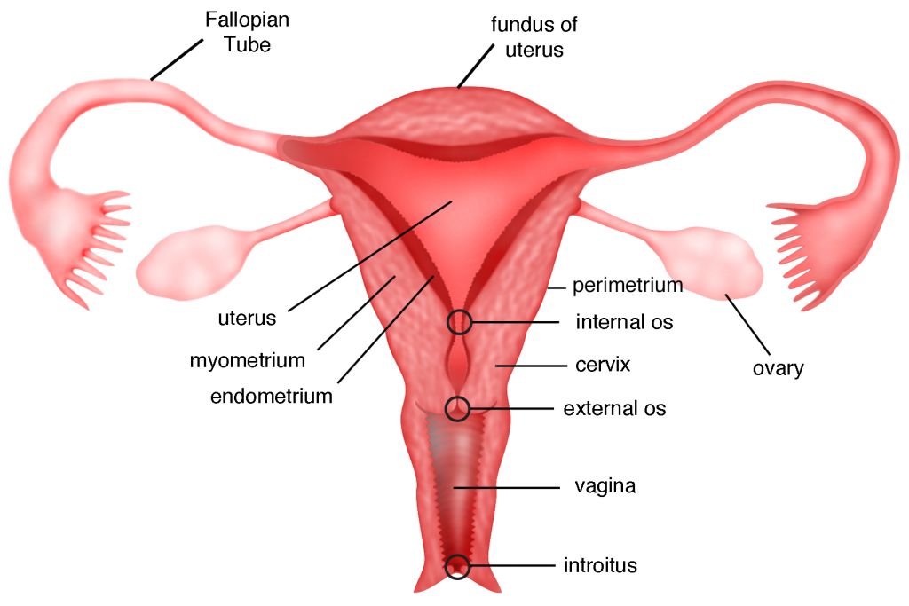
Perimetrium layer…
It forms the outermost wall of the uterus. It is the serous layer. It is attached to the peritoneum layer on the fundus side of the uterus and also to the urinary bladder. There forms a vesicouterine pouch. This layer forms the rectouterine pouch below the body of the uterus and the cervix where it joins the rectum. It is also called Pouch of Douglas. Areolar connective tissue and epithelium tissue are present in its wall.
Myometrium layer.
It forms the middle layer of the uterus. Muscles are located in this layer. These muscles are smooth muscles. The process of child birth is seen due to the contraction of these muscles during child birth.
Endometrium layer.
This is the innermost layer of the uterus.
Its inner wall is known as mucus membrane. It is composed of epithelium tissue and secretes mucus. During pregnancy this inner layer is known as decidua.
Ligaments of the uterus.
The uterus lies in the pelvic cavity. The following ligaments are present to hold and support it in its normal position.
Broad ligament
Round ligament
Utero sacral ligament
Transverse cervical ligament etc.
Blood supply of uterus.
Blood supply to the uterus is through branches of the uterine artery and the internal iliac artery. Venus return also occurs through its own Venus branches.
Nerve supply to the uterus is by sympathetic and parasympathetic nerves.
Functions of the Uterus..
Uterus helps in fertilization of ovum and sperm.
After fertilization, it helps the zygote to implant in the inner wall of the uterus and maintain the pregnancy.
In pregnancy, the content inside the uterus increases, the size of the uterus also increases so that the pregnancy can continue.
It functions to provide nutrition to the fetus in the uterus during pregnancy.
The muscles of the uterus contract to help the baby come out during delivery.
The endometrium, the inner wall of the uterus, breaks down during the menstrual cycle. As this cycle continues every 26 to 30 days, the chance of infection is reduced due to influx of wbc.
e. Prevention of infection –
f. Mechanism of hearing
Mechanism of hearing i.e. physiology of hearing means act of hearing. The wavelength for hearing is 20 to 20,000 Hz. The human ear is capable of frequencies between 500 and 5,000 hz. The frequency at which the sound waves vibrate is known as the pitch, as the vibration increases, the pitch increases.
Every sound produces sound waves which strike the outer part of the auricle and from there enter through the external auditory canal, these sound waves vibrate the tympanic membrane i.e. the ear drum which is the junction between the external ear and the middle ear.
The sound waves are connected to the tympanic membrane by the malleus bone to the incus and incus to the stapes and the stapes bone is further connected to the oval window. Goes and from there goes to the endolymph and the round window vibrates and the vestibule goes to the cerebrum through the cochlear nerve and the sound is recognized.
Q – 4. Answer the following questions (ANY FOUR) 12
a. Which factors affect the growth and development of microbes.
What factors affect the growth and development of microorganisms? Write it down
1) Moisture
Like nourishing food, every bacteria needs water for growth. In fact, bacteria cannot get food in the absence of water, because every food element needs to be in a liquid state to pass through the bacterial wall. All types of bacteria grow well in liquid medium (Aqueous medium), an environment without complete moisture prevents its growth. or destroys.
In addition, cells cannot live in low or high humidity
2) Light
Most bacteria are destroyed by direct exposure to ultraviolet rays in sunlight.
3) Temperature :-
Temperature is a very important factor affecting the growth of bacteria. Optimal temperature with food, water is necessary for bacteria growth.
Different bacteria have different optimal temperatures.
37°C is the optimal temperature for bacteria growing in the human body.
However, many bacteria are mesophilic (meso = middle, phille = loving). The optimum temperature for it is 25 to 39* C.
Most bacteria grow this way.
Whereas psychrophilic (psychro = cold) bacteria grow better between 4°C to 10°C, some
Thermophilic (Therma – Heat) is also found. Its growth is best between 55°C to 75°C.
Temperature above 75 C is fatal for bacteria. In fact high temperatures are created to kill bacteria in different ways.
Like moist heat (steam), boiling water, pasteurization & autoclaving.
Many species can survive even at very low temperatures. Like yeast, mould, viruses & Rickettsia, spirochetes (76* C can survive for years).
(4) Oxygen
O2 also plays an important role in the life of bacteria. Many types of bacteria can only survive, or grow, in the presence of O2. They are called Aerobes (EX.Sarcina).
Conversely Anaerobes can live or grow in the absence of 02. E.g. Closteridium tetani-
Apart from this there are also bacteria. which can survive in the presence or absence of 02. They are known as facultative anaerobes. E.g. Salmonella typhi.
Microaerophils grow more in less oxygen than is present in air. (5)Hydrogen Ion Concentration: (Acidity and Alkalinity) PH medium
The acid or alkaline concentration of the liquid in which the bacteria grow affects the growth.
This is seen from the hydrogen ion concentration index.
PH – 0 (Zero) is the most acidic,
PH – 14 shows the lowest acidic concentration.
PH – 7.) A nutral (neutral),
pH < 7 is acidic
and alkaline at pH >7
Most bacteria grow best between pH 5.0 to 8.5. There are some exceptions to this too.
6) Osmotic pressure :-
The life of bacteria also depends on high or low osmotic pressure. If the bacteria are immersed in a liquid whose osmotic pressure is very high or very low, the bacterial cell collapses or becomes dormant due to the leakage of liquid.
Carbon Dioxide is also necessary for the growth of bacteria.
b. Explain the chain of infection. –.
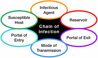
Infectious agent (pathogen)
Pathogens like bacteria, viruses, fungi etc. are present for infection and spread further through different channels.
Reservoir (the normal location of the pathogen) Reservoir :-
That is, the environment where the microorganism grows, be it humans, animals or water, and forms a colony there.
Portal of exit from the reservoir
It exits the reservoir through various roots. D. T :- Sneezing, cuffing, skin, body fluid etc
Mode of transmission Mode of transmission
Microorganisms that leave the reservoir but do not spread through it require another medium to spread. D. T. Direct contact, air, droplet etc
Portal of entry into a host Portal of entry into a host
Infection through different transmission media through different portals of entry into the host. Eg :- Respiratory tract, skin, alimentary canal etc
Susceptible host Susceptible host
Those with low immunity, old age or small children can get infections more quickly.
c. What is the role of nurse in prevention of infection?
As a nurse, we must know how to protect ourselves and patients from exposure to pathogens.
It is very important for the nurse to understand and follow the infection control policy of the organization such as use of personal protective equipment, environmental sanitation, etc.
Standard Precautions :-
To break the chain of infection, standard precautions should be taken to prevent transmission from body fluids containing pathogens.
- Hand Hygiene :-
This is the number one weapon in preventing cross-infection. Most of the spread of micro-organisms occurs through the hands, so hand washing should be done before and after each procedure. It is very important to wash hands after touching every contaminated object
- Aseptic technique
In which to use contact precaution and aseptic technique during the procedure. Use only sterile items in invasive procedures
- Environmental infection control measures
The equipment used in the hospital and the surrounding environment should be kept clean and the floor of the hospital should be cleaned with anti-septic solution. Bio-medical waste generated during procedures or work should be properly disinfected and disposed of.
- Droplet pre-caution Mask should always be worn while working in the hospital and infected patients should also wear it, precautions should be taken while cuffing, sneezing etc.
- P.P.E
Cross-infection can be prevented from personal protective equipment to the nurse and the patient, whose cap, mask, gown are used. which should be worn during the care of highly infectious patients.
- Health Education :-
Provides health education to patients and staff working under them to prevent the spread of infection in the hospital
d. What is the importance of microbiology in nursing?
1) Microorganisms have the potential to cause disease in humans so their characteristics and behavior can be known through this study.
(2) Study of Microbiology how disease occurs. We can learn how it spreads and how to stop it.
(3) Need to check laboratory to identify bacteria.
(4) Understand the importance of personal health and immune measures for resistance to microbial diseases.
(5) The prevailing superstition, ignorance and confusion among the people about diseases caused by microorganisms can be removed and correct understanding can be given. Social Stigma
Precautions can be taken while treating the patient.
(7) Microbial disease can be recognized and measures can be taken to diagnose and prevent its spread.
e. What is passive immunity? How can one get it?
Antibodies produced in one person are transferred (ready made) to another person to protect the person against disease, it is called passive immunity.
In other words, the body does not produce antibodies. But depends on readymade antibodies. Passive immunity comes in the following ways.
Injection of antisera.eg. ATS (for protection against tetanus)
Injection of gamma-globulin.
Maternal Immunity – Antibodies placenta transfer from mother to fetus. Maternal antibodies protect the baby for a few months after birth. Like against diphtheria measles etc.
Q-5 a. Write the meaning (ANY FOUR) 08 Write the meaning of the following. (any four)
- Epidemic
If there are many cases of the same disease in a specific geographical area at the same time, it is called an epidemic.
D. T. Dengue
2.Infection-
Microorganisms (bacteria, viruses, fungi) enter the human or animal body and multiply, which are not normally found in the body, which is called infection. Its signs and symptoms are visible or subclinical
3.Tendon –
A band-like structure of connective tissue in the body is called a tendon. It acts to connect the muscles to the bones in the body. This also works for movement due to attachment. Tendons function to provide structural support to the body. It is a band of white glowing connective tissue.
4.Membrane-–
The tissue layer spread over the surface of any organ is called membrane. This membrane covers the organ and provides protection. The following membranes are found in the body.
Mucus Membrane: Which is spread over the under wall of any organ. It secretes mucus. It works to provide protection to internal organs.
Synovial Membrane: It is located in the lining of the joint and secretes a fluid around the joint.
Cirrus Membrane: It is a double layered membrane surrounding the organ in which the outer layer is called parietal and the inner layer is called visceral layer.
- Afferent Neuron –
The small functional cells of the nervous system are called neurons. Afferent neuron i.e. sensory neuron which helps to carry the nerve impulses from different parts of the body to the brain. These types of nerve cells join together to form a sensory nerve. From this we get information from different senses.
B. Write the difference between W.B.C. & R.B.C. 04
(Write the question as difference. Both are given here for the sake of brevity.)
RBCs appear red in color while WBCs are white in color or colorless.
The shape of RBC is circular biconcave disk shape.. while WBC is round shape.
Nucleus is absent in RBC.. whereas nucleus is present in WBC..
RBCs are involved in the transportation of oxygen while WBCs are involved in maintaining immunity and defense mechanisms in the body.
The life span of RBC is 90 to 120 days while the life span of WBC is 5 to 21 days.
Its function is linked to the cardiovascular system while the function of wbc is linked to both the cardio vascular system and the lymphatic system.
RBCs constitute 40 to 45% of the total blood while WBCs constitute one percent of the total blood.
Only one type of RBC is found in blood whereas five types of WBC are found in blood.
RBCs have the property of circulating only in the blood circulation while WBCs can travel beyond the blood circulation to the connective tissue and lymphatic system when needed.
Decreased RBC than normal results in anemia while decreased in WBC results in leukopenia.
Q-6 a. Fill in the blanks:- Fill the empty space:- 05
Patella is a type of bone.
What type of bone is the patella? (sesamoid)
- artery contains deoxygenated blood. Deoxygenated blood flows in arteries. (pulmonary artery)
Bile is necessary for digestion of
Bile is necessary for digestion. (fate)
is 11th cranial nerve
It is the eleventh cranial nerve. (accessory nerve)
- Gives the color to the eye. Gives color to the eyes. (Iris)
b. State whether the following statements are true or false 05
Write whether the following statements are true or false,
- Alpha cells of pancrease secrete insulin.
The alpha cells of the pancreas secrete insulin. (false) - Ovum survives 72 hours after its release.
After the ovum is released it lives for 72 hours. (false) - There are 12 pairs of cranial nerves
Canial nerves have twelve pairs. (correct) - First stage of Mitosis is anaphase.
Anaphase is the first stage of mitosis. (false)
5.Process of development of RBCs is called erythropoiesis.
Erythropoiesis carries out the process of RBC development. (correct)
C. Match the following :- Join the following pairs: 05
‘a’ ‘b’
1.Outer layer of heart 1.Dermis Dermis outer layer of heart
2.Outer layer of brain 2.Periosteum Periosteum outer layer of brain
3.Outer layer of bone 3.Epidermis Epidermis out layer of bone
4.Outer layer of Uterus 4. Peri cardium
- Outer layer of Skin 5. Dura meter Dura meter outer layer of skin 6.Perimatrium
1- 4
2- 5
3-2
4- 6
5-3