ENGLISH-General Nursing & Midwifery (First Year)-BIO-SCIENCES-23/09/2024-UPLOADPAPER SOLUTION NO.9
BIO-SCIENCES-23/09/2024-PAPER SOLUTION NO.9
Q-1a. List out the organs of Respiratory system.
List of Organs of the Respiratory System
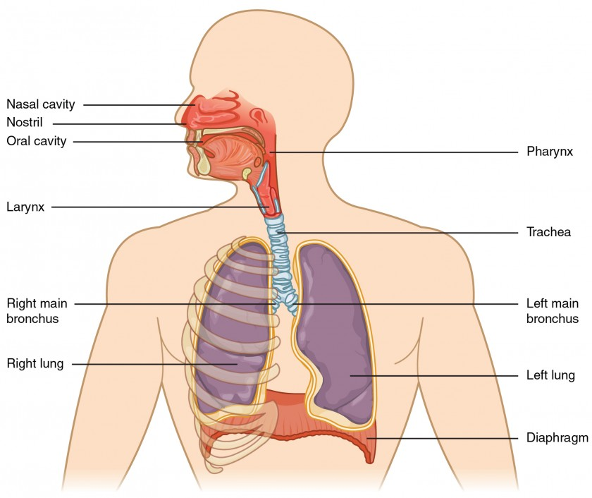
The organs located above the thoracic cavity in the respiratory tract are called the organs of the upper respiratory tract, which include the following:
- Nose
- Pharynx
- Larynx
The organs located inside the thoracic cavity in the respiratory tract are called the organs of the lower respiratory tract, which include the following:
- Trachea
- Bronchi (Right and Left)
- Bronchioles
- Terminal Bronchioles
- Alveoli
- Lungs (Right and Left)
b. Explain the physiology of Respiration.
Respiration
Respiration is the process of gas exchange between two surfaces. In this process, the air from the atmosphere enters the lungs. Gas exchange occurs between the lung tissues and the blood, which is known as external respiration. The gas exchange between the body’s cells/tissues and the blood is called internal respiration.
During the process of respiration, oxygen enters the lungs through inhalation, and carbon dioxide is expelled from the body through exhalation.
Normally, respiration occurs 16 to 18 times per minute.
Mechanism of the Respiratory Cycle:
The respiratory cycle consists of the following phases:
- Inspiration
- Expiration
- Pause
Inspiration:
The process of taking air from the atmosphere into the lungs through inhalation is called inspiration.
When the brain sends nerve impulses to the diaphragm and intercostal muscles to contract, the diaphragm and intercostal muscles contract, causing an increase in the size of the thoracic cavity. As a result, the air pressure inside the thoracic cavity decreases, allowing air to enter the lungs from the atmosphere due to the pressure difference. This process is known as inspiration.
As the diaphragm receives contraction impulses, it flattens and moves downward. The intercostal muscles contract, causing the ribs and muscles to move upward and outward, increasing the size of the thoracic cavity and creating negative pressure inside the cavity. Since the air pressure outside is higher than inside, air flows into the lungs. Inspiration is an active process.
Expiration:
The process of expelling air from the lungs into the atmosphere is called expiration. It is a passive process that begins after the completion of inspiration.
During expiration, the contracted diaphragm and intercostal muscles relax. As a result, the diaphragm returns to its original dome shape, and the ribs move downward and inward, reducing the size of the thoracic cavity, which causes the air to be pushed out of the lungs into the atmosphere.
During expiration, the air pressure in the lungs is higher than the atmospheric pressure, which facilitates the expulsion of air.
Pause:
This is the resting stage of the lungs where neither inspiration nor expiration occurs. This resting period is called the pause phase
c. Describe gross structure of lungs.
Lungs
The lungs are vital organs of the respiratory system. They are located in the thoracic cavity, with one lung on each side of the mediastinal space, making a total of two lungs.
The primary function of the lungs is to bring oxygen from the atmosphere into the body and remove carbon dioxide from the body.
There are two lungs located in the thoracic cavity, and they are cone-shaped.
The lungs are separated in the thoracic cavity by the heart and the mediastinal space.
The lungs are made up of spongy tissue that contains many air-filled cavities. The color of the lungs is usually brown or gray.
The weight of the right lung is approximately 625 grams, while the left lung weighs about 575 grams. The right lung is heavier and larger in structure compared to the left lung.
The lungs are divided into lobes. The right lung has three lobes:
- Superior lobe
- Middle lobe
- Inferior lobe
The left lung has two lobes:
- Superior lobe
- Inferior lobe
These lobes are separated by fissures.
The right lung has two fissures, while the left lung has one fissure.
The lungs are classified into the following parts:
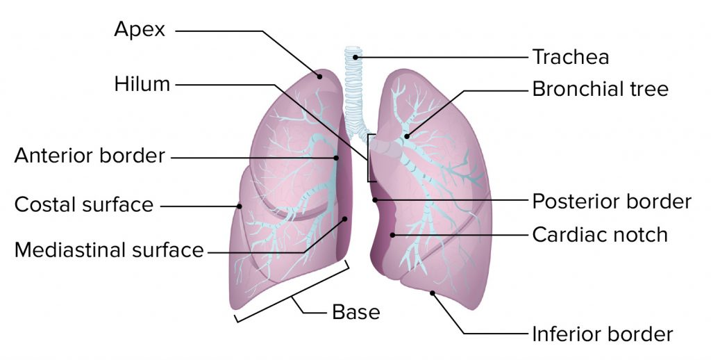
Parts of the Lung
- Apex:
The triangular and rounded upper part of the lung is called the apex. It extends up to the level of the clavicle (collar bone). - Base:
The broad lower part of the lung is called the base. This part is attached downward to the diaphragm and is concave in shape. - Anterior Border:
This border is thin and shorter than the posterior border. It contains a cardiac notch, which accommodates a portion of the heart. - Posterior Border:
This border is thicker, and it extends from the 7th cervical vertebra to the 10th thoracic vertebra. - Inferior Border:
This is located at the bottom of the lung, separating the costal surface and the medial surface.- The costal surface is large and convex, and is in contact with the costal pleura.
- It is connected to the ribs and intercostal muscles through costal cartilage.
- Medial Surface:
- This surface is concave and has a central groove called the hilum.
- The hilum is located at the level of the 5th, 6th, and 7th thoracic vertebrae.
- Through the hilum, the bronchi, pulmonary blood vessels, lymphatic vessels, and nerves enter and exit each lobe of the lung.
- The mediastinal space is located in the middle of this surface, separating the two lungs.
- This space contains the heart, great vessels, trachea, bronchi, esophagus, and other structures, separating both lungs.
Structure of the Lobes of the Lung
Each lung lobe is made up of many lobules.
- One lobe is separated from another by fissures.
- In the central part of the lung is a groove called the hilum, from where the bronchus enters each lobe.
- After entering, the bronchus divides into:
- Secondary bronchi
- Tertiary bronchi
- Terminal bronchioles
- Alveolar sacs
- And finally into small, grape-like structures called alveoli.
This creates a tree-like structure inside the lobes, known as the bronchial tree or respiratory tree.
Surrounding the alveoli is a capillary network of the pulmonary artery and pulmonary vein.
- Oxygen from inspired air in the alveoli and carbon dioxide in the capillary blood are exchanged here.
- This process is known as external respiration.
Thus, each lung lobe contains:
- The bronchial tree
- Pulmonary vessels and their capillaries
- Lymphatic capillaries
- Nerves
- Parenchymal tissue of the lung
Pleura
The pleura is a serous membrane surrounding both lungs. It has two layers:
- The outer layer is called the parietal pleura
- The inner layer is called the visceral pleura
Between these two layers is a space called the pleural cavity, which contains serous fluid, also known as pleural fluid.
This pleural fluid:
- Prevents friction between the two layers
- Allows free expansion of the lungs during breathing
- Acts as a lubricant due to its slippery nature
The visceral pleura is closely attached to the lung surface, while the parietal pleura is connected to the ribs and muscles.
OR
a. List out the organs of female reproductive system.
Organs of the Female Reproductive Tract
The female reproductive tract is divided into two parts:
- External Organs
- Internal Organs
External Organs:
- Mons pubis
- Labia majora
- Labia minora
- Hymen
- Clitoris
- Bartholin’s glands
- Vestibular glands
- Perineum
Internal Organs:
- Ovaries (2)
- Fallopian tubes (2)
- Uterus (1)
- Vagina and Vaginal cavity
b. Explain the process of menstrual cycle.
Menstrual Cycle
The menstrual cycle begins in females after the puberty phase, during which functional changes are observed in the ovaries and uterus.
The menstrual cycle typically occurs every 26 to 30 days, and it is regulated by hormonal fluctuations in the blood.
The onset of the menstrual cycle is known as menarche.
After puberty, the menstrual cycle continues regularly, temporarily stopping during pregnancy and completely ending after menopause.
The onset of menstruation occurs due to the degeneration of the corpus luteum layer of the uterus, which results in bleeding through the vaginal cavity.
The menstrual cycle is divided into the following phases:
1. Menstrual Phase
- This phase occurs once every 28 days and generally lasts for about four days.
- When fertilization of the egg does not occur, the levels of supporting hormones (estrogen and progesterone) decrease, and the level of oxytocin hormone increases.
- This rise in oxytocin stimulates uterine contractions, leading to degeneration of the corpus luteum layer of the uterine wall.
- Blood is then expelled through the vagina.
- This phase usually lasts from Day 1 to Day 4 of the cycle.
The menstrual flow contains:
- Endometrial glands
- Endometrial cells
- Blood
- Unfertilized ovum
Approximately 100 to 200 ml of blood is lost during 3 to 5 days of this phase.
2. Proliferative Phase
- The menstrual phase ends by Day 5.
- From Day 6 to Day 14, the proliferative phase begins.
During this phase:
- The Follicle Stimulating Hormone (FSH) stimulates ovarian follicles, which leads to an increase in estrogen production.
- Estrogen stimulates the proliferation of the endometrium (uterine lining).
Starting from Day 6:
- The endometrium begins to develop, with cell multiplication, increased mucus-secreting glands, and more blood capillaries.
- This makes the endometrium thicker and more vascular, preparing it for possible implantation of a fertilized egg.
By the end of this phase:
- The uterine lining becomes ready for implantation.
- This phase ends with ovulation, and estrogen levels begin to decline.
3. Secretory Phase
- After the proliferative phase ends, the secretory phase begins, which lasts from Day 15 to Day 28.
This phase is also called the progesterone phase, as progesterone is the dominant hormone.
During this phase:
- Due to ovulation, a mature egg is released by the ovary.
- Although estrogen and progesterone initially decrease, the corpus luteum secretes progesterone to help maintain pregnancy.
If the mature ovum is not fertilized by a sperm:
- The corpus luteum reduces progesterone secretion.
- As progesterone levels drop, oxytocin hormone increases, stimulating contractions in the uterine muscles.
Due to the absence of a fertilized egg and increased uterine contractions, the next cycle begins at the end of this phase.
c. Describe gross structure of uterus. –
Uterus
The uterus is a hollow, muscular organ located in the pelvic cavity. It is pear-shaped and lies behind the urinary bladder and in front of the rectum within the pelvic cavity.
- Its approximate dimensions are: 7.5 cm in length, 5 cm in width, and 3 cm in thickness.
- The uterus weighs about 30 to 40 grams, but during pregnancy, its weight increases significantly, reaching approximately 900 to 1000 grams.
In women, the uterus is positioned in a normal anatomical posture:
- Anteversion: Tilted forward
- Anteflexion: Bent forward
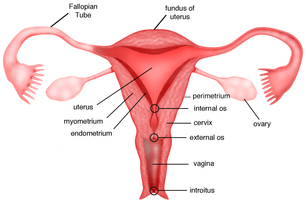
Anatomical Parts of the Uterus
The uterus is divided into three main parts:
- Fundus:
The dome-shaped upper part of the uterus is called the fundus.
From each side of the fundus, one fallopian tube arises. - Body:
The main central part of the uterus is called the body.
It resembles a narrow canal-like structure. - Cervix:
The lower, narrow, rounded portion of the uterus is called the cervix.- The internal opening of the cervix (towards the uterus) is known as the internal os.
- The external opening (opening into the vaginal cavity) is called the external os.
Structure of the Uterine Wall
The wall of the uterus consists of three layers, classified from outer to inner as follows:
Layers of the Uterus
1. Perimetrium Layer
- This is the outermost wall of the uterus.
- It is a serous layer that connects with the peritoneum on the side of the fundus, and is also in contact with the urinary bladder.
- At this point, it forms a pouch called the vesicouterine pouch.
- On the back side, at the level of the uterine body and cervix, it connects with the rectum, forming the rectouterine pouch, also known as the pouch of Douglas.
- This layer consists of areolar connective tissue and epithelial tissue.
2. Myometrium Layer
- This is the middle layer of the uterus.
- It consists of muscle fibers, specifically smooth muscles.
- During childbirth, contractions of these muscles help in the process of labor.
3. Endometrium Layer
- This is the innermost layer of the uterus.
- Its inner lining is a mucous membrane, made of epithelial tissue, which secretes mucus.
- During pregnancy, this layer is known as the decidua.
Ligaments of the Uterus
The uterus is held in place in the pelvic cavity and supported in its normal anatomical position by the following ligaments:
- Broad ligament
- Round ligament
- Uterosacral ligament
- Transverse cervical (cardinal) ligament
Blood Supply of the Uterus
- The uterus receives blood through the uterine artery, a branch of the internal iliac artery.
- Venous return occurs through branches of the uterine veins.
Nerve Supply of the Uterus
- The uterus is supplied by both the sympathetic and parasympathetic nervous systems.
Functions of the Uterus
- The uterus facilitates fertilization of the ovum and sperm.
- After fertilization, it helps in the implantation of the zygote into the uterine wall and maintains pregnancy.
- As the contents increase during pregnancy, the uterus expands in size to accommodate the growing fetus.
- It provides nourishment to the fetus during pregnancy.
- Contractions of the uterine muscles help in expelling the baby during delivery.
- The endometrium (inner wall) of the uterus sheds during menstruation.
- This menstrual cycle occurs every 26 to 30 days, and the presence of white blood cells (WBCs) in menstrual discharge reduces the risk of infections.
Q-2 a) Write the gross structure and functions of the heart.
Heart
The heart is one of the most important organs of the circulatory system. Throughout human life, the heart beats continuously, and due to its pumping action, blood circulates constantly through the blood vessels.
The heart is a hollow, muscular organ.
- In males, it weighs approximately 310 grams, and in females, around 250 grams.
- The heart beats approximately 100,000 times per day.
Structure of the Heart
The heart is composed of hollow muscles, and its wall consists of three tissue layers:
1. Epicardium or Pericardium
- This is the outermost layer of the heart.
- It is thin and transparent, made of fibrous connective tissue.
- On the outer side, there is a fibrous tissue layer, and inside that, there is a serous membrane made up of two layers:
- The outer layer is called the parietal pericardium.
- The inner layer is called the visceral pericardium.
- The space between these two layers is known as the pericardial cavity, which contains serous fluid (also called pericardial fluid).
- This fluid helps prevent friction between the two layers.
- The outer pericardial layer provides protection to the heart and also covers the major blood vessels emerging from the heart.
2. Myocardium
- The myocardium is the middle layer of the heart wall, located beneath the pericardium.
- It is made of specialized cardiac muscle tissue, responsible for the pumping action of the heart.
- This layer is thicker at the apex and thinner at the base.
- The left ventricular wall is thicker than the right ventricular wall.
- These muscles have involuntary contractions, and their activity is controlled by the autonomic nervous system and the cardiac conducting system.
3. Endocardium
- This is the innermost layer of the heart wall.
- It is in direct contact with blood.
- Made of epithelial and connective tissue, this layer is smooth and shiny, allowing unobstructed blood flow inside the heart.
- It also covers the heart valves and continues along the inner lining of the blood vessels emerging from the heart.
Functions of the Heart
- The heart supplies oxygenated blood to all organs and tissues of the body.
- It is a vital organ of the cardiovascular system, without which life cannot be sustained.
- The heart circulates blood to the lungs for oxygenation and purification.
- It regulates both pulmonary circulation and systemic circulation.
- The heart regulates heart rate based on body needs and temperature.
- By maintaining blood circulation to all parts, the heart helps regulate body temperature.
- It sends blood to excretory organs, allowing filtration and removal of waste products from the blood.
b) Explain the physiology of vision.
Physiology of Vision
- Vision is a complex physiological process involving coordination between the eyes and the brain.
- Light enters the eye through the transparent cornea, which helps to focus the light.
- This light then passes through the pupil, whose size is controlled by the iris to regulate the amount of light entering the eye.
- After the pupil, light reaches the lens, which adjusts its shape to focus the light precisely on the retina.
- The retina is the innermost layer at the back of the eye, containing millions of photoreceptor cells, mainly rods and cones.
- Rods are responsible for vision in low light, while cones function in bright light and color vision.
- These photoreceptor cells convert light into electrical signals, a process known as transduction.
- The electrical signals travel to the brain via the optic nerve.
- In the brain, the visual cortex located in the occipital lobe analyzes these signals.
- The brain combines input from both eyes to create depth perception, distance, and three-dimensional (3D) vision.
- The brain further processes the visual information to recognize color, shape, motion, and depth.
- Ultimately, vision is a process formed through the coordination of various parts of the eye and the brain, allowing us to perceive the world around us.
Key Points:
- The transparent cornea and lens help to focus light.
- Photoreceptor cells convert light into electrical signals.
- Signals reach the brain through the optic nerve.
- The brain interprets visual information and helps form images.
OR
a) Write the gross structure and functions of brain.
Gross Structure and Function of the Brain
Gross Structure of the Brain:
The brain is the control center of the human body. It is divided into three major parts:
- Cerebrum
- Cerebellum
- Brainstem
1) Cerebrum:
- It is the largest part of the brain and is divided into two hemispheres – the right and left.
Lobes of the Cerebrum:
- Frontal Lobe: Responsible for voluntary movement, reasoning, problem-solving, decision-making, and planning.
- Parietal Lobe: Processes sensory information.
- Temporal Lobe: Important for memory, hearing, and language comprehension.
- Occipital Lobe: Processes visual information.
2) Cerebellum:
- Located at the back of the brain, it is responsible for:
- Body balance
- Coordination of movement
- Muscle tone
- Fine motor control
3) Brainstem:
- Connects the brain to the spinal cord.
- Controls vital functions like respiration, heart rate, digestion, and blood pressure.
Components of the Brainstem:
- Midbrain: Controls visual and auditory reflexes.
- Pons: Regulates sleep, breathing, facial movements, and posture.
- Medulla Oblongata: Controls automatic functions such as breathing, heart rate, blood pressure, and swallowing.
Functions of the Brain:
Cerebrum:
- Responsible for thinking, memory, emotions, sensory perception, and voluntary movement.
Cerebellum:
- Plays a vital role in balance, movement coordination, muscle tone, and fine motor skills.
Brainstem:
- Regulates automatic functions like breathing, heart rate, blood pressure, digestion, and the sleep-wake cycle.
Key Parts and Their Functions:
- Cerebral Cortex: Controls language, reasoning, problem-solving, decision-making, and sensory perception.
- Thalamus: Relays sensory information to various parts of the brain.
- Hypothalamus: Regulates body temperature, hunger, thirst, sleep, and hormonal activities.
- Amygdala: Controls emotions, especially fear and pleasure.
- Hippocampus: Essential for memory formation and learning.
Main Functions of the Brain:
- Sensory Perception: Processes information from the senses – vision, hearing, touch, taste, and smell.
- Motor Control: Controls voluntary and involuntary body movements.
- Thinking and Decision-Making: Responsible for problem-solving and making decisions.
- Emotions and Behavior: Regulates emotions, mood, and behavior.
- Homeostasis: Maintains body temperature, hunger, sleep, and hormonal balance.
Important Automatic Functions of the Brain:
- Respiration
- Heart rate regulation
- Blood pressure control
- Digestion
- Sleep-wake cycle
Conclusion:
The brain is the control center of the human body.
It is responsible for vital functions, sensory perception, thoughts, emotions, and physical movement.
All parts of the brain work together to maintain the body’s functions in harmony.
Without the brain, human life is not possible.
b) What is skeletal system? Write down functions of seletal system.
Skeletal System
Terminology:
The skeletal system is an important system of the human body, consisting of bones, cartilage, ligaments, and tendons. This system provides structural support, protection to internal organs, and necessary support for body movement.
Functions of the Skeletal System:
- Structural Support:
The skeletal system provides a strong and stable structure to the body, determining its shape and size.
Bones support all internal organs.
Example: The spine supports the upper part of the body. - Protection of Internal Organs:
Bones protect internal organs from external injuries.
Example: The rib cage protects the heart and lungs, while the skull protects the brain. - Movement:
Bones are connected to muscles, and when muscles contract, they pull the bones, resulting in movement.
Example: Muscles attached to the bones of the arms and legs help in walking, running, and other movements. - Production of Blood Cells:
Bone marrow is the site where blood cells are produced, including red blood cells (RBCs), white blood cells (WBCs), and platelets.
Example: The femur (a long bone) contains red bone marrow. - Storage of Minerals:
The skeletal system stores essential minerals like calcium and phosphorus, which are needed for various bodily functions.
When required, these minerals are released into the bloodstream. - Regulation of Hormones:
The skeletal system produces hormones such as osteocalcin, which plays a role in metabolism and mineral balance in the body. - Maintaining Posture:
The skeletal system helps maintain body balance and alignment.
The spine maintains the proper posture of the body.
Components of the Skeletal System:
- Bones: Provide structural support to the body
- Cartilage: Provides flexibility between bones
- Ligaments: Connect bone to bone
- Tendons: Connect muscles to bones
Types of Bones:
- Long Bones – e.g., Femur, Humerus
- Short Bones – e.g., Carpals
- Flat Bones – e.g., Skull, Ribs
- Irregular Bones – e.g., Vertebrae
Conclusion:
The skeletal system is crucial for providing structural support, protecting internal organs, enabling movement, and performing metabolic functions in the human body.
Its proper functioning is essential for maintaining overall health and well-being.
Q-3 Write short answer (any two)
a) Write down functions of the skin.
Functions of the Skin
The skin forms a continuous outer covering of the body and acts as the first line of protection.
1. Protection Against Microorganisms and Injuries:
- The skin prevents microorganisms from directly entering the body.
- It blocks harmful substances and external injuries from affecting internal organs.
2. Regulation of Body Temperature:
- The skin plays a crucial role in maintaining normal body temperature through sweating and blood vessel dilation/constriction.
3. Structural Framework and Shape:
- The skin provides an outer framework for the body.
- It is the largest organ and covers all internal organs, thereby giving shape to the body.
4. Synthesis of Vitamin D:
- The skin contains a chemical called 7-dehydrocholesterol, which, when exposed to ultraviolet (UV) rays from sunlight, gets converted into Vitamin D3 (cholecalciferol).
- Hence, the skin is responsible for the synthesis of Vitamin D.
5. Excretion of Waste Products:
- The skin helps in the excretion of waste products and harmful substances from the body.
- Through the process of perspiration (sweating), it removes certain waste materials like urea and salts.
6. Absorption of Substances:
- Some substances can be absorbed through the skin, making it a useful route for medication.
- Certain ointments and topical medicines are absorbed through the skin and enter the systemic circulation.
7. Sensory Function:
- The skin contains sensory nerve endings that help transmit impulses related to touch, temperature, and pain to the brain.
- This allows the brain to interpret external stimuli effectively.
8. Storage of Nutrients:
- The skin stores certain nutrient materials, such as fats, which provide energy and insulation.
9. Wound Healing:
- The skin plays an important role in wound healing, helping to repair tissue after an injury.
b) Explain mitosis cell division in detail.
Mitosis (Cell Division)
Mitosis is a process in which a single cell divides into two identical daughter cells. This process is extremely important for growth, development, tissue repair, and asexual reproduction in living organisms. During mitosis, the genetic material (DNA) is precisely divided, ensuring both new cells receive the same genetic information.
Phases of Mitosis:
Mitosis is divided into four main phases:
- Prophase
- Metaphase
- Anaphase
- Telophase
1. Prophase
Features:
- Chromosomes condense and become visible under a microscope.
- Each chromosome consists of two chromatids joined at the centromere.
- The nuclear membrane begins to break down.
- Centrosomes move to opposite poles of the cell.
- Spindle fibers (microtubules) begin to form, helping in separating the chromosomes.
Importance:
Prepares for the accurate division of genetic material.
2. Metaphase
Features:
- All chromosomes line up at the center of the cell on the metaphase plate.
- Spindle fibers attach to the centromeres of each chromosome.
- This alignment ensures equal distribution of genetic material.
Importance:
Ensures proper arrangement for accurate chromosome separation.
3. Anaphase
Features:
- Spindle fibers separate the chromatids of each chromosome.
- The chromatids become independent chromosomes and move toward opposite poles of the cell.
- Ensures equal genetic distribution in both new cells.
Importance:
Critical phase for creating identical genetic copies.
4. Telophase
Features:
- Separated chromatids re-condense inside newly forming nuclei.
- Nuclear membranes reform, creating two new nuclei.
- Spindle fibers disintegrate.
Importance:
Completes the process of genetic information distribution.
5. Cytokinesis
After mitosis, cytokinesis occurs—this is the division of the cytoplasm, resulting in two separate daughter cells.
Importance of Mitosis:
- Growth and Development – Necessary for the development of living organisms.
- Tissue Repair – New cells are produced for wound healing and repair.
- Asexual Reproduction – Some organisms reproduce through mitosis.
- Genetic Stability – Maintains stable genetic information by creating identical copies.
- Biological Research – Essential in understanding cell division and life processes in biology.
Real-Life Importance of Mitosis:
- In Humans: Important for body development and healing wounds.
- In Plants: Helps in growth and formation of new leaves and tissues.
- In Animals/Insects: Required for regeneration and reproduction.
Conclusion:
Mitosis is a crucial cell division process that plays a vital role in the life of living organisms. It is essential for growth, development, repair, and genetic stability.
Through precise division of genetic material, mitosis ensures that all new cells have identical genetic information.
Without mitosis, life would not be possible.
c) Explain the process of formation of urine.
Process of Urine Formation
Urine formation takes place in the kidneys and involves three main steps:
- Filtration
- Reabsorption
- Secretion
These steps occur in the nephrons, the functional units of the kidneys.
1. Filtration
Location: Glomerulus
Process:
In this step, blood enters the glomerulus. Due to blood pressure, water, salts, glucose, amino acids, and waste substances (like urea) are filtered from the blood into the Bowman’s capsule.
Importance:
Large particles such as proteins and blood cells are not filtered, ensuring only necessary substances pass through.
Detail:
Blood flows through the capillaries of the glomerulus. The high pressure acts as a filter, allowing small molecules and water to pass through. The filtered fluid, called glomerular filtrate, enters the Bowman’s capsule.
2. Reabsorption
Location:
- Proximal Convoluted Tubule (PCT)
- Loop of Henle
- Distal Convoluted Tubule (DCT)
Process:
From the filtered fluid, essential substances like water, glucose, amino acids, sodium, and potassium are reabsorbed back into the blood.
Importance:
This step helps regulate the body’s water and salt balance.
Detail:
- Around 65–70% of the filtered water and salts are reabsorbed in the PCT.
- The Loop of Henle creates a concentration gradient, helping further water reabsorption.
- The DCT, under hormonal control, reabsorbs additional substances as needed.
3. Secretion
Location:
- Distal Convoluted Tubule (DCT)
- Collecting Duct
Process:
Additional waste products like hydrogen ions (H⁺), potassium ions (K⁺), and drugs are secreted from the blood into the tubules.
Importance:
Helps maintain the acid-base balance and remove toxins from the body.
Detail:
Secretion is an active transport process, moving substances from blood into the tubule.
Unwanted ions, metabolic waste, and drugs are eliminated this way.
4. Excretion
Location: Collecting Duct
Process:
The final filtrate, now called urine, flows from the collecting duct into the ureter, and then to the bladder.
Importance:
This is the final step, where urine is eliminated from the body.
Detail:
In the collecting duct, urine concentration is finalized.
Urine then flows to the bladder via the ureter, gets stored, and is eventually expelled through the urethra.
Main Components of Urine:
- Water (95%)
- Urea
- Creatinine
- Uric Acid
- Salts (Sodium, Potassium, Chloride)
- Hormones and other substances
Summary of Urine Formation Steps:
- Filtration: Filters out waste and useful substances from the blood.
- Reabsorption: Reabsorbs useful substances back into the blood.
- Secretion: Removes extra toxins and ions.
- Excretion: Final urine is expelled from the body.
Q.4 Write short notes.
a) Factors affecting on microbial growth.
Factors Affecting Bacterial Growth
1) Moisture (Water Content)
Every bacterium, like any living organism, requires water along with nourishing food for growth.
In the absence of water, bacteria cannot absorb nutrients, as all food elements must be in a liquid (aqueous) form to pass through the bacterial cell wall.
- Most types of bacteria grow well in moist environments.
- An environment completely lacking moisture inhibits or kills bacterial growth.
- Cells cannot survive in too high or too low humidity.
2) Light
- Ultraviolet (UV) rays present in sunlight are harmful to most bacteria.
- Direct exposure to UV rays destroys many bacteria.
3) Temperature
Temperature is a critical factor affecting bacterial growth. Along with food and water, bacteria require an optimal temperature for growth.
- Different bacteria have different optimal temperature ranges.
- For bacteria that grow in the human body, the optimal temperature is 37°C.
- Many bacteria are mesophilic (meso = middle, philic = loving), growing best at 25°C to 39°C.
- Psychrophilic bacteria (psychro = cold) grow well between 4°C to 10°C.
- Thermophilic bacteria (thermo = heat) grow between 55°C to 75°C.
Temperatures above 75°C are usually fatal to bacteria.
High temperatures are intentionally used to kill bacteria — e.g., moist heat, boiling, pasteurization, autoclaving.
- Some organisms like yeast, mold, viruses, rickettsia, and spirochetes can survive even at very low temperatures. (Some may survive up to 76°C for years.)
4) Oxygen (O₂)
Oxygen plays a significant role in bacterial survival and growth.
- Aerobes: Bacteria that can only live and grow in the presence of oxygen.
Example: Sarcina - Anaerobes: Bacteria that can live and grow in the absence of oxygen.
Example: Clostridium tetani - Facultative anaerobes: Bacteria that can survive both with or without oxygen.
Example: Salmonella typhi - Microaerophiles: Bacteria that grow better in less oxygen than atmospheric levels.
5) Hydrogen Ion Concentration (pH level)
The acidity or alkalinity of the medium affects bacterial growth.
This is measured using the pH scale:
- pH 0 – Strongly acidic
- pH 14 – Strongly alkaline
- pH 7 – Neutral
- pH < 7 – Acidic
- pH > 7 – Alkaline
Most bacteria grow best in a pH range of 5.0 to 8.5.
However, some exceptions may exist.
6) Osmotic Pressure
Bacterial survival is also influenced by osmotic pressure.
- If a bacterium is placed in a medium with too high or too low osmotic pressure, water flows out of the bacterial cell, causing it to collapse or become dormant.
7) Carbon Dioxide (CO₂)
- Carbon dioxide is also essential for the growth of certain bacteria, especially capnophilic bacteria that thrive in CO₂-rich environments.
b) Role of nurse in control of spread of infection.
Infection Prevention and Control for Nurses
As a nurse, it is essential to know how to protect both ourselves and our patients from exposure to pathogens.
It is very important for nurses to understand and follow the infection control policies of the organization, including the use of Personal Protective Equipment (PPE) and maintaining environmental sanitation.
1. Standard Precautions:
To break the chain of infection, standard precautions must be taken to prevent the transmission of pathogens through body fluids.
2. Hand Hygiene:
This is the number one weapon to prevent cross-infection.
Since most microorganisms are spread through hands, proper handwashing before and after every procedure is essential.
Handwashing is also crucial after touching any contaminated surface.
3. Aseptic Technique:
Use aseptic techniques during procedures, especially those involving contact.
Use only sterile items during invasive procedures to prevent infection.
4. Environmental Infection Control Measures:
Keep hospital equipment and surroundings clean.
Hospital floors should be cleaned with antiseptic solutions.
Proper disinfection and disposal of bio-medical waste generated during procedures or nursing care must be ensured.
5. Droplet Precaution:
Always wear a mask while working in a hospital.
Infected patients should also wear a mask.
Educate them to take precautions during coughing or sneezing to prevent the spread of infections.
6. Personal Protective Equipment (PPE):
PPE helps prevent cross-infection between the nurse and the patient.
PPE includes caps, masks, gowns, and gloves, especially when caring for highly infectious patients.
7. Health Education:
To prevent the spread of infection in the hospital, health education should be provided to both patients and the staff working under the nurse’s supervision.
c) Immunity
Immunity
Immunity is the defense mechanism shown by the host body against conditions caused by microorganisms or their toxins (antigens).
In simple terms, when an antigen or microorganism enters the body, and the body becomes resistant and provides protection, it is referred to as immunity.
Immunity is a type of resistance activated by the host body.
When any foreign body (antigen) enters the host, the body produces antibodies to fight against it.
The ability of the body to fight against antigens is known as immunity.
Types of Immunity:
Immunity is mainly classified into two types:
- Innate Immunity (Natural / Inborn)
- Acquired Immunity (Developed After Birth)
1. Innate Immunity (Inborn Immunity)
- This is the immunity present from birth.
- It is a natural form of immunity that does not require prior exposure to pathogens.
Types of Innate Immunity:
- Species Immunity – Different species have different levels of immunity.
- Racial Immunity – Different races may have different levels of immunity.
- Individual Immunity – Every individual may have a unique level of natural immunity.
2. Acquired Immunity (Developed Immunity)
- Immunity that is developed after birth during one’s lifetime.
- It is formed when the body is exposed to diseases or through vaccinations.
Types of Acquired Immunity:
- Active Immunity – The body itself produces antibodies against the antigen.
- Passive Immunity – The body receives ready-made antibodies from another source.
1) Active Immunity
Active immunity develops after the entry of an antigen into the body and the body produces its own antibodies.
Types of Active Immunity:
A) Active Natural Immunity:
- Occurs when a person is exposed to a natural infection, and the body produces antibodies.
- It provides long-lasting or lifelong protection.
- Involves two types of responses:
- Humoral Immunity – Develops from plasma and involves B-lymphocytes producing antibodies.
- Cell-Mediated Immunity – Involves T-lymphocytes, which protect against chronic bacterial infections and intracellular pathogens.
B) Active Artificial Immunity:
- Developed through vaccination.
- Vaccines stimulate the immune system to produce antibodies.
Examples:
- BCG
- Hepatitis vaccine
- DPT (Diphtheria, Pertussis, Tetanus)
- DT
- Pentavalent vaccine
2) Passive Immunity
Passive immunity occurs when ready-made antibodies are transferred from another source into the body.
Types of Passive Immunity:
A) Passive Natural Immunity:
- Antibodies are transferred from mother to fetus.
Also called Congenital Immunity.
Examples:
- IgG from placenta
- IgA from breast milk
B) Passive Artificial Immunity:
- Man-made immunity, where antibodies are administered directly to the recipient for immediate protection.
Sources:
- Antitoxins
- Antibacterials
- Antivirals
Examples:
- Tetanus Toxoid (TT)
- Gas Gangrene Antitoxin
- Anti-venom Serum
- Anti-lymphatic Serum
These provide immediate but temporary protection.
Uses of Passive Immunization:
- Provides immediate protection in emergencies.
- Suppresses active immune response in certain cases.
- Useful in serious infections requiring urgent treatment.
d) Importance of microbiology in nursing
Importance of Studying Microorganisms (Microbiology)
- Microorganisms have the ability to cause diseases in humans, so through this study, we can understand their characteristics and behavior.
- By studying microbiology, we can learn how diseases occur, how they spread, and how they can be prevented.
- Laboratory testing is essential for identifying bacteria and confirming diagnosis.
- To prevent diseases caused by microorganisms, the importance of personal hygiene and immunization measures can be understood and practiced.
- This study helps remove superstitions, myths, ignorance, and social stigma related to infectious diseases by creating awareness among people.
- While treating patients, precautionary measures can be taken to prevent cross-infection.
- Diseases caused by microorganisms can be recognized, and proper diagnosis and prevention strategies can be implemented to stop their spread.
Q-5 Define following (any six)
a) Sterilization –
Sterilization
Sterilization is the process by which all microorganisms—such as bacteria, viruses, fungi, and their spores—are completely destroyed.
Sterilization is especially used in medical, pharmaceutical, laboratory, and food processing fields.
Main Methods of Sterilization:
1. Heat Sterilization:
- Autoclaving:
This method uses high temperature and pressure (steam under pressure) to achieve sterilization. - Dry Heat Sterilization:
In this method, high temperature without moisture (dry heat) is used to sterilize materials.
2. Chemical Sterilization:
- Ethylene Oxide:
A gas used to sterilize medical instruments and equipment, especially heat-sensitive items. - Hydrogen Peroxide:
Used to sterilize instruments and surfaces, especially in healthcare settings.
3. Filtration:
Used to remove microorganisms from liquids and gases without killing them—common for heat-sensitive solutions.
4. Radiation:
- Gamma Radiation:
Used to sterilize food products and medical instruments by destroying microbial DNA. - UV Light (Ultraviolet):
Used to sterilize air and surfaces, especially in laboratories and hospital environments.
Purpose of Sterilization:
The main aim of sterilization is to prevent contamination and the spread of infection, thereby ensuring health and safety in clinical and public environments.
b) Nosocomial infection
Nosocomial Infection (Hospital-Acquired Infection or Healthcare-Associated Infection – HAI)
A nosocomial infection, also known as a hospital-acquired infection or healthcare-associated infection (HAI), is an infection that a patient acquires after being admitted to the hospital, which was not present at the time of admission.
These infections typically occur within healthcare settings and may spread through contact with patients, healthcare staff, visitors, medical instruments, or the hospital environment.
Common Types of Nosocomial Infections:
- Urinary Tract Infection (UTI):
Often caused by the use of urinary catheters. - Surgical Site Infection:
Occurs at the site of surgery after a surgical procedure. - Respiratory Infections:
Common in patients on ventilators (e.g., ventilator-associated pneumonia). - Bloodstream Infections:
Caused by the use of IV catheters or central lines.
c) Incubation period
Incubation Period
The incubation period is the time between the entry of an infection and the appearance of the first signs or symptoms of the disease.
During this period, the infectious agent (such as a virus or bacteria) is actively replicating inside the host, but the host does not yet show any signs of illness.
The length of the incubation period can vary depending on the specific infectious agent, and it may range from a few hours to several months.
d) Basal Metabolic rate
BMI (Body Mass Index)
BMI = Weight in kilograms / (Height in meters)²
BMI Categories:
- Underweight: < 18.5 kg/m²
- Normal weight: 18.5 – 24.9 kg/m²
- Overweight: 25.0 – 29.9 kg/m²
- Obese: ≥ 30 kg/m²
e) Stroke Volume
Stroke volume is the amount of blood ejected from the heart with each contraction.
The normal stroke volume is approximately 70 mL.
f) Osmosis
Osmosis is a process in which fluid moves from an area of lower concentration to an area of higher concentration through a semipermeable membrane, and continues to move until the concentration on both sides of the membrane becomes equal.
g) Human Physiology
Human physiology is the scientific study of the functions and mechanisms in the human body.
It focuses on how organs, tissues, and systems of the body work together to maintain life, support growth, and respond to environmental changes.
It includes processes like:
- Circulation
- Respiration
- Digestion
- Metabolism
- Neural signaling
In short, the study of how different parts of the human body function is called human physiology.
h) Tidal Volume
Tidal volume is the amount of air that enters the lungs during normal inspiration and exits during normal expiration in a single respiratory cycle.
Normally, the tidal volume is about 500 mL.
Q-6(A) Fill in the blanks
1.Human anatomical waste should be collected in …….color bags. ✅ Answer: Yellow
2.The father of antiseptic surgery is………… ✅ Answer: Joseph Lister
3.The color of the eye is due to ………. ✅ Answer: Iris
4……….. is the longest and strongest bone in human body. ✅ Answer: Femur
5.The lobes of liver are divided by……. ligaments ✅ Answer: Falciform
(B) True or False
1.The pituitary gland is the master gland of the body. ✅
2.Sensory nerves are called efferent nerves. ❌
3.Homeostasis of internal environment is maintained by autonomic nervous system and endocrine system. ✅
4.Moving away from the median plane is called adduction ❌
5.Blood is the fluid connective tissue of the body. ✅
(C) Multiple Choice Questions –
1.Spleen is called a “graveyard” of the
✅ Correct answer: (a) Erythrocytes
b) Deucocyte
c) Lymphocytes
d) Reticulo endothelial system
2. Insulin causes
a) Increase blood glucose
b) Lipolysis
✅ Correct answer: (c) Decrease blood glucose
d) Increase protein breakdown
3. The cell that has phagocytic nature is
a) RBC
✅ Correct answer: (b) Neutrophil
c) Platelet
d) Astrocyte
4.Permanent stoppage of menstruation is called
✅ Correct answer: (a) Menopause
b) Menarche
c) Reproduction
d) Oogenesis
5.The only movable bone in the skull is
a) Maxilla
b) Frontoparietal bone
✅ Correct answer: (c) Mandible
d) Nasal bone