—ENGLISH-ANATOMY UNIT 9 ENDOCRINE SYSTEM
ENDOCRINE SYSTEM (Endocrine System):
Endocrine system is the endocrine system of the body. Which secretes a special type of secretion, i.e. hormones, through different glands, keeping the internal environment of the body stable and performing many vital functions.
In the body Glands that are made up of secretory epithelium cells and are mainly divided into two parts.
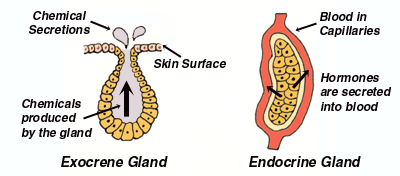
Endocrine Glands Glands):
- These types of glands in the body are ductless glands. These types of glands secrete their secretions directly into the blood or lymph. Therefore, they are called endocrine glands.
- The secretions of these glands are called hormones. Any endocrine gland secretes one or more hormones.
Exocrine Glands:
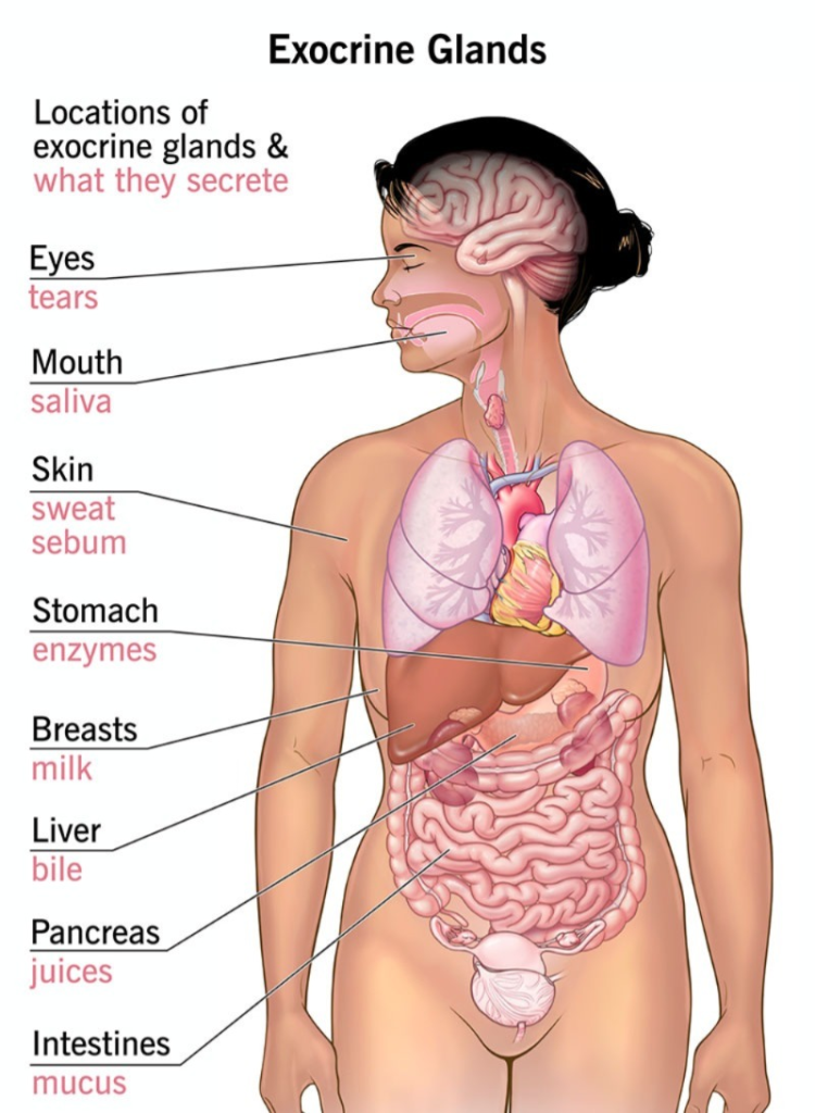
- These types of glands secrete their secretions directly onto the surface of any organ or tissue. The secretions of these glands are secreted onto the surface through ducts in the gland and do not mix directly with the blood, hence they are called exocrine glands.
- The secretions of these glands can be identified as juice, liquid or chyme. The size and shape of these glands vary. Mainly these types of glands are associated with the digestive tract in the function of digestion.
Here in this unit we will look at the study of endocrine glands. In which the study of the structure, function and related problems of these types of glands is known as endocrinology.
Endocrine glands do not have a duct. Hence they are also known as ductless glands. With the help of all these glands, the majority of the body’s functions are controlled.
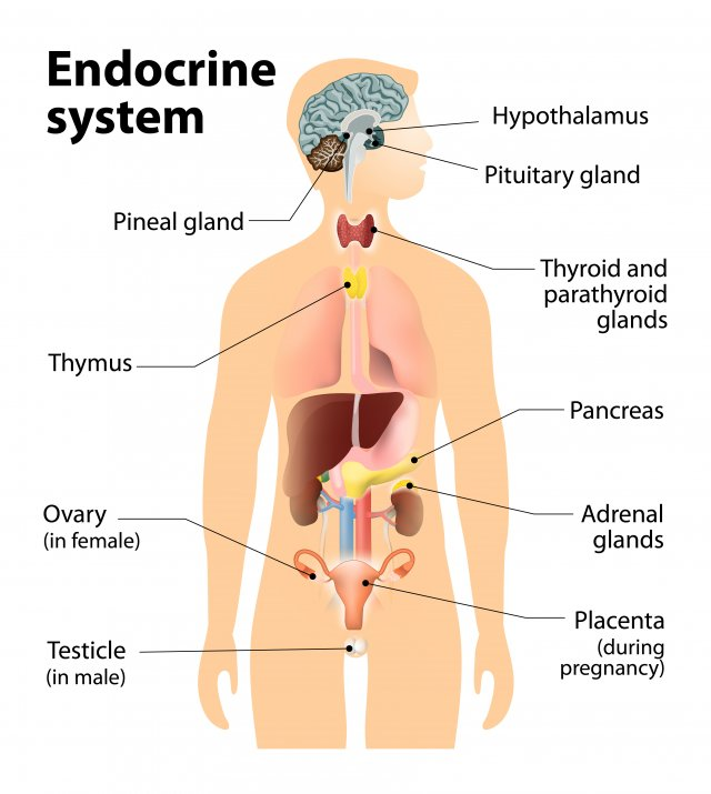
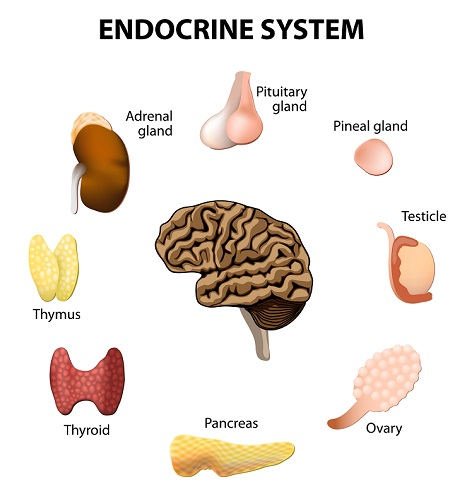
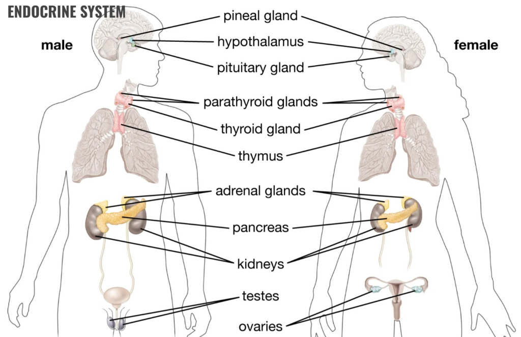
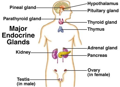
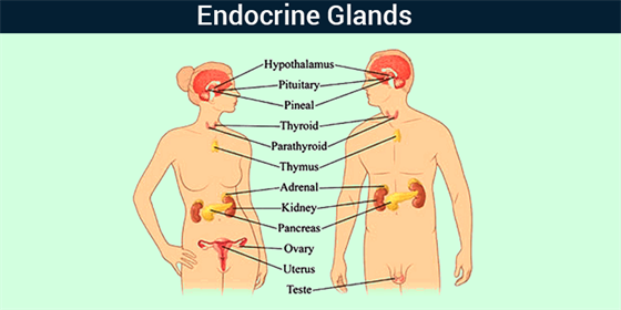
List out the endocrine glands in the body:
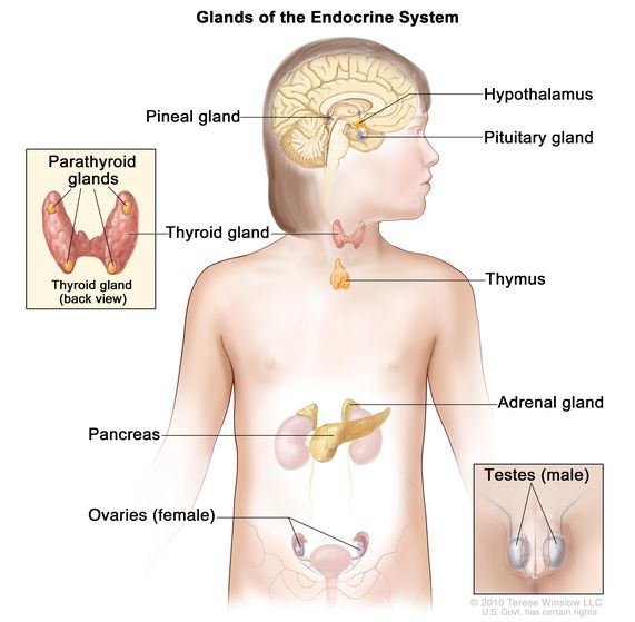
The following endocrine glands are present in the body. Here the glands and their classification can be given as follows.
Pituitary gland:
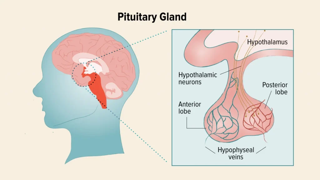
- The pituitary gland is known as the master gland in the body because the functions of other glands are regulated by the hormones of this gland.
- The pituitary gland is located in the hypophyseal fossa of the sphenoid bone of the cranial cavity. The pituitary gland is also known as the hypophysis. The pituitary gland is strongly connected to the hypothalamus, so many functions are regulated due to this interconnection.
- The size of the pituitary gland is 1.2 to 1.5 cm long. Its weight is approximately 0.5 grams. It is a peanut-shaped gland.
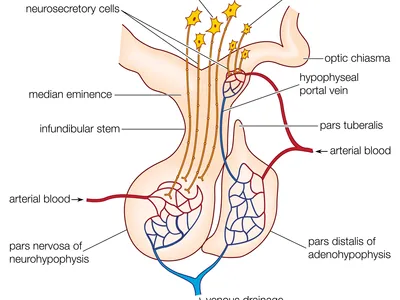
- The pituitary gland mainly has 3 lobes. Each lobe secretes a different hormone, which can be described as follows. The hormones secreted from each lobe are affected by the releasing and inhibiting hormones secreted from the hypothalamus. Releasing hormones secreted by the hypothalamus increase the secretion of the pituitary gland while inhibiting hormones decrease the secretion of the pituitary gland.
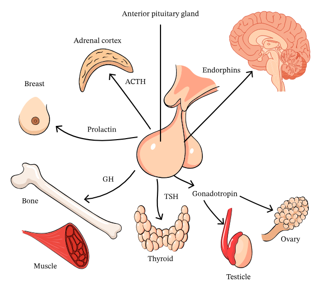
Anterior Lobe (Anterior Lobe).
- This lobe is also known as the adenohypophysis. It is composed of connective tissue fibers.
- Hypothalamus Due to the releasing and inhibiting hormones secreted from it, the following hormones are secreted from the anterior lobe of the pituitary gland.
- The hypothalamus is connected to the anterior lobe of the pituitary gland through blood and the hypothalamus has control over the hormones secreted from the anterior lobe.
- The following hormones are secreted from the anterior lobe.
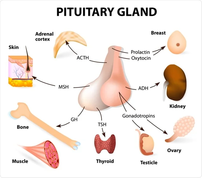
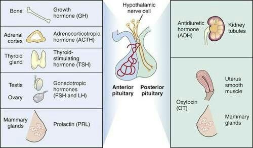
A. Growth Hormone
Growth hormone is also called another somatotrophic hormone. The main function of this hormone in the body is to maintain the growth of the body. If this hormone is secreted normally, then the normal growth of the body is maintained. If the amount of this hormone is secreted in excess, then overgrowth is seen in the body which is called gigantism and if the amount of this hormone in the gland decreases, then the growth in the body is seen less than normal which is also called dwarfism. The secretion of this hormone depends on the growth hormone releasing and inhibiting hormone secreted from the hypothalamus.
B. Thyroid Stimulating Hormone (TSH)
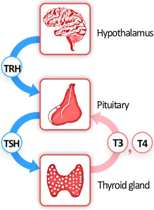
This hormone is secreted from the lobe of the anterior pituitary gland. Which stimulates the thyroid gland and the thyroid gland produces its normal hormone.
Ç. Adreno Corticotropic Hormone (ACTH)
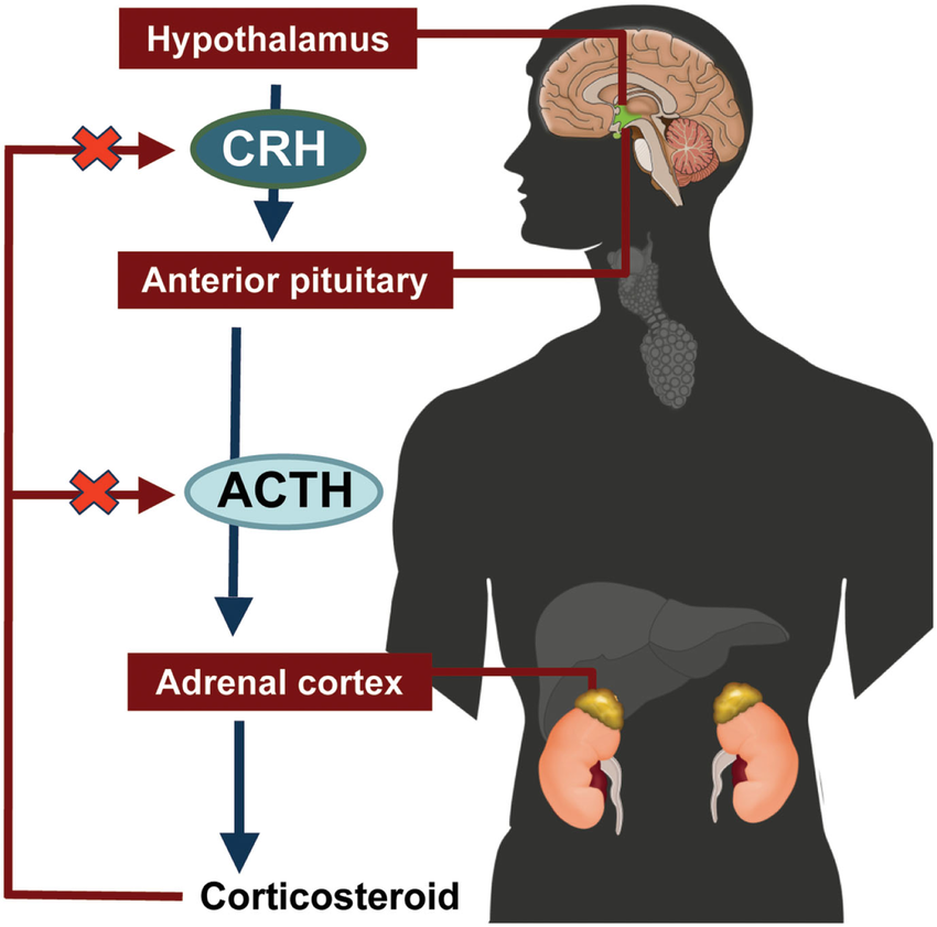
Corticotrophic releasing hormone and inhibitory hormone are secreted by the hypothalamus. Through which a hormone called ACTH is secreted from the anterior lobe of the pituitary gland.
This hormone increases the production of hormones secreted from the cortex of the adrenal gland. The adrenal gland is associated with the secretion of steroid hormones.
D. Prolactin Hormone
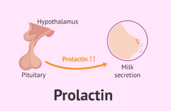
This hormone is secreted by the anterior lobe. It is the hormone responsible for the production of breast milk. The releasing and inhibiting hormones of the hypothalamus secrete prolactin hormone from the anterior lobe after childbirth, which leads to milk production.
This hormone is especially useful for the production of breast milk after childbirth.
E. Gonadotropic Hormone
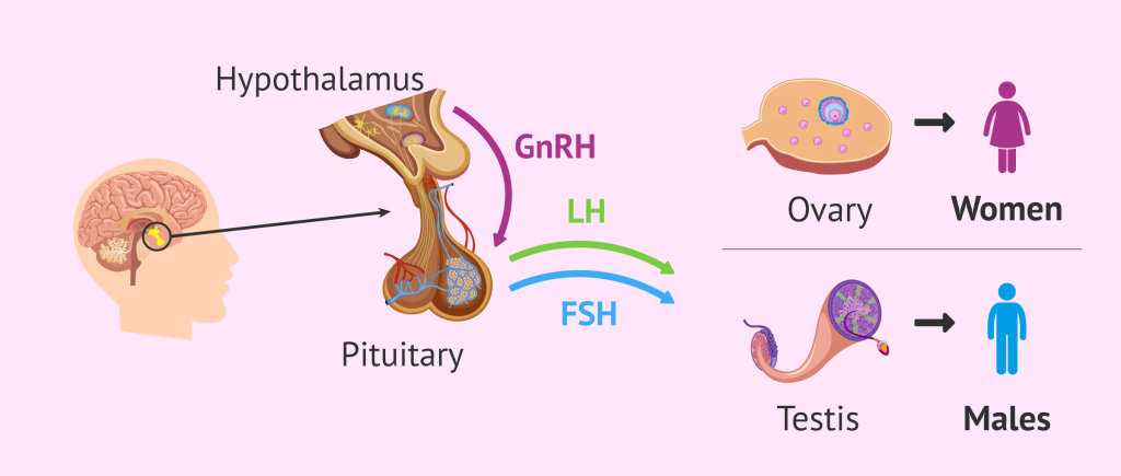
This hormone is also called the sex hormone. Which secretes gonadotrophinous hormones in males and females.
The secretion of this hormone is also affected by the releasing and inhibiting hormones of the hypothalamus.
This hormone is secreted in two main ways
1. Follicle Stimulating Hormone (FSH)
This hormone produces estrogen and progesterone. Which functions for ovum production in females and spermatozoa production in males.
2. Luteinizing Hormone (LH)
This hormone produces the hormone progesterone which is responsible for the characteristics of menstruation in females and helps in the production of testosterone from the testes in males
Intermediate Lobe:
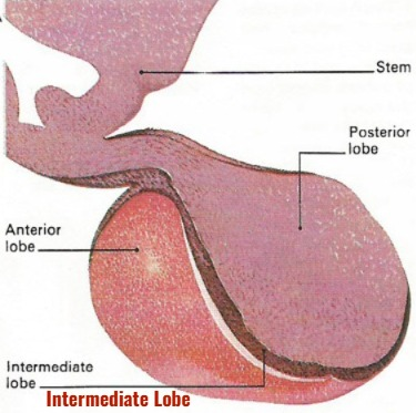
The pituitary gland has an intermediate lobe between the anterior lobe and the posterior lobe. The function of this lobe in the human body is not yet known for sure.
Posterior Lobe:
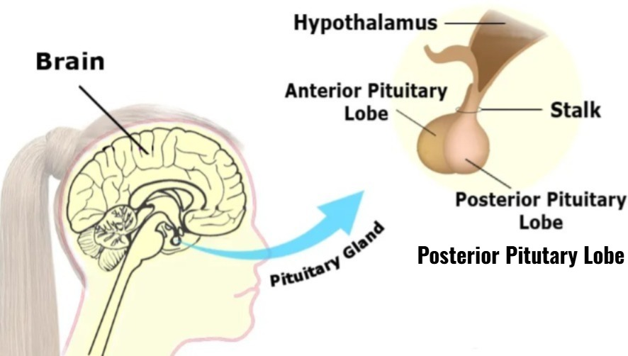
This is a lobe in the posterior pituitary gland.
It is known as the neurohypophysis.
This lobe of the pituitary gland is connected to the hypothalamus by nerves and nerve fibers. Because of which it is known as the neurohypophysis. The following hormones are secreted by this lobe.
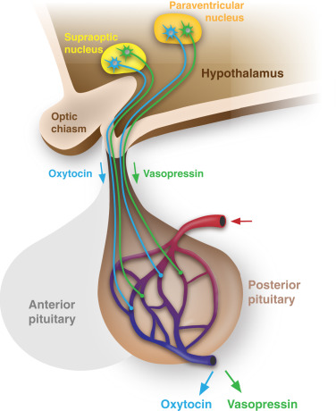
À. Oxytocin:

This is a hormone secreted by the posterior lobe. It is a hormone that starts normal labor pains during delivery and causes contraction of the myometrium muscles in the uterus. Due to which the process of childbirth can be done well.
When the child sucks the mother’s breast milk through the oxytocin hormone, this hormone also works to eject the milk from the breast.
This hormone is also responsible for the contraction of smooth muscles, which contract during sexual intercourse, due to which the sperm can reach the fallopian tube from the vaginal cavity and uterus.
B. Anti Diuretic Hormone (ADH):
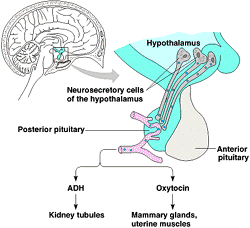
This hormone is also known by another name Vasopressin. This hormone prevents the body from excreting large amounts of urine, which causes the absorption of water in the renal tubules, due to which urine excretion is controlled.
It plays a very important role in maintaining the amount of water in the body and in fluid balance.
It causes contraction of smooth muscles, due to which it plays an important role in increasing blood pressure.
Functions of the Pituitary gland:
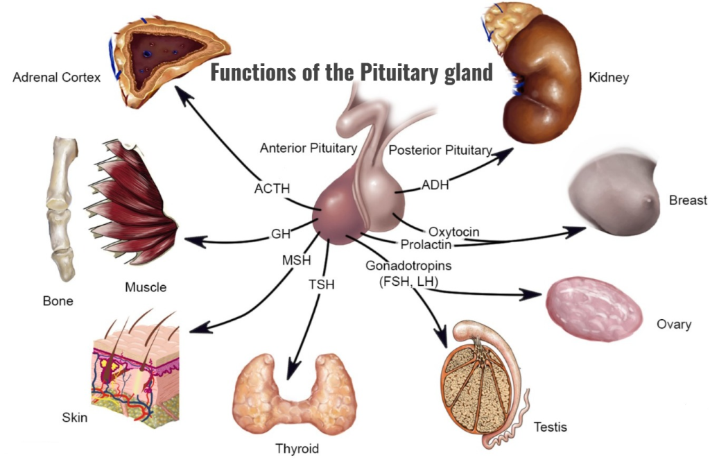
- The pituitary gland acts as the master gland in the body. Due to which it helps in maintaining the normal function of many other glands.
- The growth hormone of the pituitary gland maintains the normal growth of the body.
- The pituitary gland regulates steroid hormones in the body.
- The prolactin hormone of the pituitary gland plays an important role in milk production.
- The hormones of the pituitary gland play important roles in maintaining fertility. is.
- Hormones of the pituitary gland play important roles in maintaining normal delivery, breast development, and breastfeeding.
- Hormones of the pituitary gland play important roles in maintaining water balance and blood pressure in the body.
Relationship of hypothalamus and pituitary gland:
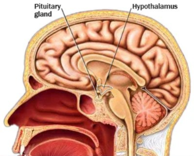
- The Pituitary Gland is located below the Hypothalamus. It is connected to a structure called the infundibulum.
- The releasing and inhibitory hormones secreted from the hypothalamus affect the pituitary gland. They stimulate the pituitary gland to secrete its own hormones.
- The anterior lobe of the pituitary gland is connected to the hypothalamus through a blood connection. This is called the Hypophyseal Portal System. Due to which the hormones of the anterior lobe are secreted.
- The posterior lobe of the pituitary gland is connected to the hypothalamus through a nerve connection. Due to which the hormones of the posterior lobe are secreted.
Thyroid Gland Gland):

- The thyroid gland is an important gland of the endocrine system. It is located in the soft tissue of the neck. This gland is a butterfly-shaped gland.
- The weight of this gland is approximately 30 grams. Its length is 5 centimeters and width is 3 cm.
- This gland is located from the level of the fifth cervical vertebra to the level of the first thoracic vertebra.
- The thyroid gland has one lobe on each side. Which is covered with fibrous tissue around it. The middle part connecting the two lobes is called the isthmus. The lobes of the thyroid gland are pyramidal in shape.
- The tissue in the thyroid gland is made up of small structures called follicles. Each follicle is made up of simple cuboidal glandular epithelium tissue. Which is associated with secretion.
- The function of the thyroid gland is regulated by the hormone thyroid stimulating hormone released from the pituitary gland.
The thyroid gland secretes the following hormones.
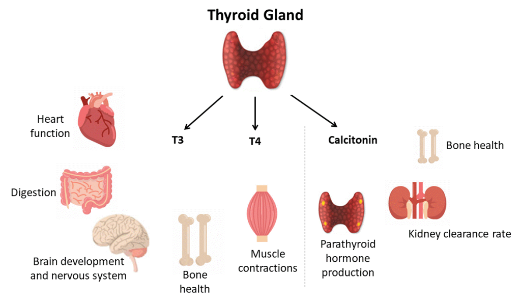
1. Triiodothyronine (Triiodothyronine) T3:
The main function of this hormone is to maintain normal physical growth and development in the body. This hormone also plays a useful role in maintaining heart rate and some metabolic activities.
2. Thyroxin (Thyroxin) T4:
This hormone also functions similarly to the T3 hormone. That is, it maintains metabolic activity in the body and works to maintain normal physical growth and development. This hormone increases the basal metabolic rate.
3. Calcitonin:
This hormone is secreted by the parafollicular cells of the thyroid gland. Which affects the metabolism of calcium in the blood.
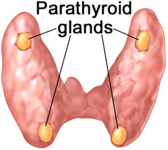
Parathyroid glands:
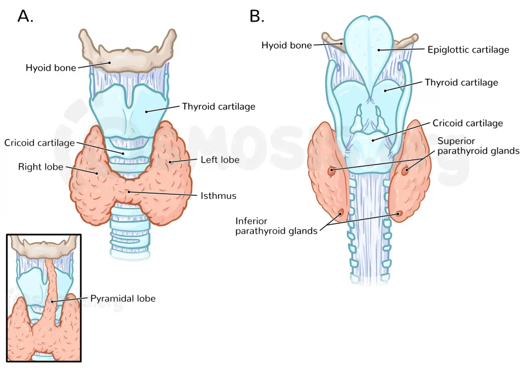
- The parathyroid glands are located behind the thyroid gland in the body. They are 4 in number.
- These parathyroid glands are arranged in pairs, superior and inferior.
The effect of the hormones of these glands is opposite to the calcitonin hormone of the thyroid gland. Which reduces the level of calcium in the blood. This gland works to maintain the balance of calcium.
- Parathyroid hormone stimulates the kidneys and is released from the kidneys. It releases a hormone called calcitriol, which promotes the absorption of calcium from food in the gastrointestinal tract. This increases the amount of calcium in the blood.
- The hormone of this gland releases calcium stored in the bones into the blood. This gland also works to maintain the balance of calcium as well as phosphorus and magnesium.
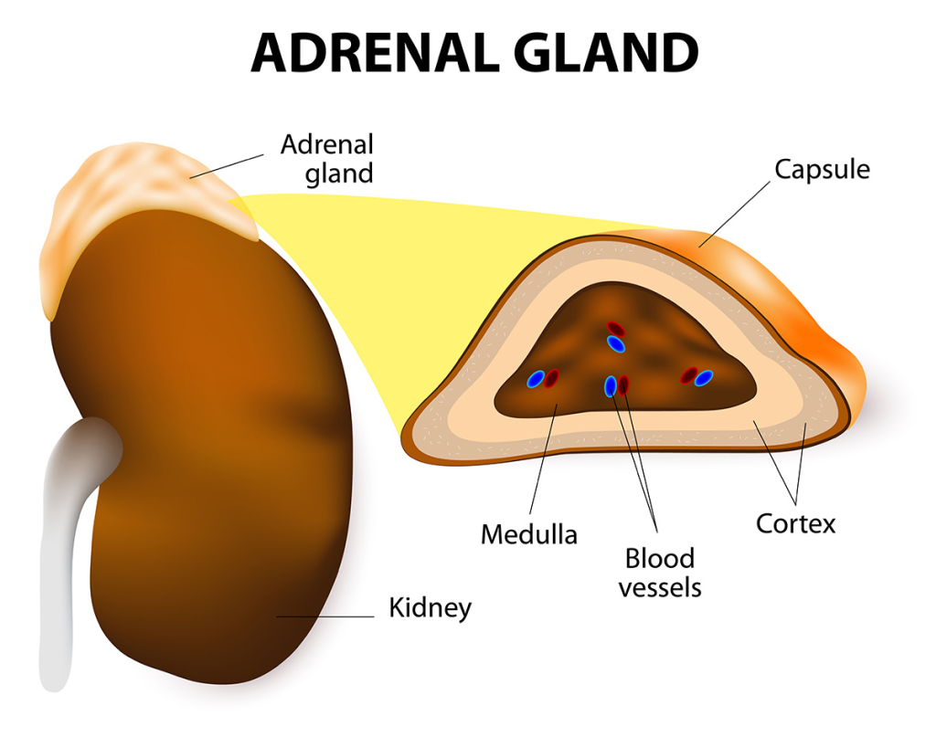
Adrenal glands:
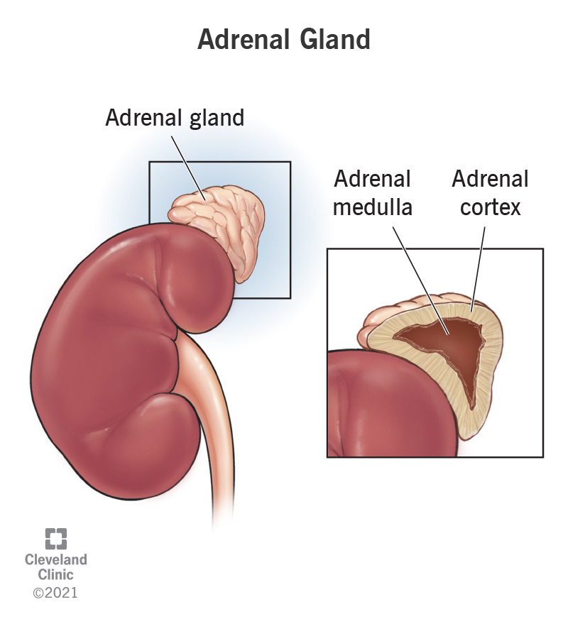
- This gland is also called the suprarenal gland because it is located above the kidneys on both sides. It is found in the number of 2. Its size is 3 to 5 centimeters long, 2 to 3 centimeters wide and 1 centimeter thick. Its weight is found to be approximately 3 to 5 grams.
- The outer part of the adrenal gland is known as the adrenal cortex and the inner part is known as the adrenal medulla.
Adrenal cortex (Adrenal Cortex).
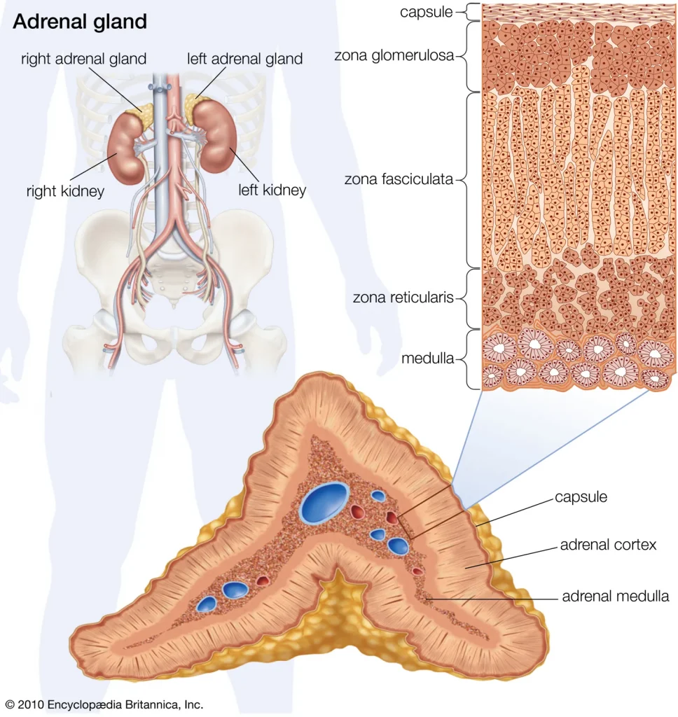
- This part is located on the outside of the adrenal gland. The cells in this part secrete steroid hormones.
- The following steroid hormones are secreted by the cortex of the adrenal gland.
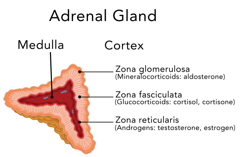
A. Glucocorticoids.
- This hormone is secreted by the Zona Fasciculata cells in the adrenal gland.
- ACTH, secreted from the anterior lobe of the pituitary gland, stimulates the adrenal cortex to secrete glucocorticoid hormones.
- This hormone helps in the metabolism of protein, glucose and fat in the body.
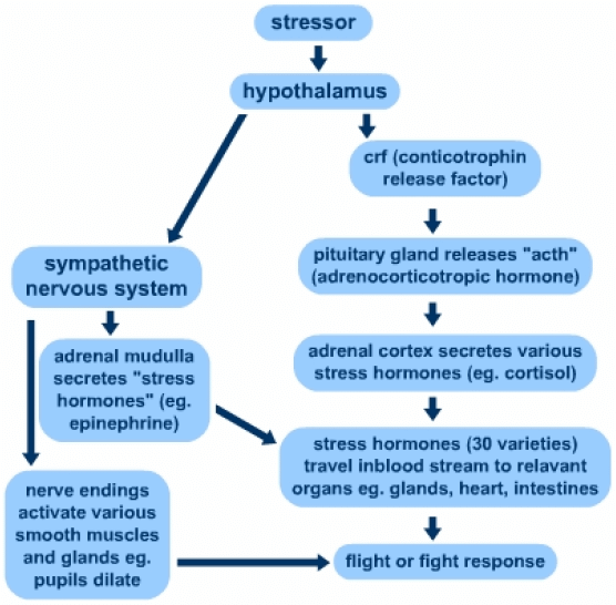
- It provides resistance to stress in the body. Due to which heart rate and blood pressure increase during stress. Also, there is an increase in blood sugar.
- The effect of this hormone is seen in every cell of the body. Its highest level in the blood is seen in the morning and its level gradually decreases till night.
- This hormone also gives anti-inflammatory effect in the body. It also depresses the immune system of our body. So our immune response decreases.
- They include cortisol and cortisone hormones.
B. Mineralocorticoid (Mineralocorticoids).
- This hormone is secreted by the Zona Glomerulosa cells in the adrenal gland.
- This hormone secreted from the adrenal cortex mainly includes the aldosterone hormone. This hormone works to maintain the amount of mineral salts in the body. Due to which the fluid and electrolyte balance is maintained in the body.
- Aldosterone hormone reabsorbs sodium and excretes potassium through urine. Due to which water retention occurs in the body and due to body fluid maintenance, blood pressure is maintained.
- This mechanism is maintained by the renin angiotensin aldosterone system (RAAS).
C. Androgens.
- This hormone is secreted by the Zona Reticularis cells in the adrenal gland.
- It is also known as sex hormone. Testosterone in males and estrogen in females are considered androgen steroid hormones.
- Which performs important functions in developing sex characteristics, hair growth and muscular structure in males and females.
Adrenal Medulla.
- The middle part of the adrenal gland is known as the adrenal medulla. This part secretes catecholamine hormones. Among them, adrenaline and noradrenaline, i.e. epinephrine and norepinephrine, are mainly secreted.
- These hormones maintain the fight or flight response of the autonomic nervous system and the sympathetic nervous system during stress.
These hormones perform the following functions in the body.
- Increases blood pressure and increases heart rate
- Constricts the diameter of blood vessels
- Increases blood sugar and increases the rate of cellular metabolism Is
- Dilates the pupil
- Dilates the airways of the lungs, etc. functions are seen through these hormones.
Response to stress
Thymus gland:
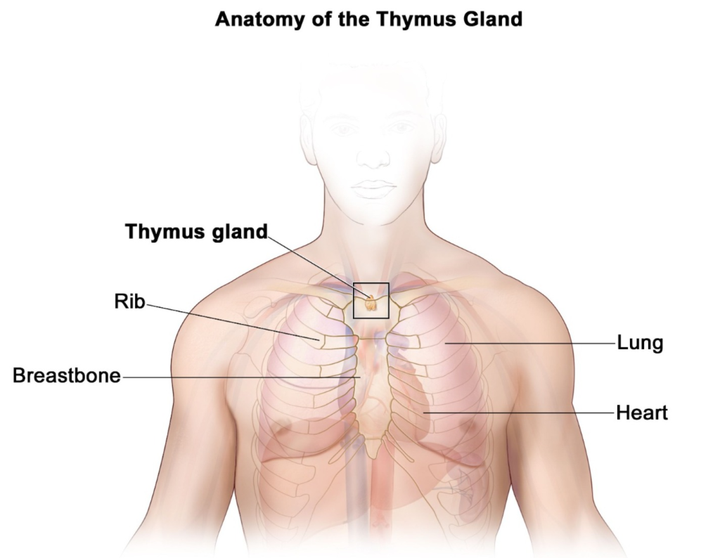
This gland is located in the mediastinum below the sternum in the thoracic cavity. This gland is larger in size in children and after puberty it becomes smaller in size as they grow older. Its weight at birth is approximately 10 to 15 grams. This gland secretes a hormone called thymosin.
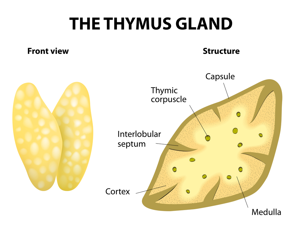
Structure:
It has two lobes. It has a capsule. Each lobe is divided into lobules, with the outer part called the cortex and the inner part called the medulla.
Function (Function) :
- B-lymphocytes produced in the bone marrow are activated as T-lymphocytes after entering the thymus gland.
- Thymosin stimulates the thymus gland and other lymphoid tissues to mature. Which is secreted by the epithelial cells of the hormone gland.
Pineal gland :
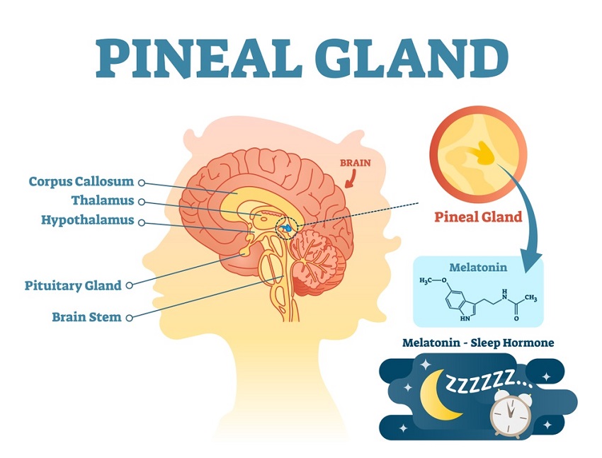
- This gland is attached to the roof of the ventricle in the cranial cavity. Its shape is pine cone shaped.
- It is 10 mm long and appears reddish brown in color.
- This gland secretes a hormone called melatonin. The secretion of this hormone is seen during dark light. Sunlight or light reduces the secretion of melatonin hormone. This results in decreased sleep.
- In the absence of light, norepinephrine stimulates the secretion of melatonin. which causes sleep.
Pancreas:
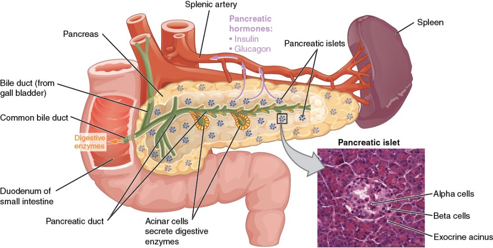
- The pancreas functions as both an endocrine and an exocrine gland. The part of the pancreas that secretes hormones is called the endocrine gland, and some of the cells in the same pancreas that produce digestive juices are called the exocrine pancreas.
- The pancreas is an organ 12 to 15 cm long. It weighs approximately 100 grams. The pancreas is divided into three parts: head, body, and tail.
- The pancreatic cells in the pancreas are arranged in clusters. These millions of small pancreatic endocrine tissues are called pancreatic islets or islets of Langerhans.
These pancreatic islets contain the following specific cells that are involved in the secretion of specific hormones.
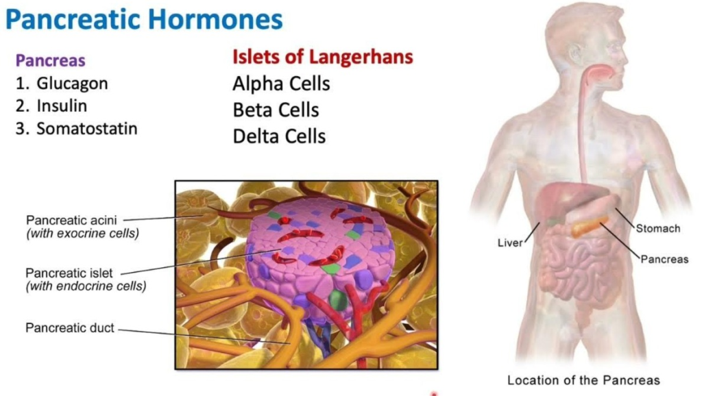
- Alpha cells secrete glucagon.
- Beta cells secrete insulin Secretes.
- Delta cells secrete somatostatin.
Blood glucose levels in the human body are controlled by the hormones insulin and glucagon.
Insulin
- Insulin is secreted by the beta cells in the pancreas. It works to reduce the blood glucose level in the body.
- It works to convert the excess glucose in the blood into glycogen. This process is known as Glycogenesis.
- This hormone synthesizes proteins and also helps in the synthesis of fatty acids.
Glucagon (Glucagon)
- It is a hormone secreted by the alpha cells in the pancreas. It works opposite to insulin, that is, when the blood sugar level drops below normal, the alpha cells of the pancreas release glucagon and it converts sugar from glycogen to glucose.
- In this way, it maintains the glucose level in the blood. It prevents hypoglycemia in the body.
Somatostatin
- This hormone is secreted by the delta cells in the pancreas.
- It reduces the secretion of insulin and glucagon and reduces the absorption of nutrient materials through the gastrointestinal tract.
Other hormones of the Human Body:
Many tissues in the body function like endocrine glands and secrete local hormones. These locally secreted hormones are as follows.
Erythropoietin (Erythropoietin)
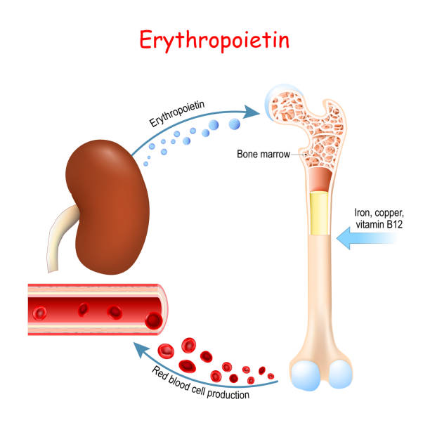
Erythropoietin is a hormone secreted by the kidneys that helps in the formation and production of RBCs. This hormone stimulates erythropoiesis.
Histamine.
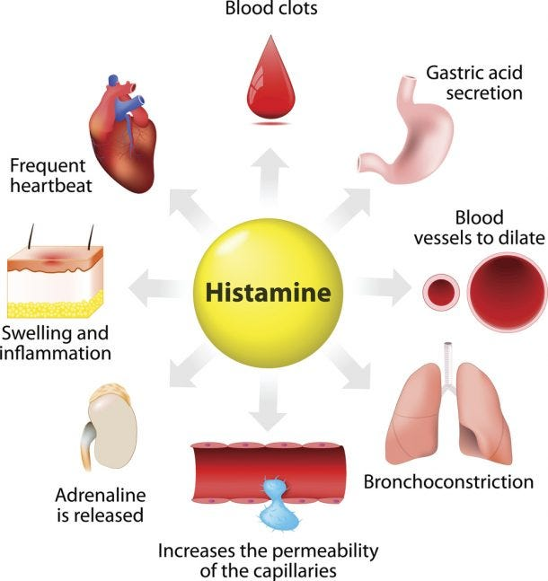
This is a local hormone secreted by mast cells of connective tissue in the body. This hormone prevents infection, that is, it acts as an anti-inflammatory.
Prostaglandins.
- This is a hormone that acts as a lipid substance in many tissues in the body. Which has many physiological functions.
- It causes the feeling of pain
- Responsible for fever
- Regulates blood pressure
- Also important for inflammatory response
- Contracts the muscles of the uterus during labor pain
- Plays an important role in the blood clotting mechanism.
Ovaries:
- The ovary is a female gonad. It is located on either side of the uterus in the ovarian fossa. It is located behind the broad ligament.
- They are 2 in number (pair of ovaries). It is homologous to the testis.
- It is attached to the pelvic wall by ovarian wall ligaments.
Location:
The structure surrounding the ovarian fossa (a fossa is a pit-like structure that contains an organ) is supported by the ureters (the back of the urinary tract), the obliterated umbilical artery (an artery that disappears shortly after birth), and the obturated intercostal muscles, vessels, and nerves in front and below.
Shape and Size:
The ovary is oval in shape. It is 3 cm long, 1.5 cm wide and 1 cm thick.
Surface And Colour:
The ovary of young adults is pink in colour and smooth. In older women, it is rough, irregular, and gray in color because they have ovulated frequently.
Attachment:
Both ovaries are attached to the uterus at the top by the ovarian ligament and at the back by the broad ligament. It is called the mesovarium, blood vessels and nerves pass through the mesovarium.
Blood Supply:
It is supplied by the ovarian artery and is a branch of the abdominal aorta.
The vein is called the pampiniform plexus. It emerges from the ovary itself.
The right vein drains into the inferior vena cava and the left vein drains into the left renal vein.
Nerve Supply:
Sympathetic fibers that emerge from the T10 and T11 spinal ligaments.
Parasympathetic nerves emerge from the vagus nerve.
Histology Of Ovary (Histology (study of tissue is called histology) of ovary):
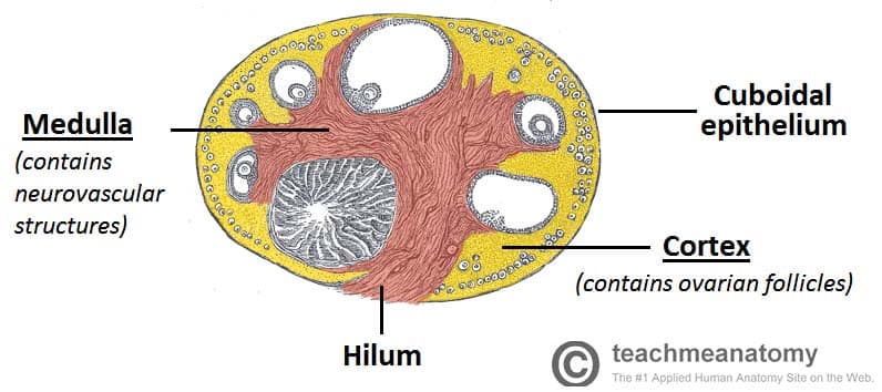
- The ovary is oval in shape.
It is lined with a single layer of cuboidal epithelium cells. It is called the germinal epithelium. - It contains dense tissue. It is called the tunica albinia and is located inside the germinal epithelium.
It has a total of 2 parts.
- Cortex:- It is made up of the stoma and follicles of the ovary.
- Medula:- It is made up of connective tissue and is connected by a network of blood vessels and is made up of elastic fibers.
Oogenesis:
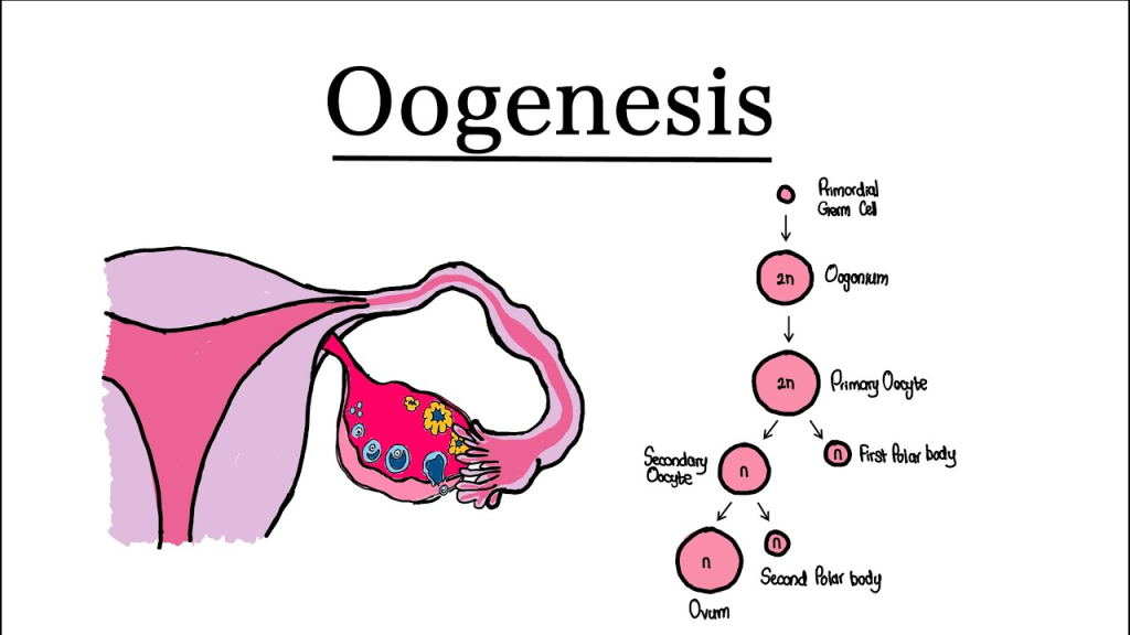
This is a process of producing female gametes. The process of ova from primary germ cells is called oogenesis.
By the 6th month of the intrauterine period, about 6 million oogonia are present. The oogonia undergo meiotic division and convert into primary oocytes and are arrested in prophase.
The 1st meiotic division is arrested at puberty. When puberty begins, menstruation begins. The first menstrual cycle is called menarche.
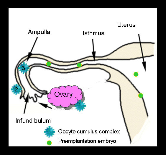
When the first menstrual cycle begins, ovulation occurs and the egg is released and ovulation occurs, the first egg is released.
When ovulation occurs, follicles are formed before it. It happens with the help of FSH and then it is seen due to estrogen. Those follicles mature and become Graafian follicles.
A mature ovum is formed from the Graafian follicle. The ovum is released on the 14th day of the menstrual cycle and if it is fertilized, it becomes a fetus and if it is not fertilized, menstruation occurs.
Testes:
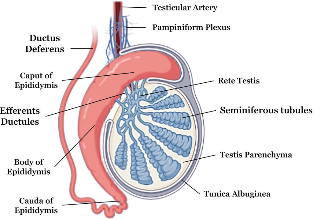
They are oval-shaped and are 2.5 cm long and 2.5 cm wide. They weigh 10 to 15 grams.
The fetal period means that the testicles are in the abdomen when the baby is in the uterus. At the 32nd week, these testicles descend into the scrotum.
The testicles have three layers:
1.Tunica Vaginalis:- It is made up of 2 layers. Parietal and visceral and there is a cavity between these. It covers the testis from all sides but does not cover the back.
2.Tunica Albuginea:- This is a fibrous covering layer, and lies beneath the tunica vaginalis and it in-growing and separates the glandular lobes of the testis.
3.Tunica Vasculosa:- As its name suggests, it is vascular, meaning that The relationship is with blood as this layer has a network of capillaries and is made up of connective tissue.
HORMONS: Male hormones:
MEN’S HORMON Male hormones:
Female Like the testes, endocrine secretions are also produced, but they are not repeated. Follicle stimulating hormone acts on the seminiferous tubules, which is necessary for the production of sperm. Luteinizing hormone acts on the interstitial cells, which is necessary for the production of testosterone.
Testosterone:-
This is responsible for some of the characteristics of the testes. E.g. Voice, development of reproductive organs, growth of hair on chest, beard, axilla and pubis etc.