—ENGLISH-ANATOMY (UNIT : 8) URINARY SYSTEM
URINARY SYSTEM:
KEY TERMS:
1) Renal cortex :
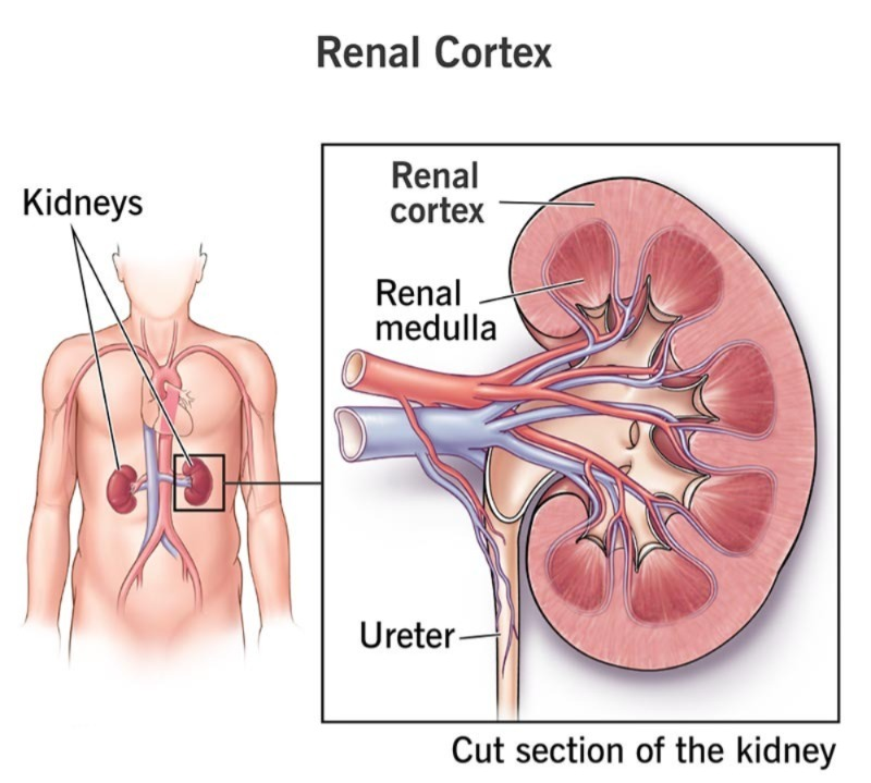
The outermost layer (part) of the kidney.
2) Bowman’s capsule:
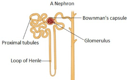
Nephron (Nephron which is the basic functional unit of the kidney) Bowman’s capsule is the most anterior cup-shaped mouth-like part of the nephron .
3)Nephron (Nephron):
It is the basic functional unit of the kidney. It is the microscopic structural unit of the kidney and the basic functional unit in the kidney.
4) Renal medulla :
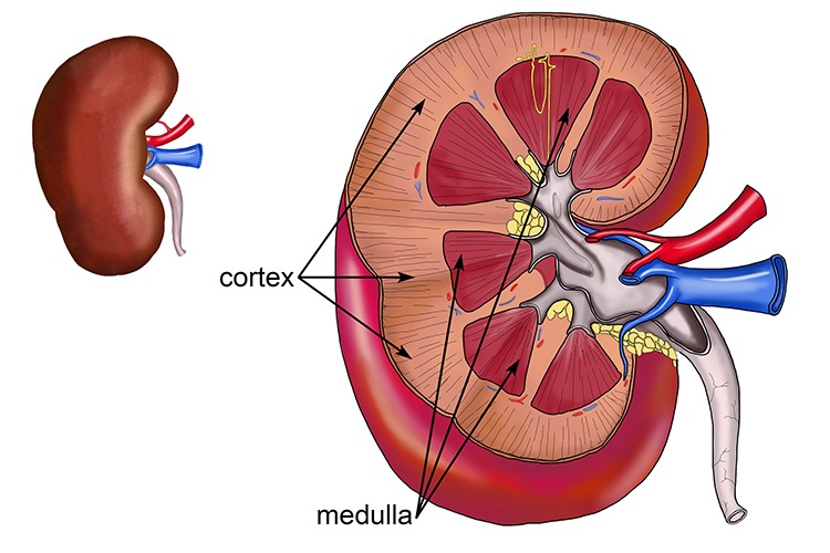
This is the innermost layer of the kidney.
5) Glomerulus:
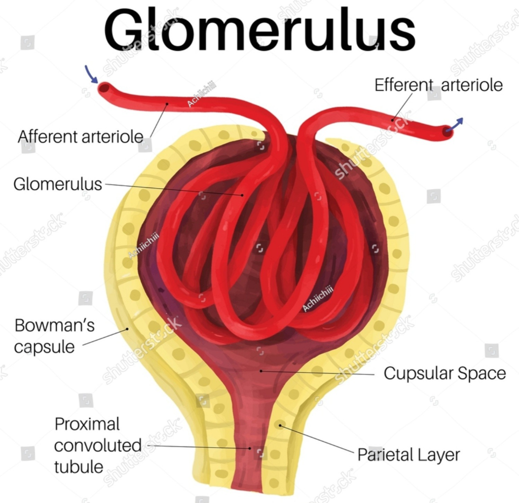
The glomerulus is a cluster of very thin capillaries. Which is located inside the Bowman’s capsule.
6) Osmolarity:
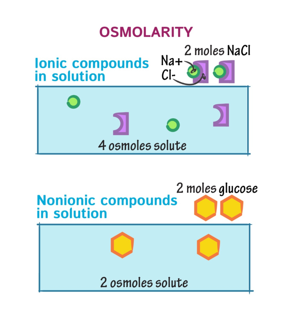
Osmolarity is the expression of the osmotic pressure of any solution (liquid). In which the number of moles of solute present in a kilogram of water.
7) Ureters:
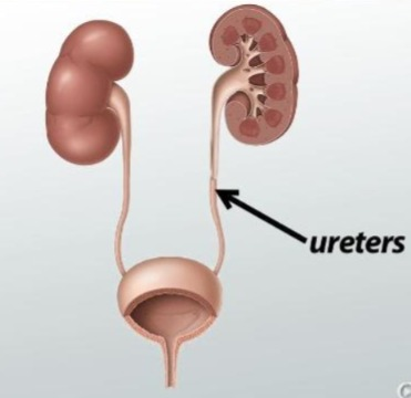
This is a type of tubular structure. The tube connects the kidney to the urinary bladder. It passes urine from the kidneys to the bladder.
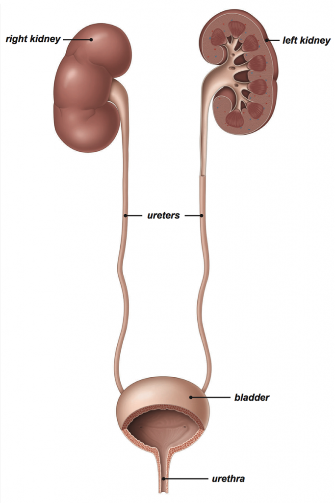
8) Urethra:
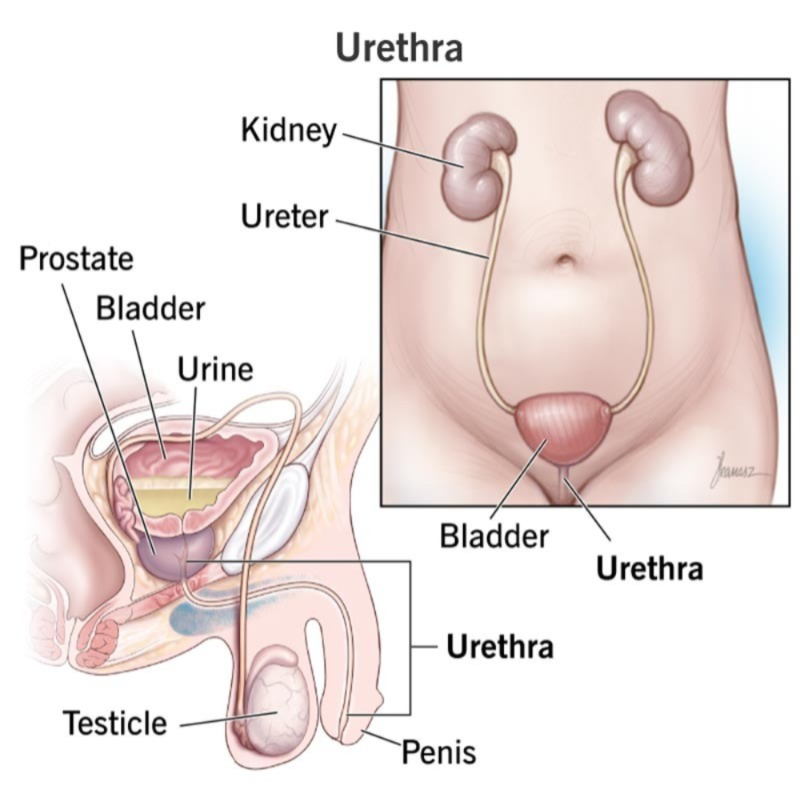
This is a type of tubular structure that connects the urinary bladder to the external environment of the body. Urine is excreted from the body through its external opening.
9) Bifurcation:
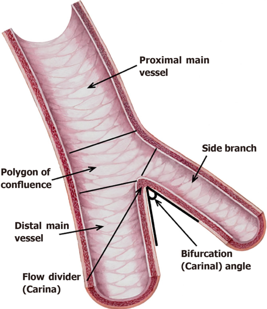
Bifurcation means the division of any branch into two parts.
10) Diuresis :
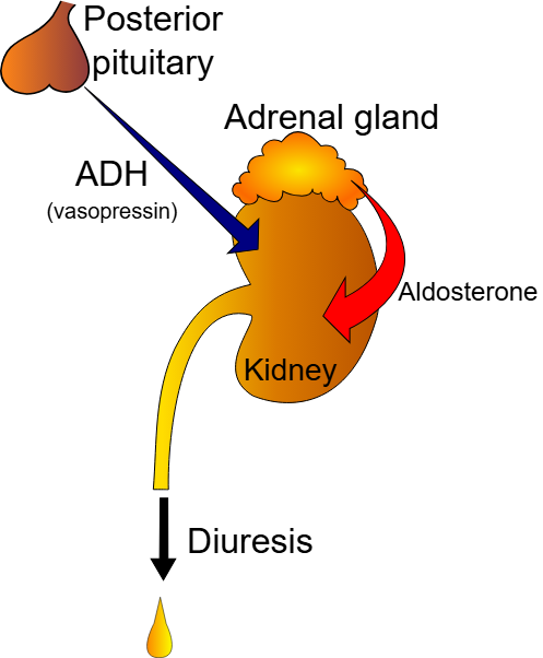
Excessive production of urine.
OR
Diuresis is a physiological process that helps remove excess water, salt, and waste products from the body, in which the kidneys produce excessive amounts of urine. is.
11) Renal artery:
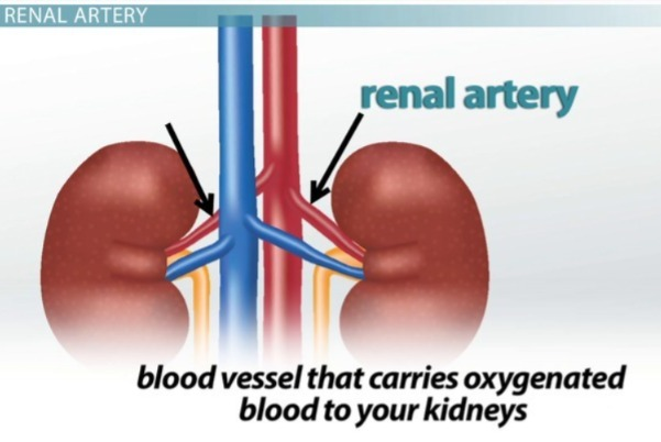
The renal artery is a blood vessel that supplies oxygenated blood to the kidneys.
12)Excretion:
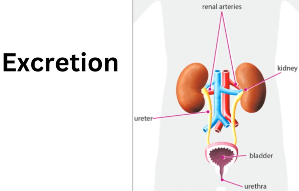
Excretion means removing any waste product of the body (urine) outside the body.
13) Renal vein :
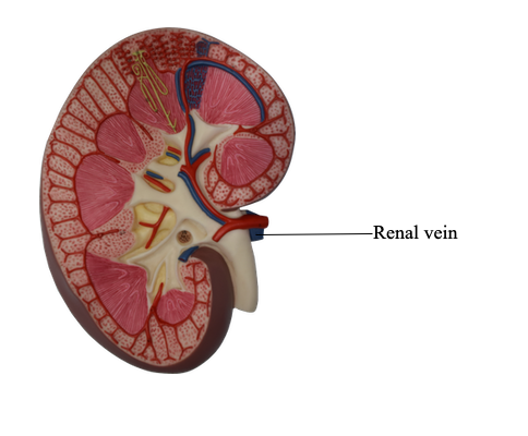
These blood vessels return the filtered blood (deoxygenated blood) to the circulation. They return the blood from the kidneys.
14) Renin :
Renin is a hormone that is secreted by the juxtaglomerular cells of the kidneys in response to low blood pressure and low sodium levels in the body and by sympathetic stimulation, which plays an important role in the regulation of blood pressure through the renin-angiotensin-aldosterone system (RAAS).
OR
Renin is a hormone secreted by the kidneys. This hormone regulates systemic blood pressure.
15) Sphincter:

Sphincters are ring-like muscle fibers that have the ability to constrict.
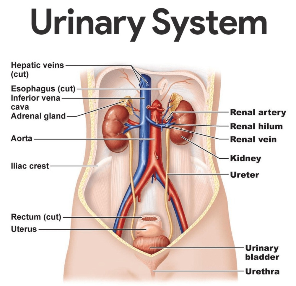
Introduction of Urinary System:
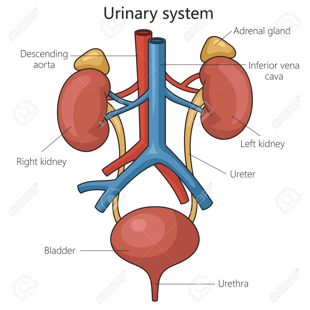
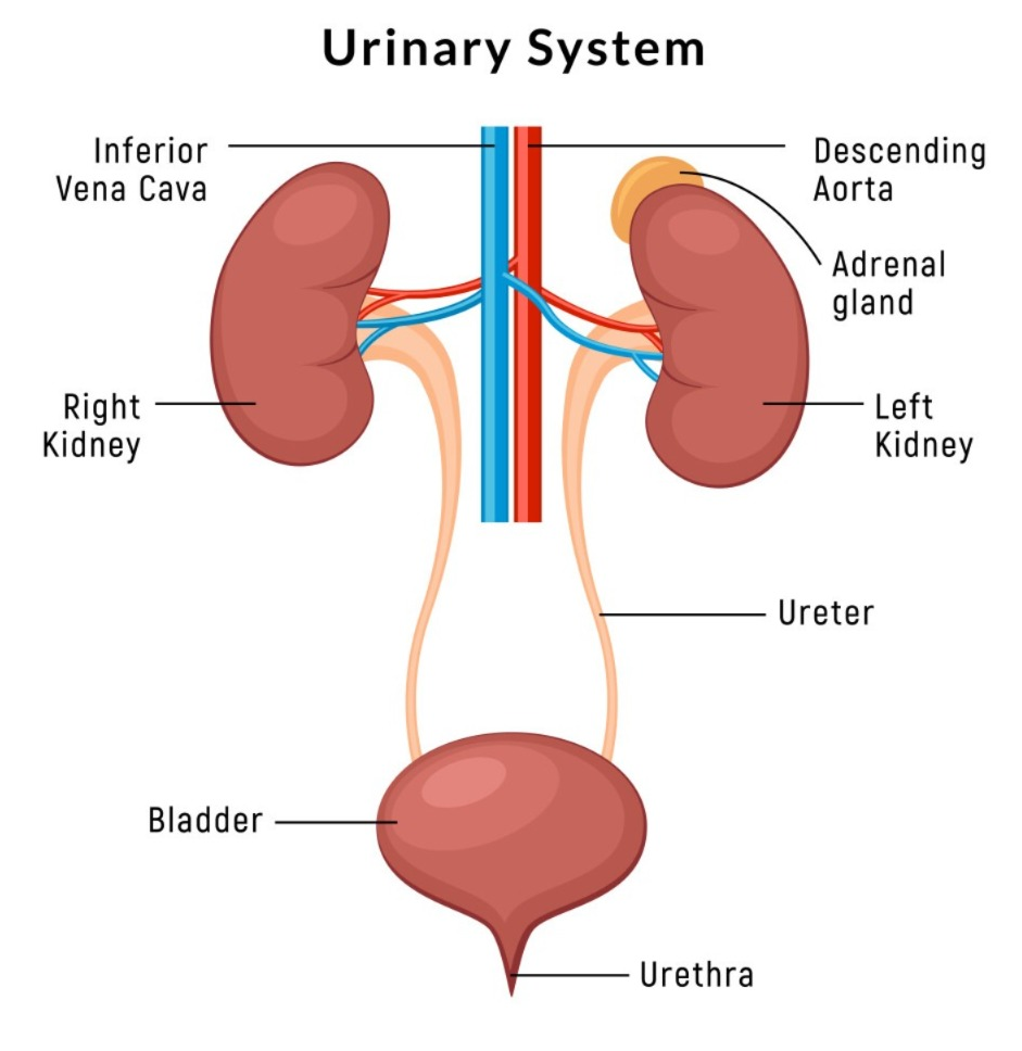
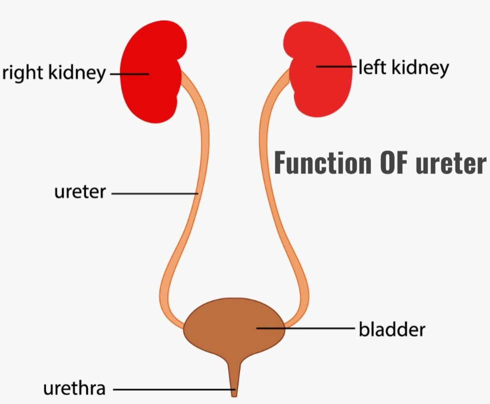
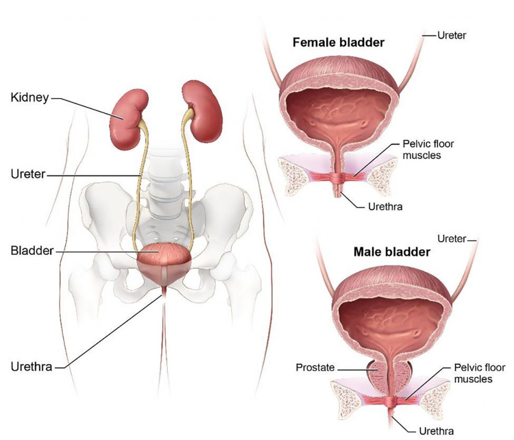
The urinary system is a type of excretory system of the body. It removes the body’s waste products from the body. It removes any substances in the body that are not used in the body (metabolic waste). It includes the following organs.
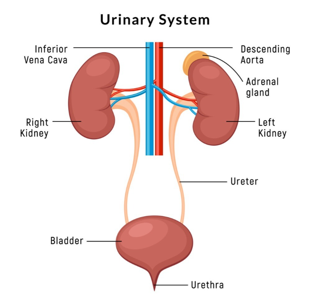
2 kidneys : Which form urine. They are right and left.
2 ureters : Which carry urine from the kidneys to the urinary bladder. There are 2 of them, right and left.
1urinary bladder : This is an organ in which urine is collected. It is 1 in number.
1 urethra : Through which the urine collected in the urinary bladder is excreted outside the body.
Gross Structure Of Kidney
- The kidneys are 2 in number in the human body. They are located in the abdominal cavity, one on each side of the vertebral column, on the right and left sides of the body, on the posterior side.
- The kidneys are bean-shaped organs. It is located from the level of the 12th thoracic vertebra to the level of the 3rd lumbar vertebra.
- The kidney is 11 centimeters long and 5 to 6 centimeters wide. It weighs approximately 150 grams. The right kidney is located slightly lower than the left kidney because the liver occupies a larger part of the right side.
Organs surrounding the kidneys :
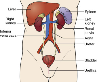
The kidneys are organs located in the abdominal cavity. They are located one on each side of the abdomen, on the right and left. The organs of the abdominal cavity such as the liver, small intestine, adrenal glands, stomach, spleen, pancreas, etc. are located around both kidneys.
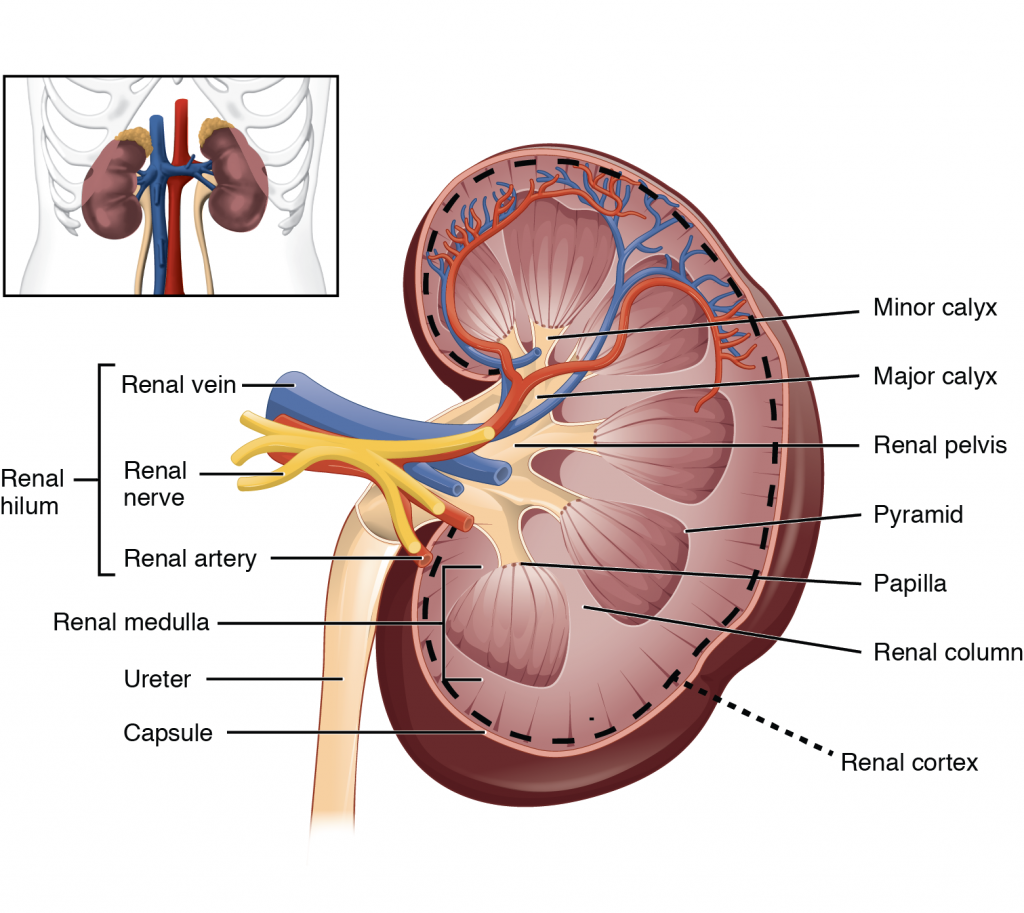
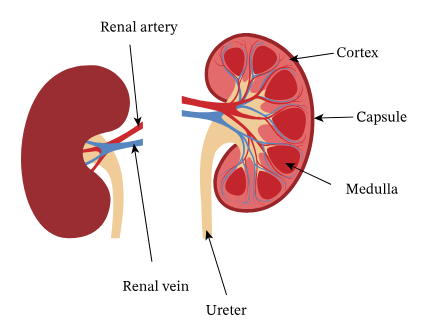
Structure of the kidney:
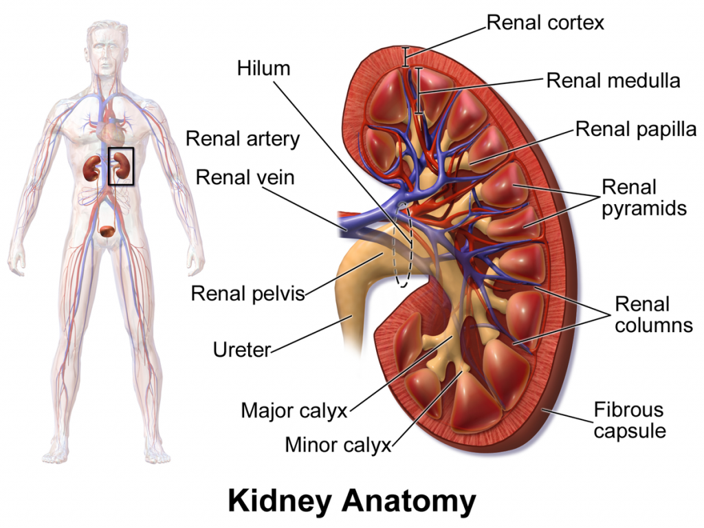
- The kidney is an irregularly shaped organ. It has a groove in the middle called the hilum or renal hilum. Through which the structures of the renal artery, renal vein, nerves, lymph vessels and ureter enter and exit.
- The inner border or hilum of the kidney is found on the side of the vertebral column. Its outer border is convex. The kidney is a hanging organ on both sides in the abdominal cavity. To keep it in its position, a network of fatty tissue and fibroelastic connective tissue called renal fascia surrounds it. With the help of this, the kidney can maintain its position and also gets protection.
When the kidney is viewed in longitudinal section It is found distributed in three structures of the kidney.
1.Fibrous Capsule:
It is a part of the fibrous tissue surrounding the kidney. This membrane is arranged around the kidney. It acts as a layer to maintain the shape of the kidney and protect it.
2.Cortex:
It is a part of the tissue of reddish brown color. Which is located under the kidney capsule.
3.Medula (Medula):
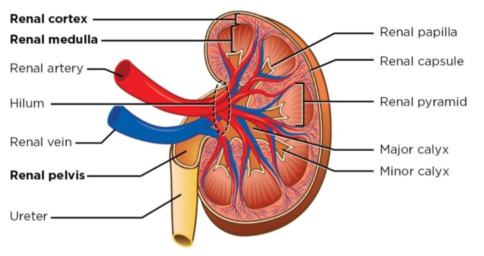
- The inner part of the kidney from the cortex is called the medulla. It is also reddish brown in color. It contains triangular pyramid-like structures called renal pyramids. The base of this renal pyramid faces the cortex and the pointed part of the pyramid, i.e. the renal papilla, faces the hilum.
- This renal papilla forms a cup-like structure on the front called the calyx. The part with the larger space is called the major calyx and the part with the smaller space is called the minor calyx. The minor calyx opens into the major calyx. From this calyx, a funnel-shaped The wide part is called the renal pelvis.
- The urine filtered by the kidney passes through this calyx and enters the wide part of the funnel shape, i.e. the renal pelvis. Here the urine is collected and then passes through the narrow structure coming out of the kidney from the renal pelvis at the front, called the ureter, through which the urine exits the kidney and reaches the urinary bladder.
- The urine filtered by the kidney passes from the minor calyx to the major calyx and from the major calyx to the renal pelvis. From there it reaches the urinary bladder through the ureter. This action is not controlled by any nervous system. The walls of the renal pelvis contain special muscles and pacemaker cells, due to whose contraction the urine flows forward.
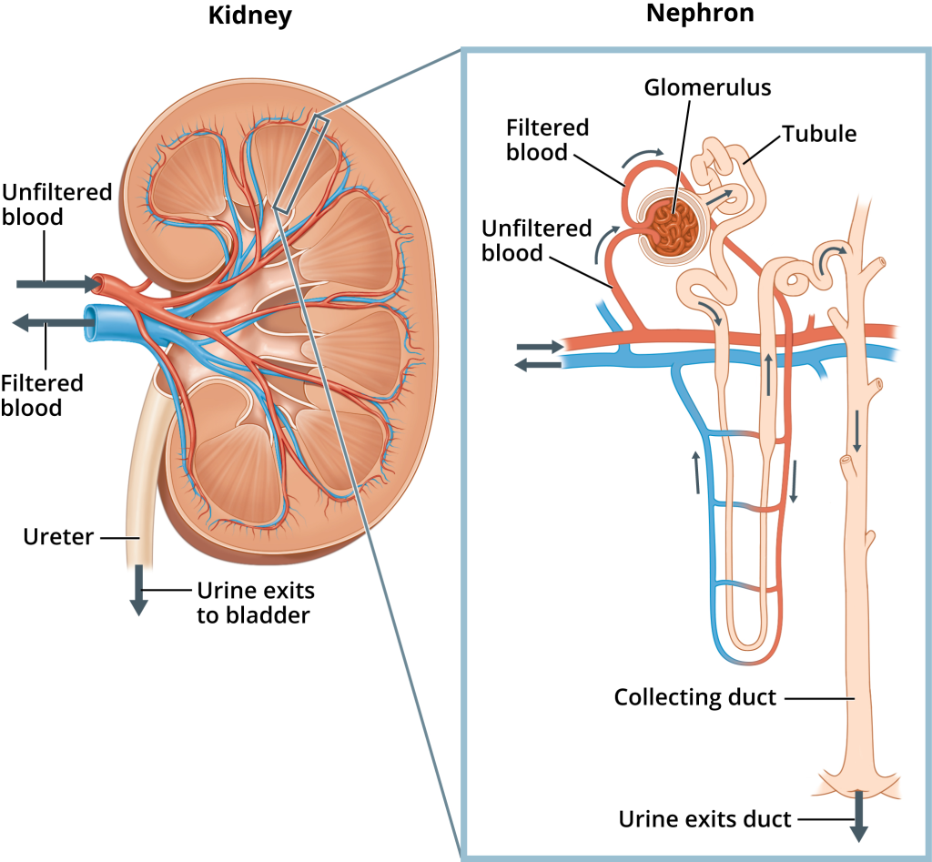
Microscopic Structure of Kidney or Structure Of Nephron:
When looking at the microscopic structure of the kidney, many compositions are seen. In which, when looking at the kidney under the microscope, the microscopic functional structure i.e. Nephron is seen, which is the main working unit of the kidney. There are millions of nephrons in the kidney.
The microscopic structure of a nephron includes the following structures.
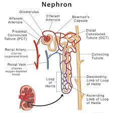
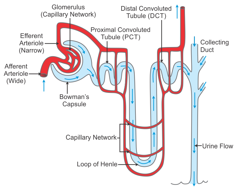
1. Bowman’s Capsule:
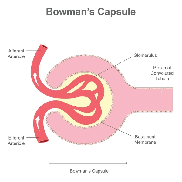
- At the front of the nephron is a cup-shaped mouth called the Bowman’s capsule. The wall of this capsule contains a special type of epithelial cells called podocytes. Both the parietal and visceral layers of this Bowman’s capsule are found.
- In the middle of the cup-shaped part of the Bowman’s capsule is a network of arterial capillaries called the glomerulus. The podocyte cells here help in the filtration process. The three-centimeter long part beyond the Bowman’s capsule is called the tubular part. This tubular part can be divided into the following parts.
2. Proximal Convoluted tubule:

- The initial tubular portion anterior to the Bowman’s capsule is known as the proximal convoluted tubule. It is the initial part of the tubules of the nephron. Its wall is lined with epithelial cells. This part is located in the cortex of the kidney.
3. Loop of Henle:

- The U-shaped part (Sharp U Turn) after the proximal convoluted tubule Loop of Henle It is called. It is also called medullary loop. The loop of Henle consists of ascending and descending loop. A sharp bend is seen in the middle part which forms a U-shaped part. The loop of Henle is located in the medulla of a kidney.
4. Distal Convoluted Tubule:
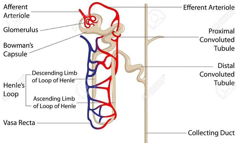
The tube-shaped part of the nephron after the loop of Henle is called the distal convoluted tubule. This tube-shaped part forms a sending limb. Which joins the collecting duct further.
5. Collecting Duct:
- Collecting duct is a straight tube. Which connects the distal convoluted tubule of many nephrons. These tubes are known as large ducts. These collecting tubes connect with each other and open into the minor calyx of the renal pyramid.
- When the renal artery enters the kidney from the hilum, it then divides into many smaller arterioles. When these arterioles enter the Bowman’s capsule, these arterioles are called afferent arterioles. The network of capillaries in the glomerular capsule is called glomerules. The arterioles of this network of capillaries, which are called Bowman’s The ones that come out of the capsule are called efferent arterioles .
- The diameter of the efferent arterioles is smaller than the diameter of the afferent arterioles, due to which pressure is created on the glomerular membrane and the filtration process is accelerated and the process of urine formation starts.
- The efferent arterioles again divide into small capillaries and absorb nutrients and water and mix them with the blood circulation. These small Vessels drain blood out of the kidney through the renal vein.
Functions of the Kidney:
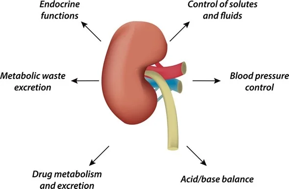
- The kidneys mainly function to form urine.
- The kidneys filter the blood and remove waste products through urine.
- The kidneys function to maintain a normal balance of electrolytes.
- They regulate the blood’s pH. It functions to maintain.
- It functions to remove the waste products accumulated at the end of metabolism from the body.
- The kidneys secrete a hormone called erythropoietin, which stimulates RBCs. It plays a very important role in the production of urine.
- The kidneys secrete a hormone called renin, which plays a very important role in maintaining blood pressure.
- The kidneys work to maintain water balance in the body.
Stages Of Urine Formation:
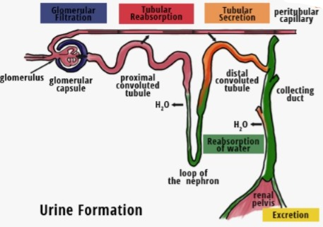
The main function of the kidneys is to excrete waste products from the body through urine. The urine produced by the kidneys is collected in the urinary bladder. It is then excreted from the body through the urethra. This urine formation is prepared through the following stages.
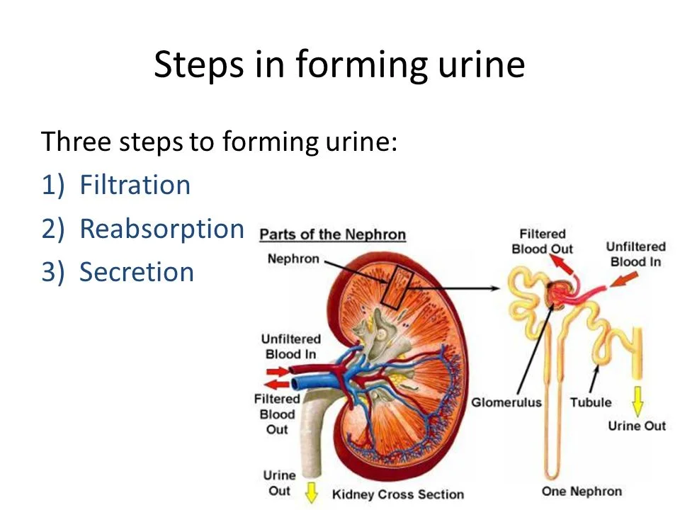
Simple Filtration:
- The function of filtration in the kidney is done by the nephron. The afferent arteriole enters the Bowman’s capsule in the structure of this nephron and its diameter is larger than the efferent arteriole. The network of these arteries is called the glomerulus.
- The pressure is created on the glomerulus membrane due to the difference in the diameter of the arterioles. Due to which the filtration process is accelerated.
- Since both the glomerular membrane and the blood capillary membrane are semi-permeable, the pressure from the blood towards the Bowman’s capsule increases and urea, uric acid, creatinine and many other substances present in the blood are filtered in the Bowman’s capsule. Large molecules and proteins such as plasma proteins present in the blood are not filtered due to which the pressure in the blood capillaries is high.
- The amount of urine filtered from the glomerular membrane in one minute is called Glomerular Filtration Rate (GFR). Which is normally 120ml.
Tubular Secretion:
- Since the blood does not remain in the blood capillaries for a long time on the glomerular membrane, some substances cannot be filtered and are secreted directly into the tubules from the peritubular capillaries surrounding the convoluted tubule.
- Some types of medicines, some toxic substances, some excess electrolytes and other excess substances present in the blood are secreted from the peritubular capillaries into the tubular area and mixed with urine. This phase is called the phase of tubular secretion.
Tubular Reabsorption:
- The urine filtered by the glomerulus enters the Bowman’s capsule. Every minute, approximately 120 ml of urine is filtered and enters this capsule. In 24 hours, 150 to 200 liters of urine is filtered through the blood and enters the Bowman’s capsule. But not all of it is excreted through urine. This fluid is mainly reabsorbed in the tubular portion and approximately one to one and a half liters of urine is excreted from the body during the day.
- The function of reabsorption mainly takes place in the proximal convoluted tubules. Glucose, sodium chloride, water etc. are absorbed through the tubular portion through the membrane of this tube.
- If any substance and water present in the body is in normal quantity, it is not excreted through filtration. But the fluid and substance in excess of the normal quantity in the body is filtered through filtration and enters the tubular portion. This tubular portion reabsorbs water and various substances in the required amount in the body.
- The aldosterone hormone present in the blood plays a very important role in the reabsorption of sodium and excretion of potassium.
- The anti-diabetic hormone plays a very important role in the reabsorption of water.
- After going through all the above stages, the fluid that reaches the collecting duct is called urine.
- This From the collecting duct, urine reaches the minor calyces and from there the major calyces, and from there it reaches the renal pelvis. From there it reaches the urinary bladder through the ureter and the urine is excreted from the body through the urethra.
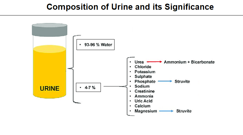
Composition of urine:
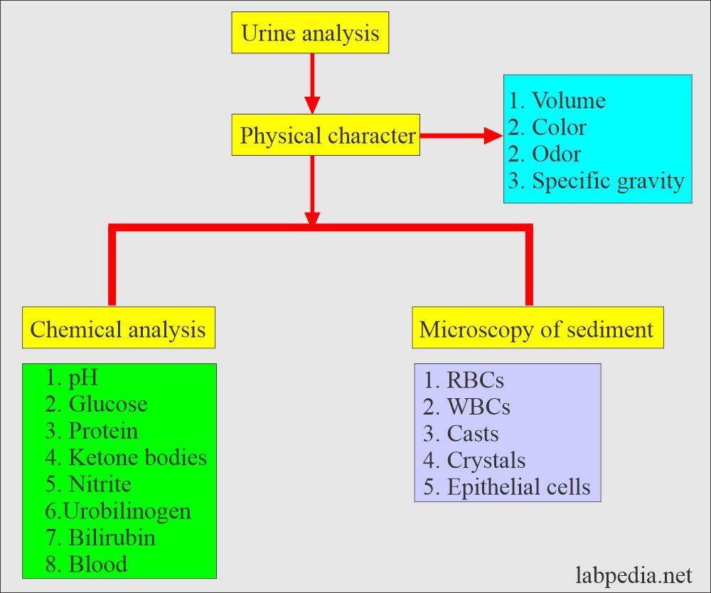
- Urine is composed of 96% water and 2% urea.
- The last 2% of urine contains creatinine, sodium, potassium, chloride, uric acid, sulfate and other waste products.
- The specific gravity of urine is 1.010 to 1.025.
- The pH of urine is slightly acidic, i.e. 4.5 to 8.
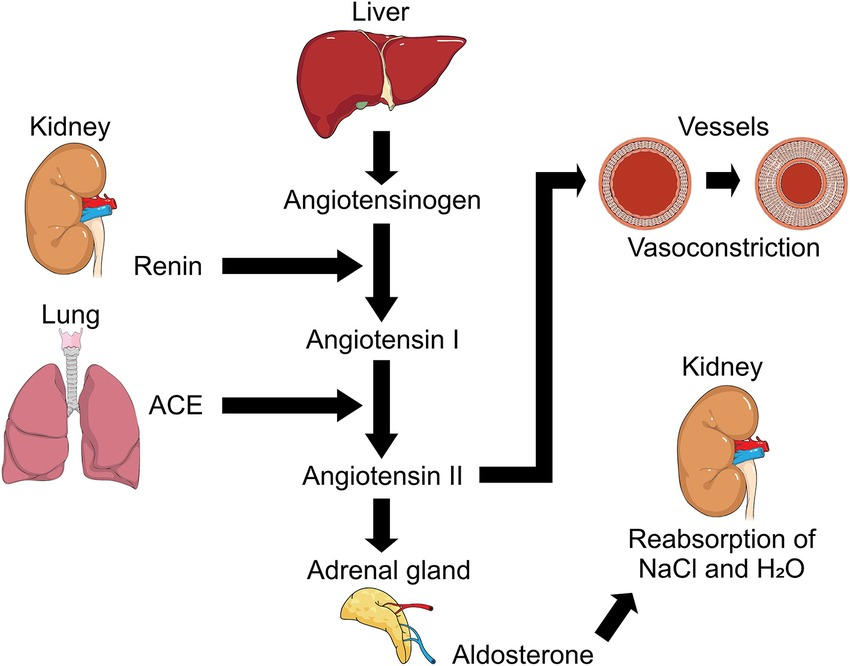
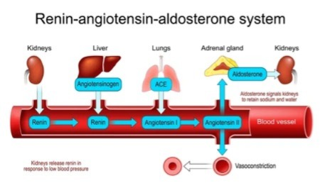
RAAS SYSTEM (RENIN ANGIOTENSIN ALDOSTERONE SYSTEM):

- This system is used to increase blood pressure or blood volume in our body when it is low.
- When blood volume or blood pressure decreases, the juxtaglomerular cells located in the glomerulus of Bowman’s capsule secrete the hormone renin.
- When the liver secretes angiotensinogen hormone, the renin hormone converts angiotensinogen to angiotensin 1.
- Lungs Angiotensin converting enzyme (ACE) is secreted from the kidney, which converts angiotensin 1 to angiotensin 2.
- Angiotensin 2 acts as a vasoconstrictor. This causes the blood vessels to constrict and blood pressure to increase.
- Angiotensin 2 activates the adrenal glands and releases the hormone aldosterone. This hormone causes the reabsorption of sodium. Due to this, the level of water also increases. Due to this, the blood volume also increases and blood pressure also increases.
- Thus, the RAAS system plays an important role in increasing blood pressure when it is low in the body.
Ureters:
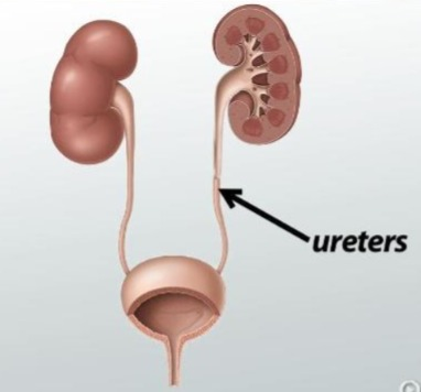
- The ureter is a tube-like structure that extends from the kidney to the urinary bladder. The major role of the ureter is to transport urine from the kidney to the bladder.
- The ureters are two ducts or tubes attached to the kidneys that pass from the kidneys to the bladder. They are located in the number of 2, left and right.
size (Size):
- 25-30cm or 10-12 inches in length. It has a 0.6cm (6mm) diameter.
Location (Location):
- The end of the renal pelvis of the kidney is narrowed to a point called the hilum. The hilum forms the beginning of the ureter. The ureter passes downward and behind the peritoneum. Which opens laterally from the posterior part of the bladder, that is, from both sides, the ureter opens into the bladder.
Structure:
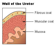
1. Mucosa:
It is the innermost layer, which is made up of mucous membrane. Its lining is made up of transitional epithelium,
2. Muscular or muscle layer:
It is the middle layer, which has an inner layer of longitudinal and an outer circular layer of smooth muscle fibers.
3. Adventitia or fibrous layer:
It is the outer layer, which is made up of areolar connective tissue. Which contains blood vessels, lymphatic vessels and nerves.
Function (Function) :
- The ureter acts as a passageway for urine from the kidneys to the bladder.
- The peristaltic contractions of the smooth muscle layer of the ureter push urine from the kidneys to the bladder.
- These contractions pass urine from the renal pelvis to the bladder.
- The rate of contraction is one to five per minute, which is how fast urine is formed It depends on it.
- The reason why urine does not go back from the bladder to the ureter is because of When the bladder fills with urine, the pressure inside the bladder increases, which puts pressure on the opening, which is a fold of the bladder mucosa that acts as a small valve, covering the opening of the ureter, preventing urine from flowing back into the ureter.
Blood supply:
Blood supply is provided by branches of the renal artery, abdominal aorta, gonadal artery, internal iliac artery, and common iliac artery. is.
Urinary bladder:
- The bladder is a hollow, muscular organ. Its function is to collect urine from the kidneys, store it, and dispose of it after a short period of time.
- It stores temporary urine.
- The bladder is roughly pear-shaped. But when it is full of urine, it becomes more oval-shaped.
Position:
- The size, shape, and position of the urinary bladder vary from person to person, depending on the amount of urine and the age of the person.
- In adults, the bladder is located in the true pelvis. When the bladder is full of urine, it passes into the abdominal cavity.
- In newborns, the bladder is in the abdominal cavity.
Capacity:
- 300 to 700 cc. When the bladder holds 300 to 400 ml of urine Then the urge to pass urine begins.
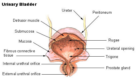
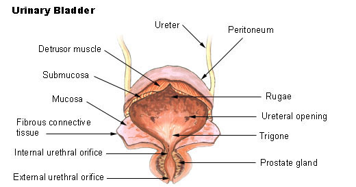
Structure:
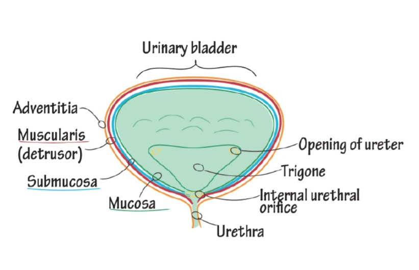
The urinary bladder is made up of three layers.
1. Mucosa:
It is the inner layer. It consists of transitional epithelium tissue. It forms folds, which are called rugae. This fold and the ability of the transitional epithelium to expand causes the bladder to distend.
2. Muscular or muscle layer:
This is the middle layer. It contains three muscle fibers.
The inner layer contains longitudinal, the middle layer contains circular, and the outer layer contains longitudinal muscle fibers.
These muscles are called detrusor muscles. In which the bladder wall is made up of three layers of smooth muscles. They are called detrusor muscles.
3. Adventitia or fibrous layer:
It is the outer layer. Which is made up of loose connective tissue. Which contains blood vessels, lymphatic vessels and nerves.
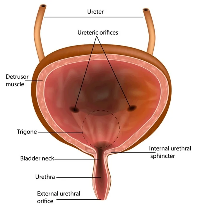
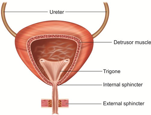
Trigone (Trigon):
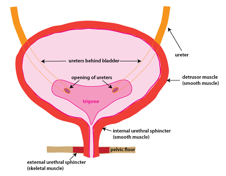
The trigon or triangle has three orifices or openings in the bladder wall. Which form a triangle or trigon. The upper two orifices are due to the openings of the two ureters in the posterior bladder wall and the lower orifice is due to the opening of the urethra.
Sphincter:
1. External Sphincter :-
2. Internal Sphincter :-
Internal urethral sphincter is not under our control (In Voluntary Control), while External sphincter is under our control (Voluntary Control) .
The internal urethral sphincter is not under our control (In Voluntary Control), while the external sphincter is under our control (Voluntary Control) .
Functions:
- The bladder acts as a collection point for urine that has not been excreted from the body.
- It helps in expelling urine from the body with the help of the urethra.
- The detrusor muscles and transitional epithelium are responsible for storing urine in the bladder.
Blood supply:
- Blood supply is provided by the internal iliac artery and drains by the internal iliac vein.
Urethra:
- The urethra is a thin-walled tube that carries urine from the bladder to the outside of the body by peristaltic movement. Urine is expelled from the urinary bladder through this tube.
- The urethra extends from the neck of the bladder to the external urethral orifice.
Female Urethra:
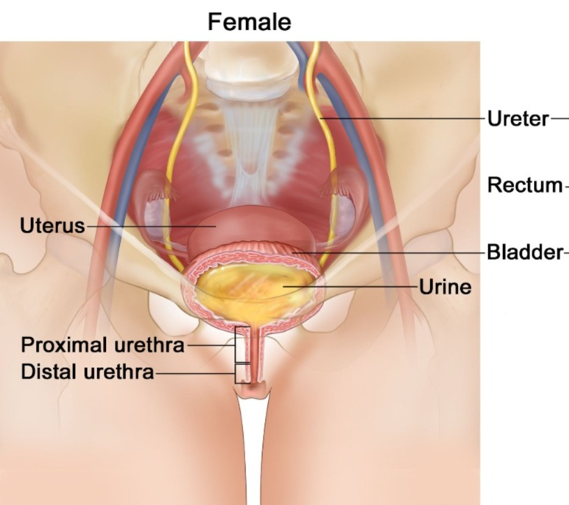
- It is 3 to 4 centimeters long. Its external orifice is located anteriorly above the vaginal opening and posterior to the symphysis pubis.
- The urethral and vaginal openings are separate.
Male Urethra:
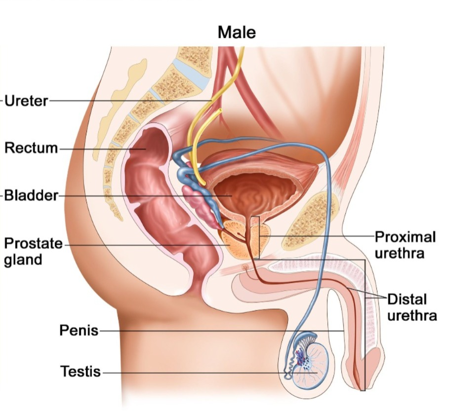
- It is 18 to 20 cm long. It has three parts. The prostatic part, the membranous part, and the spongy or penile part.
- It opens at the tip of the penis. The male urethra forms part of both the urinary system and the reproductive system.
Structure:
1.Mucosa:
The mucosa is the inner layer. Which is layered like the urinary bladder, but its end part is made of stratified squamous epithelium tissue.
2.Sub Mucosa :
It is a spongy layer. It contains blood vessels and nerves.
3. Muscularies Or Muscles Layer:
It is also similar to the layer of the urinary bladder.
Sphincter:
1.Internal Urethral Sphincter (Internal Urethral Sphincter) :
- It is located in the neck of the bladder. It contains elastic tissue and smooth muscle fibers. Which is under the control of the autonomic nervous system (In Voluntary).
- The urethral sphincter remains closed due to continuous and slow contraction.
- The internal sphincter is involuntary.
2.External Sphincter:
- It is surrounded by skeletal muscles, which are supplied by the pudendal nerve. It is voluntary control.
Micturation:
Micturation is the process by which the bladder is emptied when it is filled with urine. The process of emptying, i.e. the process of passing urine, is called micturition.
Process of Micturation:
Accumulation:
The bladder continuously collects urine until it is filled with 200 ml of urine.
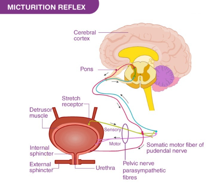
Activation:
The bladder is filled with urine. The filling causes stretching of the bladder wall, which stimulates the stretch receptors in the bladder.
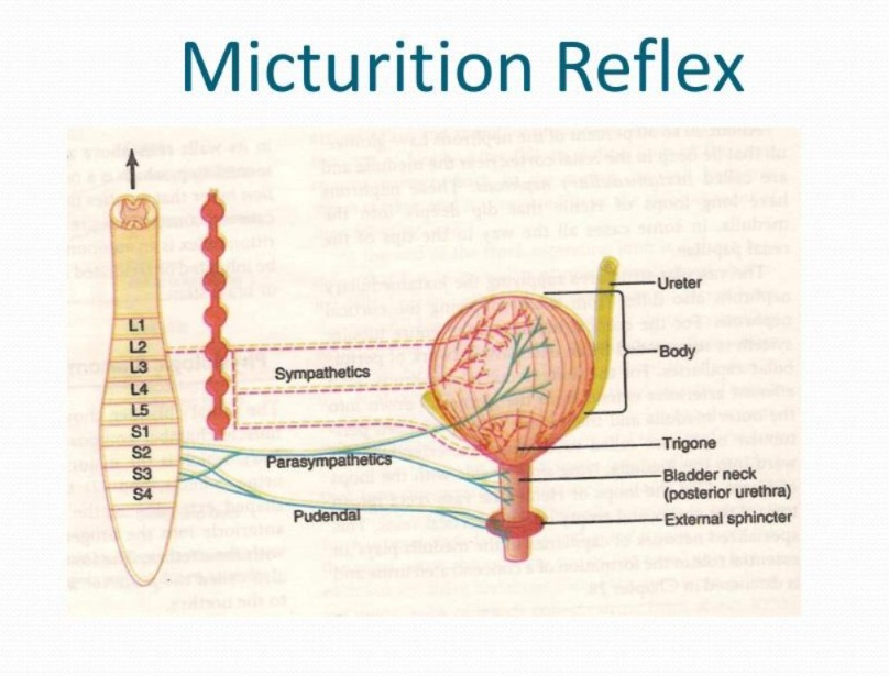
Transmission:
Impulses are transmitted from the bladder through nerves that travel from the sacral part of the spinal cord to the spinal centers (S2-3) and then back to the bladder in response to the stimulus, causing contraction of the detrusor muscles.
Passage:
Due to the strong contractions in the bladder, the urine passes the internal urethral sphincter with force. Here, the internal urethral sphincter relaxes involuntarily and the urine reaches the upper part of the urethra.
External Sphincter:
Due to the presence of skeletal muscles in the external sphincter and voluntary control, we can choose to keep the sphincter closed to hold urine or relax the sphincter to expel urine from the body, which is called micturation.