—ENGLISH-ANATOMY Unit : 5 lymphatic system
Lymphatic System:
Introduction :
- Lymphatic System contains a liquid called Lymph which is in lymph vessels Lymphatic tissue is a part of the reticular connective tissue that contains a large number of lymphocytes.
- Lymph flows in lymph vessels and enters the lymph nodes where it mixes with the blood.
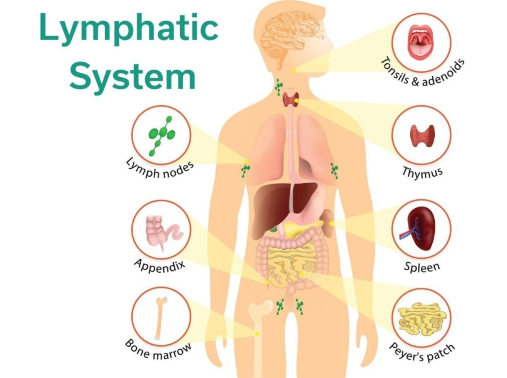
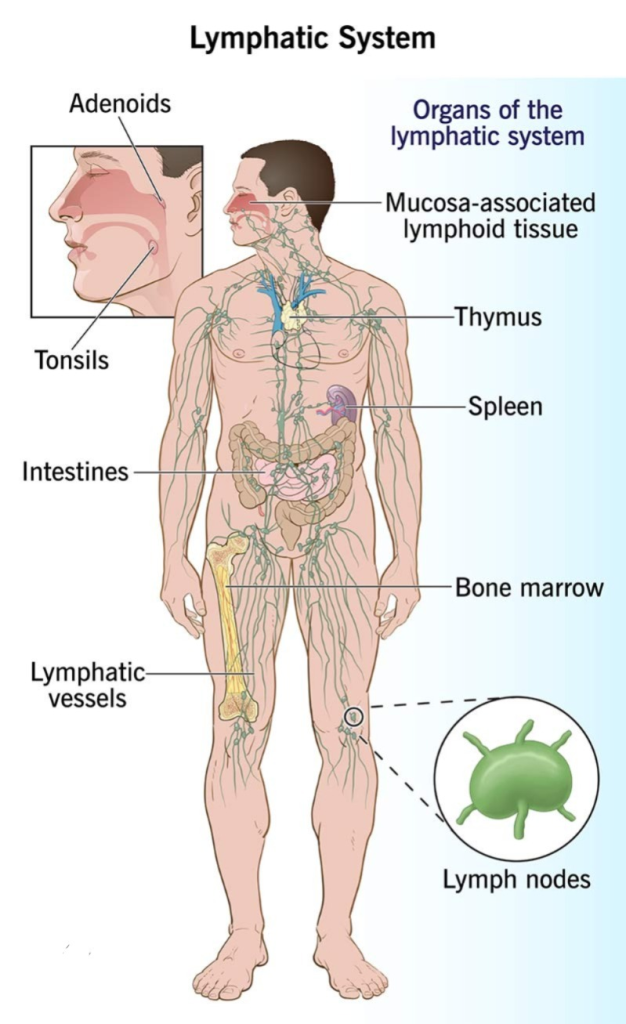
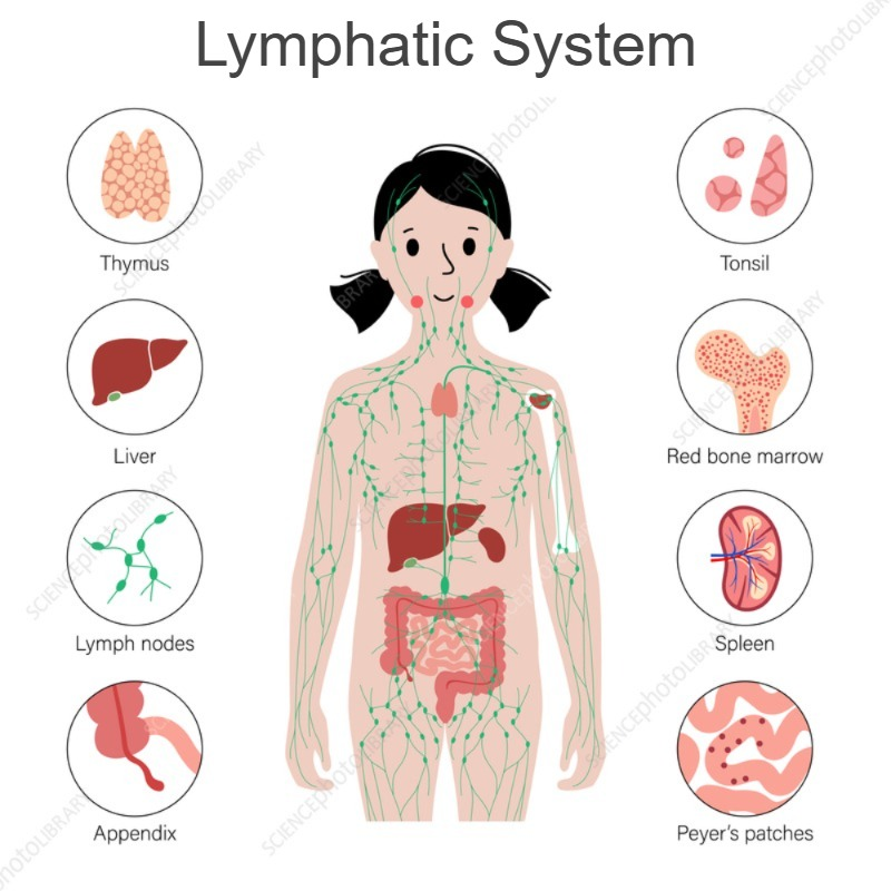
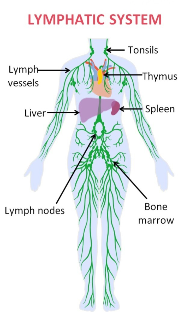
- The Lymphatic System includes the following
lymph, lymph vessels, lymph nodes, lymph organs (spleen, thyms gland), lymphoid tissue (tonsils), Bone marrow.
Functions of lymphatic system:
Drains interstitial fluid. Which absorbs tissue fluid and mixes it with the blood. Dietary lipids and fat soluble vitamins such as A,D,E,K are absorbed from the G.I. tract and mixed with the blood. Provides protection against invasion of microorganisms into the body. Some lymphocytes prevent the intrusion of microorganisms into the body and maintain antibodies in the plasma, thus playing an important role in maintaining immunity.
Work as a mediator between the body’s cells, tissues and blood.
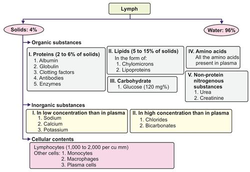
Lymph:
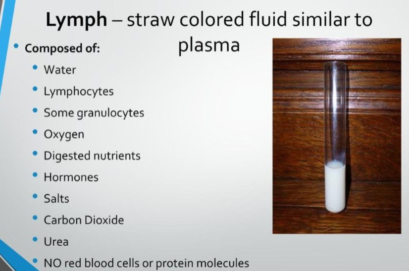
Lymph is a clear watery fluid that flows through lymph vessels. Its composition is similar to blood plasma. It has isotonic properties. With the contraction of the muscles surrounding the lymph vessels and with the help of valves in the lymph vessels, the lymph moves into the forward circulation. The lactic acid in the small intestine absorbs fat, giving it a white milky appearance.
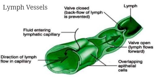
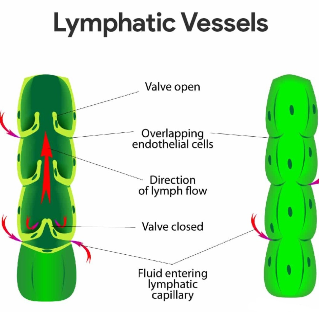
Lymph vessels (Lymph Vessels):
- The thickness of the lymph vessels resembles the structure of the vein and the structure of their walls also has layers similar to those found in the vein wall.
- Outer layer – fibrous tissue
- Middle Layer – Smooth Muscles
- Inner Layer – Endothelium
- These above lymph vessels have valves. It is useful for carrying lymph in one direction. Small lymph vessels join to form larger lymph vessels and finally they form the right lymphatic duct and thoracic lymphatic duct.
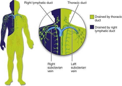
1. Thoracic Duct/Left Lymphatic Duct:
It is 38-45 cm long. It collects all the lymph from the left side of the body and then drains it into the left subclavian vein.
2. Right Lymphatic Duct:
It is 1.25 cm long. It collects lymph from the upper right side of the body. Which drains lymph into the right subclavian vein.
Lymph Capillaries :
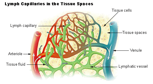
The small branches of the lymph vessels are called lymph capillaries. The diameter of which is larger than the diameter of the blood capillaries. There is a network of lymph capillaries throughout the body. Except for the central nervous system and the upper layer of the skin, the structure of the lymph capillaries resembles that of the blood capillaries. which is closely attached to the cell.
Lymph Nodes:
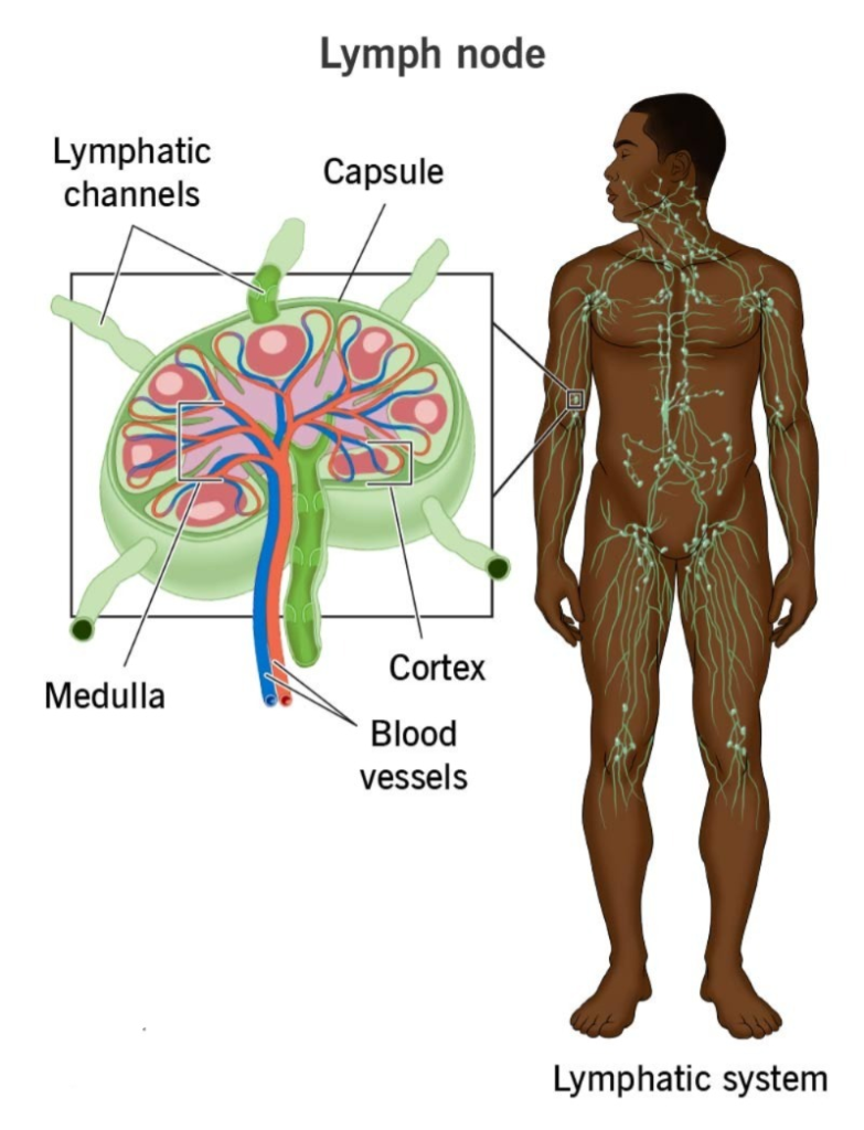
Lymph nodes are small oval or non-septate structures located along the path of lymph vessels. They work to filter lymph and are where lymphocytes are produced. Lymph nodes are mainly located in the joints and in the axilla, THORAX, ABDOMAN and groin.
Structure of lymph nodes:
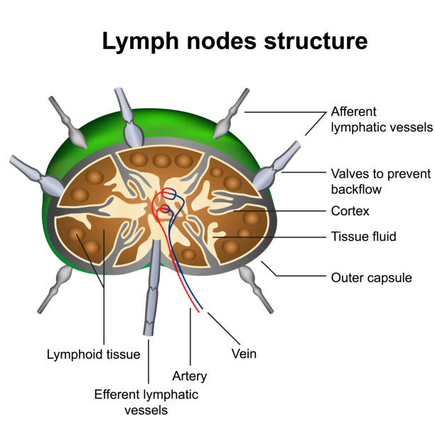
Each lymph node has an outer covering made of dense connective tissue called a capsule and an inwardly extending part called a trabeculae. Which divides the lymph node into some parts. The part of the lymph node is divided into two parts. The part towards the outer surface is called the cortex and the inner part is called the medulla. Lymph flows in a single direction in the lymph node, which is called the afferent vessels and the vessels leaving the lymph node are called the efferent vessels. The depressed part of the entering and leaving vessels is called the hilum. Blood Vessels also enter and exit from here.
Functions of lymph nodes:
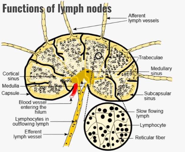
Filtering and phagocytosis:
Lymphnodes filter lymph, with lymph entering from one side and being filtered out from the other side. The macrophages present in this destroy the foreign substances present in it through the action of phagocytosis and mix with the lymph blood.
Proliferation of lymphocytes:
Plasma cells and T cells can increase in number in the lymph nodes and can also circulate from the lymph nodes to other parts of the body so that the immune response is maintained.
Hematopoiesis:
Some lymphocytes and monocytes from the bone marrow reach the lymph nodes and undergo final maturation there.
SPLEEN:
It is oval in shape. The largest lymphatic tissue in the body is the SPLEEN.
Length : 12 cm (Length : 12 cm)
Width : 7 cm (Width : 7 cm)
Thickness : 22.5 cm (Thickness : 22.5 cm)
It is located in the left hypochondriac region between the stomach and the diaphragm, lateral to the liver. Its weight is approximately 200 gm.
Organs Associated with the Spleen:
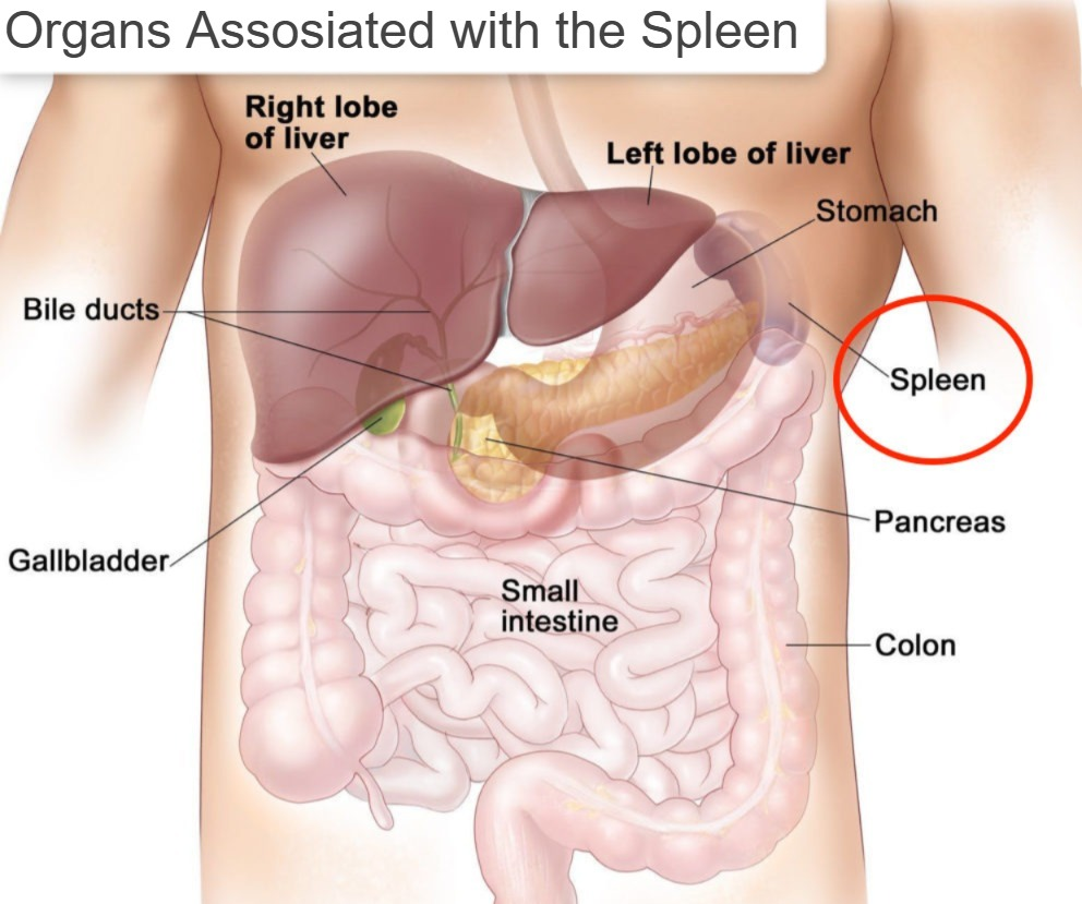
Diaphragm, Intestines, Stomach, Pancreas, Left Kidney, etc… Organs are located around the Spleen.
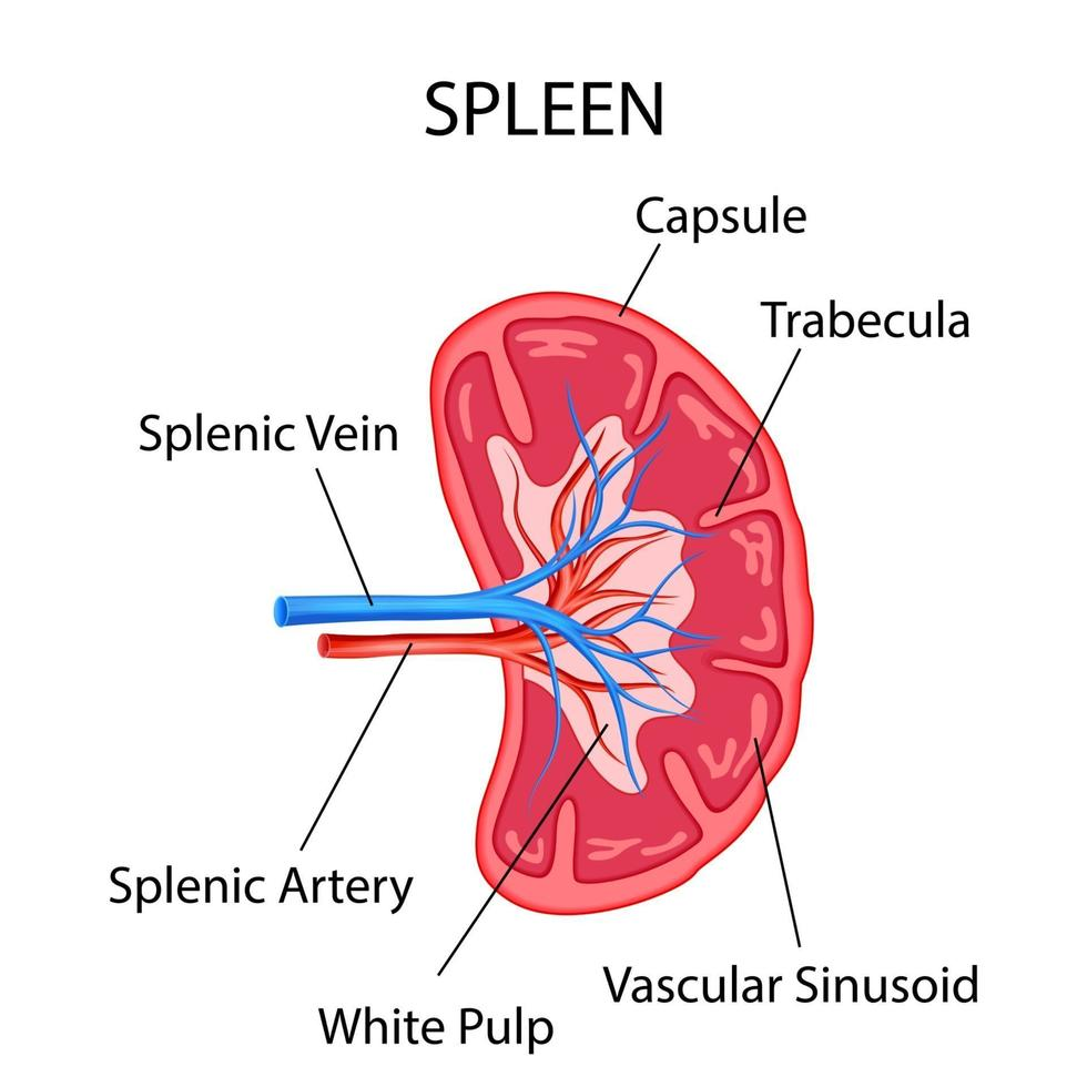
Structure :
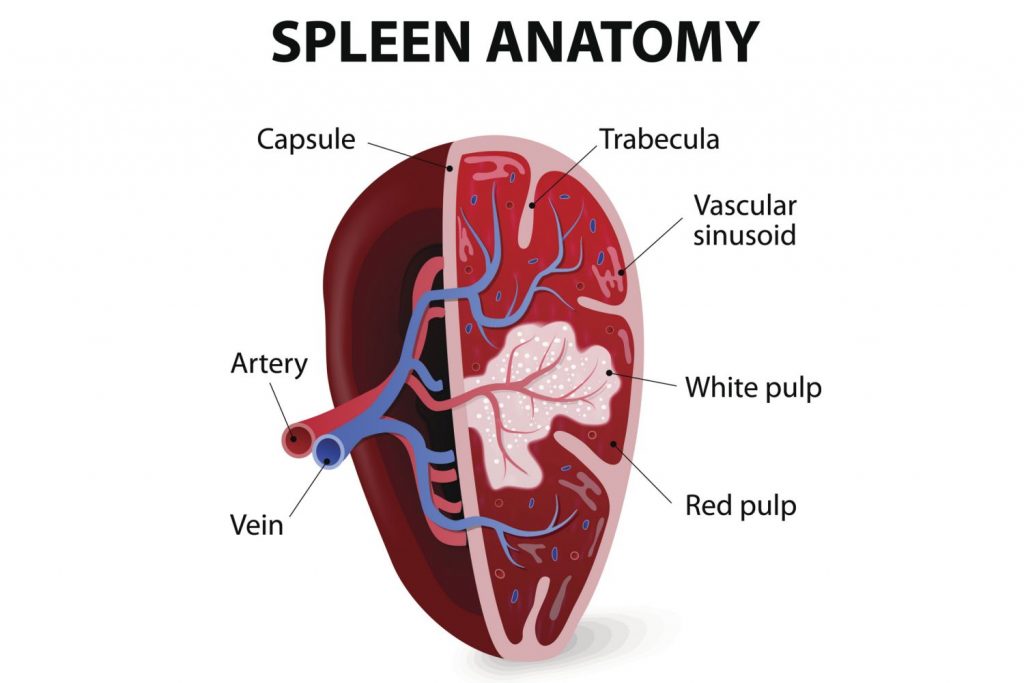
The spleenhas a network of connective tissue on its inner side called the splenic pulp. In which lymphoid tissue and some blood cells are located. A capsule made of smooth muscle fibers and elastic tissue is located around it. The tissue inside the spleen is divided into two parts. White pulp and Red Pulp. White pulp is for immunity where the number of B-cells increases and helps in making antibody plasma. Which is useful against infection. Red pulp is vascular. Phagocytosis of bacteria occurs there and RBC and platelets also work to phagocytosis of damaged ones.
Functions of Spleen:
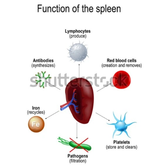
Phagocytosis: Helps destroy bacteria and removes damaged RBCs from circulation by phagocytosis.
Storage of blood: The spleen stores approximately 350 ml of blood. Which immediately returns this blood to the circulation upon receiving sympathetic stimulation in situations like hemorrhage.
Immune response: T and B lymphocytes that are activated by antigens work to maintain immunity.
Erythropoiesis: Spleen and liver are important for the production of red blood cells during fetal life. Even in adults, the spleen produces cells when needed.
Thymus gland (Thymus gland) :
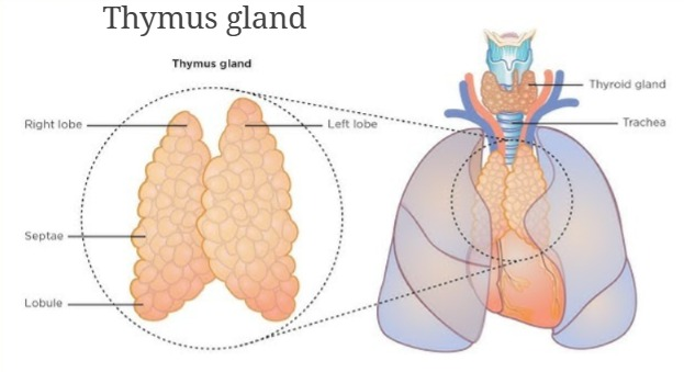
The thymus gland is a two-lobed organ of the lymphatic system. It is located in the mediastinum space. It is large in size in infants and reaches a maximum weight of 40 gr by the age of 10-12 years. Its weight at birth is 10 to 15 gr. As much as it is.
Organ Associated with the Thymus gland (Organ Associated with the Thymus gland):
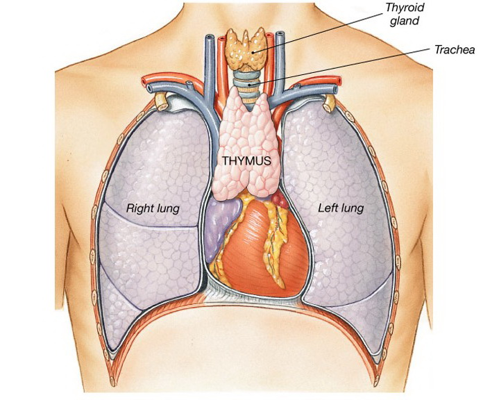
Sternum, aortic arch, trachea, lungs, heart etc organs are located around it.
Structure :
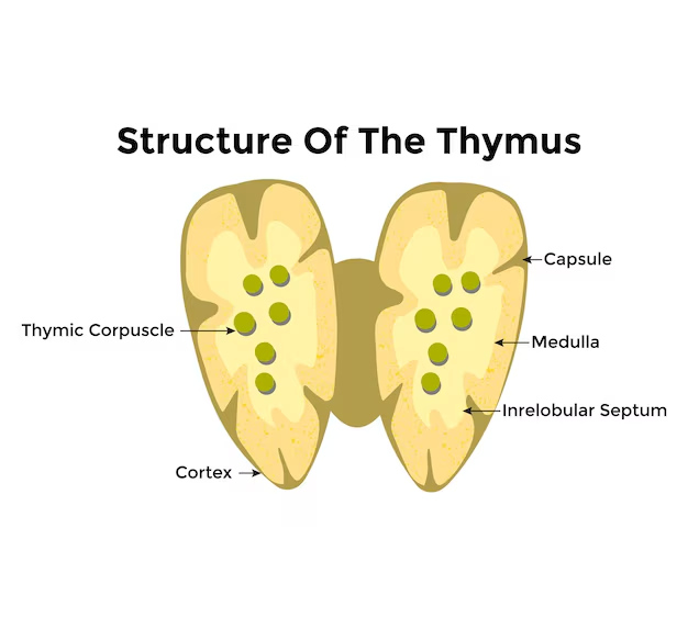
It has two lobes. It has a capsule. Each lobe is divided into lobules, the outer part of which is called the cortex and the inner part is called the medulla.
Functions:
B-lymphocytes produced in the bone marrow are activated as T-lymphocytes after entering the thymus gland.
Thymosin stimulates and matures the thymus gland and other lymphoid tissues. Which is secreted by the epithelial cells of the hormone gland.