—ENGLISH-ANATOMY UNIT 4. CVS(CARDIOVASCULAR SYSTEM) BLOOD VESSELS
CARDIOVASCULAR SYSTEM (Cardio Vascular System):
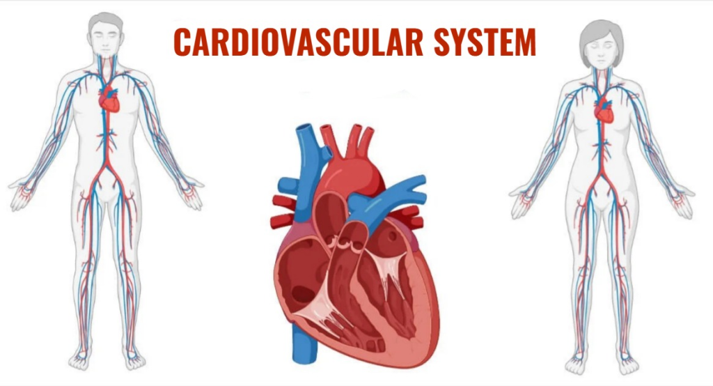
INTRODUCTION (Introduction) :
Cardiovascular System System) is a part of the Circulatory System, which includes the heart, blood, and the blood vessels that transport the blood. The heart is an important organ of the cardiovascular system that performs a continuous pumping action.
The circulatory system is a transport system of the body that performs important functions in the body by delivering nutrition and oxygen to every cell and tissue of the body through blood. To meet all these needs of the body, blood is transported throughout the body and circulates in blood vessels. The heart continuously pumps blood to circulate through the blood vessels.
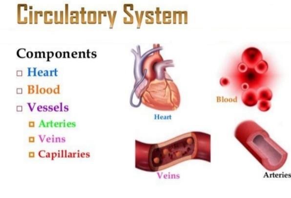
Blood vessels
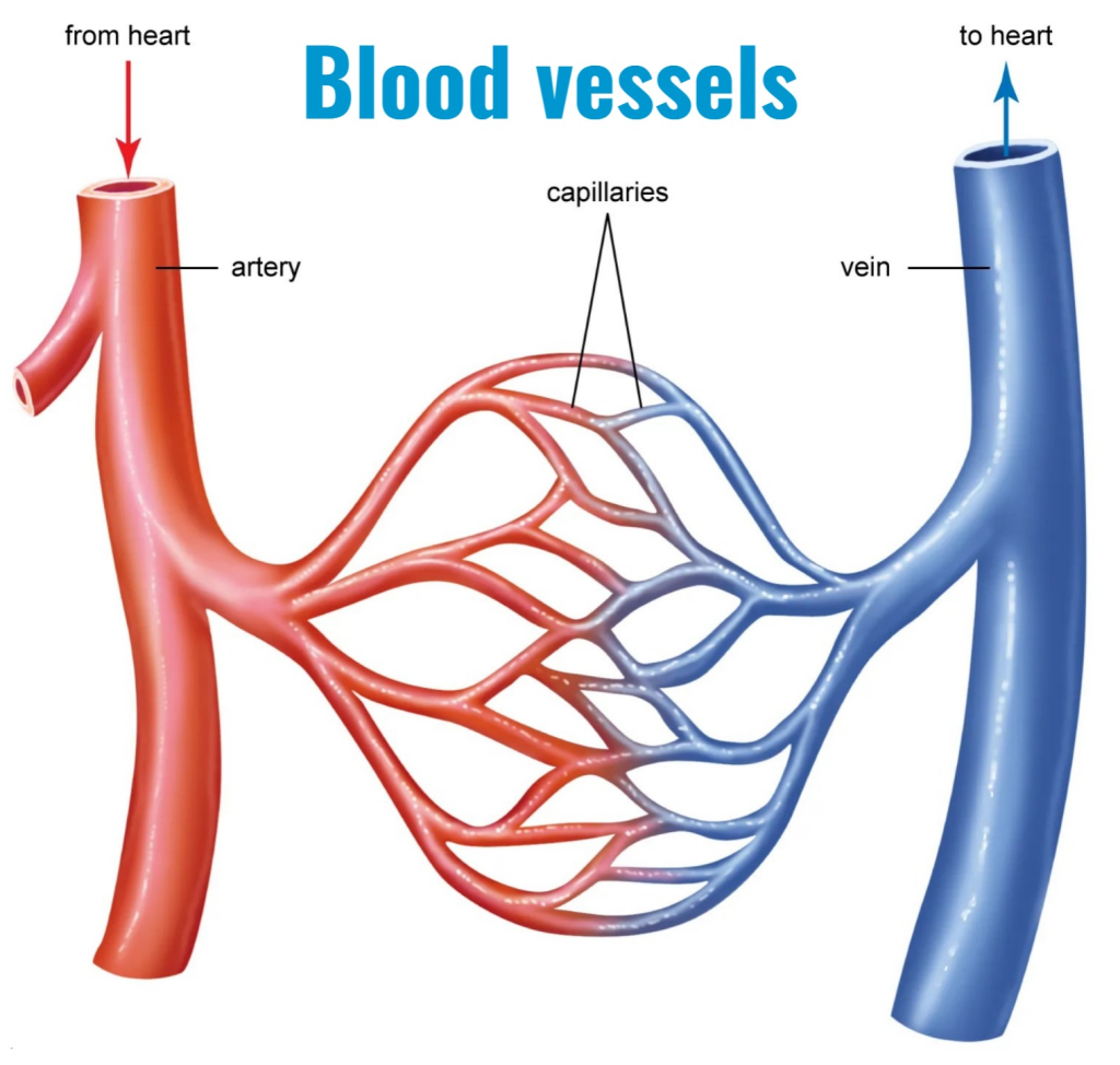
Blood circulates throughout the body through blood vessels. The heart performs a pumping action and transports blood throughout the body. Blood vessels in the body vary in size, structure and function and are known as arteries, arterioles, capillaries, venules, veins respectively.
Artery:
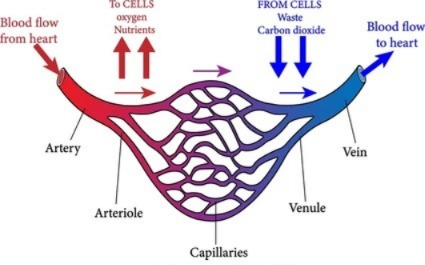
An artery is a branch of blood vessels that carries blood from the heart to the body. It is said that blood travels from the heart to the body. Mainly arteries carry oxygenated blood which reaches every cell and tissue of the body. Along with oxygen, nutrient material also reaches every cell tissue in this blood.
There are mainly two types of arteries in the body:
1. Pulmonary Artery:
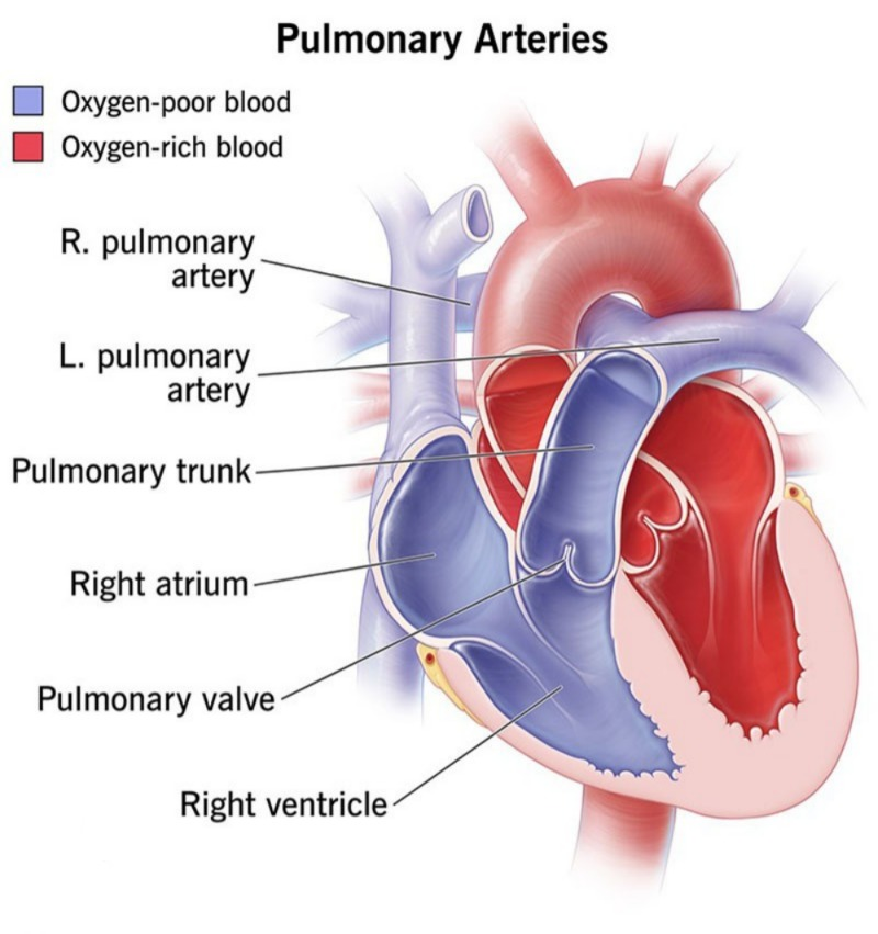
This artery carries deoxygenated blood from the heart to the lungs to be oxygenated. Deoxygenated blood flows in it. There is an exception in all the arteries of the body.
2.Systemic Artery:
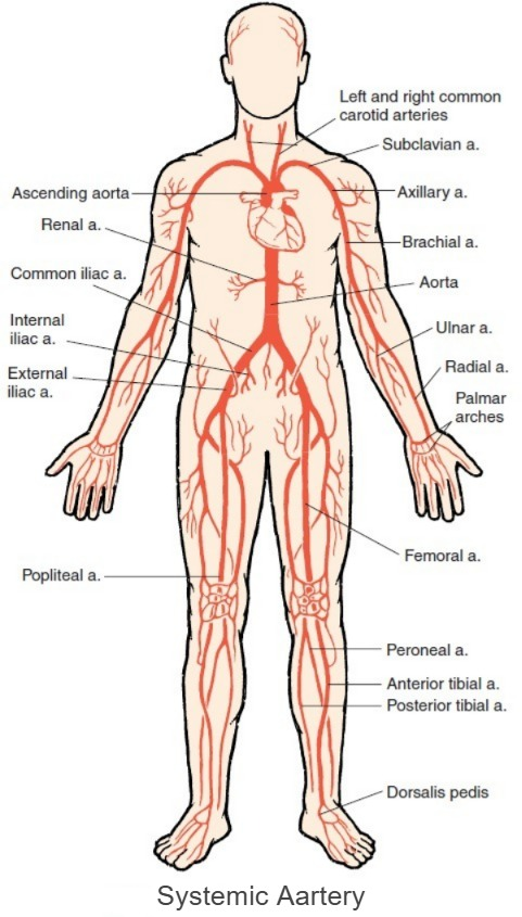
This includes all the arteries in the body. Which begin with the left ventricle of the heart. Oxygenated blood from the left ventricle circulates throughout the body through an artery called the aorta . The aorta is the largest branch of all the arteries and carries oxygenated blood through all these systemic arteries.
Arterioles:
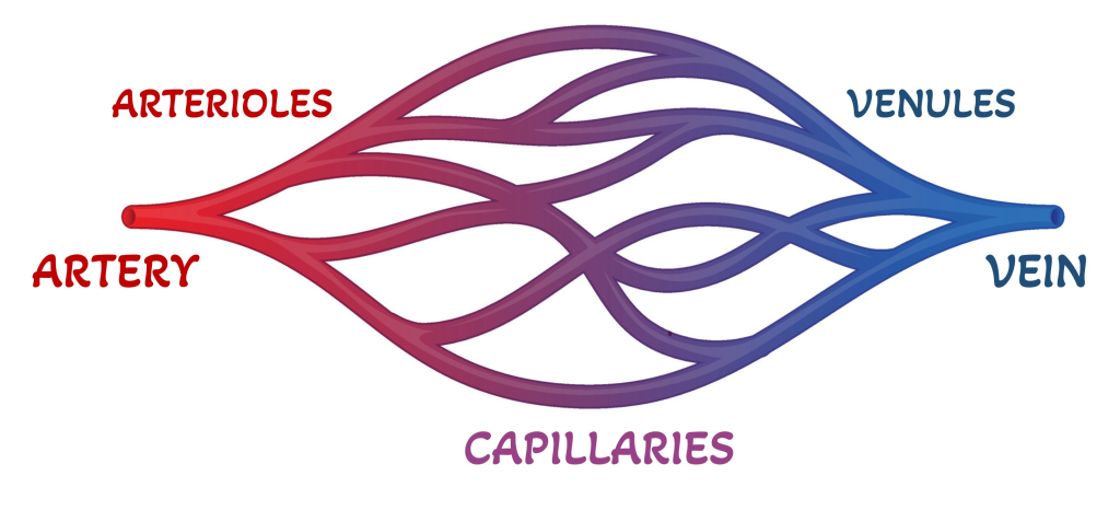
Arterioles are small branches of arteries. They are mainly formed by division from arteries. Their diameter is smaller than that of arteries and oxygenated blood mainly flows in them. Which works to deliver oxygenated blood to every cell and tissue of the body.
Capillaries:
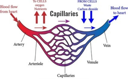
Capillaries are tiny microscopic structures of blood vessels. They are located at the ends of arterioles and venules. These capillaries are connected to cells to exchange gases and other nutritional materials. These capillaries do not have all the layers found inside blood vessels, but only a single layer of epithelium, so that the exchange of substances between blood and cells can easily take place.
Venules:
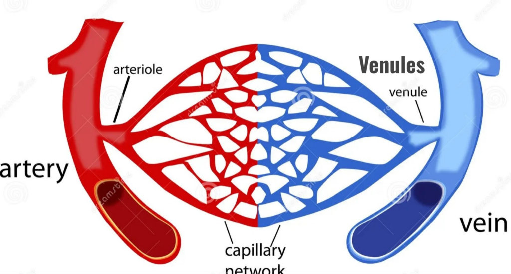
Capillaries that carry deoxygenated blood from cells and tissues join together to form smaller capillaries and venules. These venules join into smaller branches and further form larger veins which help in transporting deoxygenated blood towards the heart.
Vein:
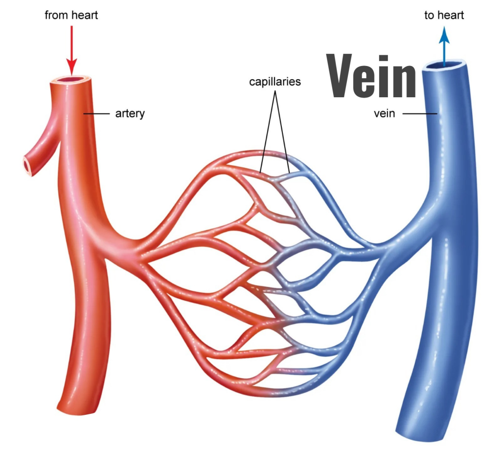
Vessels that carry deoxygenated blood from different parts of the body to the heart are called veins. Small venules join to form larger veins and carry deoxygenated blood to the right atrium of the heart. Among all the veins in the body, the superior and inferior vena cava are the main veins. Which carry deoxygenated blood from all parts of the body to the heart.
The pulmonary veins, which carry oxygenated blood from the lungs, carry oxygenated blood to the left atrium of the heart. Of all the veins in the body, this vein carries exceptionally oxygenated blood.
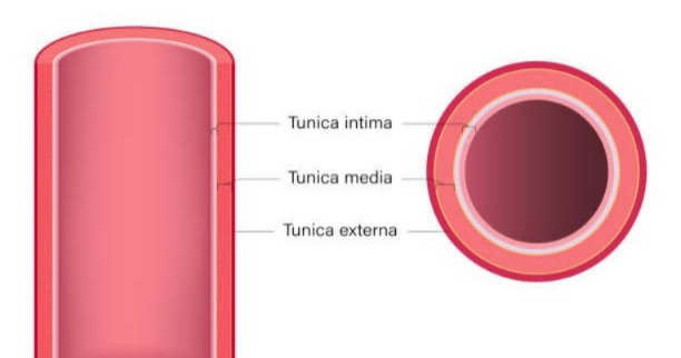
Structure of the Blood Vessel:
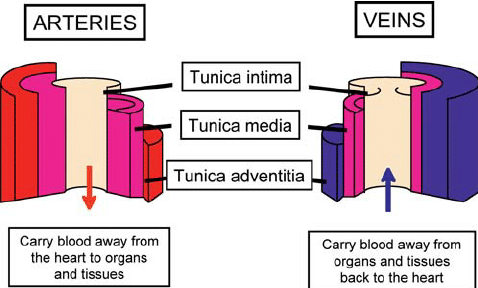
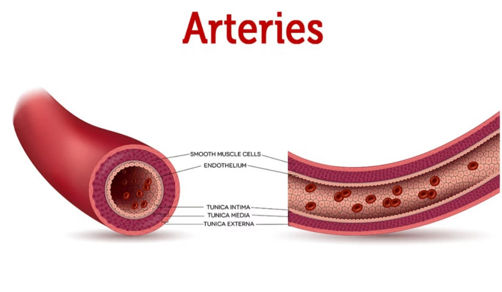
The walls of arteries and veins are made up of three types of tissue layers. The outer wall is called the Tunica Adventitia, the middle wall is called the Tunica Media, and the inner wall is called the Tunica Intima.
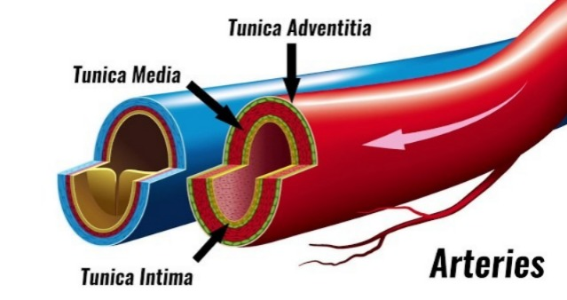
Tunica Adventitia:
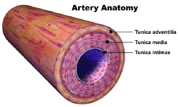
This is the outermost layer of blood vessels. It is made up of fibrous connective tissue. It works to protect the blood vessels from the outside.
Tunica Media:
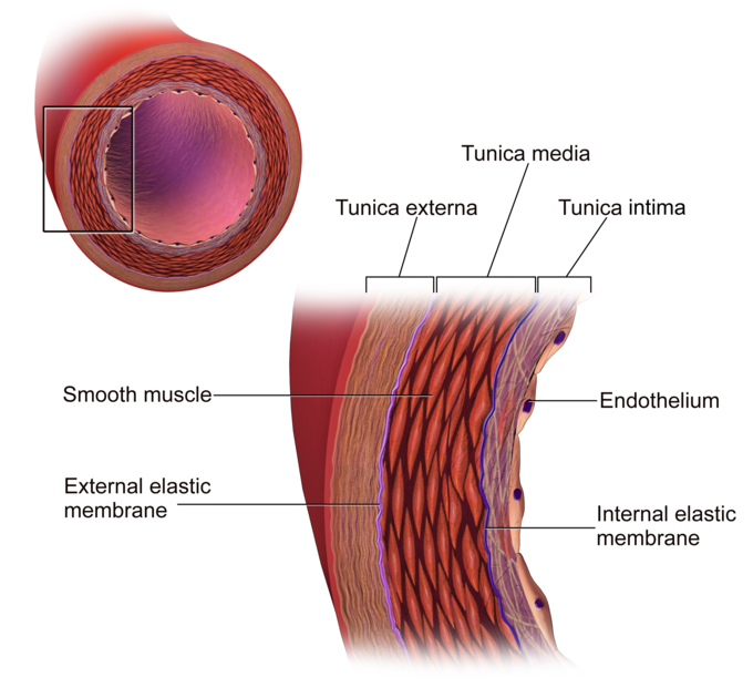
This is the middle layer of blood vessels. It is made up of smooth muscles and elastic connective tissue. Since this layer is present in greater quantities in large arteries, its dilation and constriction are easily observed. This layer is thicker in arteries compared to veins. As the diameter of the blood vessels decreases, this layer becomes thinner. This layer is not found in the wall of capillaries.
Tunica Intima:
This is the innermost layer of blood vessels. It is made up of squamous epithelium tissue and is also known as the endothelium. This layer is smooth and shiny and is in contact with blood.
Anastomoses and end arteries:
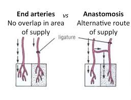
- The connection of two or more arteries is called an anastomosis. Due to this anastomosis, blood supply is provided to any region through two or more arteries.
- Alternative blood flow to any body part is seen due to anastomosis. So that the total blood circulation there does not stop due to the closure of any one branch. This additional circulation is also called collateral circulation.
- An artery that does not have the property of anastomosis and supplies only one artery to any region is known as an end artery.
- Due to obstruction in the blood flow of this artery, there is a possibility of damage to any cell and tissue due to lack of blood supply.
Control of blood vessels diameter (Control of blood vessels diameter Diameter):
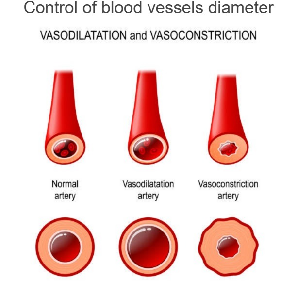
- The diameter of blood vessels is controlled by the automatic nervous system. In addition, the diameter of blood vessels can increase or decrease due to the sympathetic and parasympathetic nervous systems.
- The vasomotor center in the medulla inside the brain also controls the diameter of blood vessels.
- In addition, receptors in the artery walls also control the diameter of blood vessels.
- The process of increasing the diameter of blood vessels is called vasodilation and the process of decreasing the diameter of blood vessels is called vasoconstriction.
Blood supply to the blood vessels (Blood supply to the blood vessels)
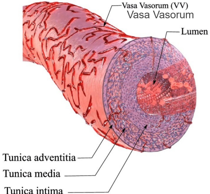
Blood is supplied to the blood vessels by a network of tiny blood capillaries called Vasa Vasorum on the outer wall of the blood vessels.
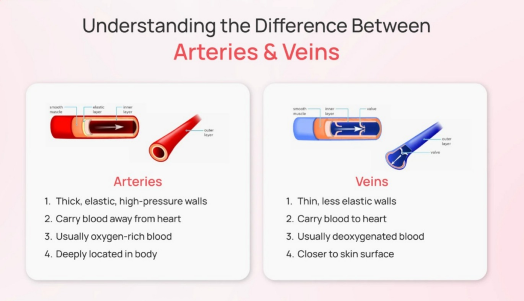
Difference between artery and vein:
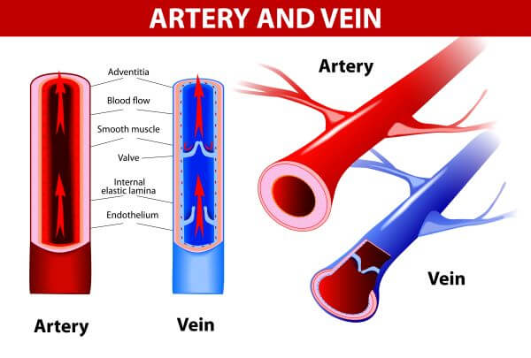
- Blood vessels that carry blood from the heart to the body are called arteries, while blood vessels that carry blood from the body to the heart are called veins.
- Oxygenated blood circulates in arteries, except for the pulmonary artery, and deoxygenated blood circulates in veins, except for the pulmonary vein.
- Arteries do not have valves while veins have valves.
- Blood in arteries appears bright red in color because it contains oxygen, while blood in veins appears dull red or less red because it contains carbon dioxide.
- Since arteries originate directly from the heart, pulsation can be heard in them while veins do not have pulses.
- Arteries are mainly located deep in the body while veins are located superficially.
- The middle layer of an artery is more elastic so that its diameter can change more while the middle layer of a vein is not as elastic as that of an artery.
- The lumen of an artery is normally narrow while the lumen of a vein is wider compared to that of an artery.
- The force of blood in arteries is greater while the force of blood in veins is less.
- When the amount of blood in the artery is less, its wall is stronger and does not shrink (collapse), while when the amount of blood in the vein is less, it shrinks