ENGLISH ANATOMY UNIT 4. CVS HEART
Cardio Vascular System
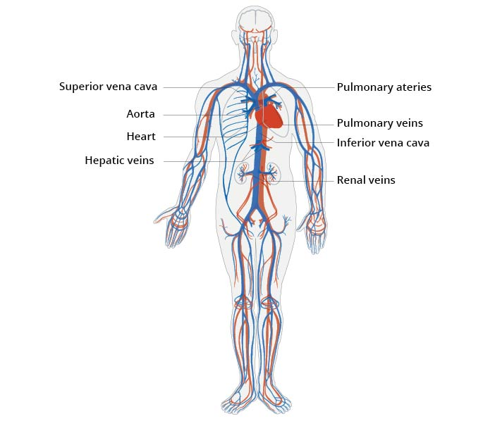
The cardiovascular system is a part of the circulatory system and includes the heart, blood, and blood vessels that transport the blood. The heart is an important organ of the cardiovascular system that performs a continuous pumping action.
Circulatory system is a transport system of the body which performs an important function in the body by delivering nutrition and oxygen to every cell and tissue of the body through the blood. To fulfill all these needs of the body, blood is transported throughout the body and circulates in the blood vessels. The heart pumps continuously to circulate the blood in the blood vessels.
- Heart…
Heart is an important organ of the circulatory system. Heart beats continuously during human life. Due to its pulsation, the blood circulates continuously in the blood vessels.
The heart is an organ made up of blood and muscles. It weighs approximately 310 grams in males and approximately 250 grams in females. The heart beats approximately one lakh times during the day.
Location of the heart..
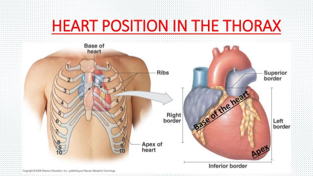
The heart lies above the diaphragm in the mediastinum space between the two lungs in the thoracic cavity.
The heart is a rough cone shape. In it, its upper broad part is known as the base and the lower angled part is known as the Apex.
The heart is the size of a man’s closed fist. It measures 12 cm in length, 9 cm in width and 6 cm in thickness.
The heart is arranged slightly to the left in the thoracic cavity between the two lungs.
The heart is found in the middle part of the thoracic cavity between the second and fifth intercostal ribs.
Organs around the heart.
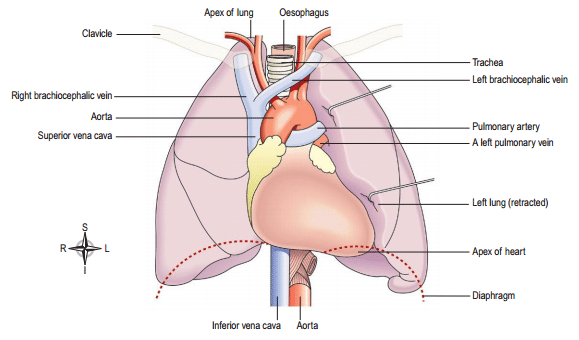
The heart is located in the thoracic cavity. It has one lung on both its left and right sides.
Underneath lies the diaphragm and central tendon.
On the upper side of the heart are the vena cava and the aorta and the pulmonary artery and pulmonary vein.
Posterior to the heart are the esophagus, trachea, bronchi and bronchioles as well as the descending aorta and thoracic vertebrae.
Anterior to the heart are the sternum bone and the ribs and intercostal muscles
Structure of the Heart..
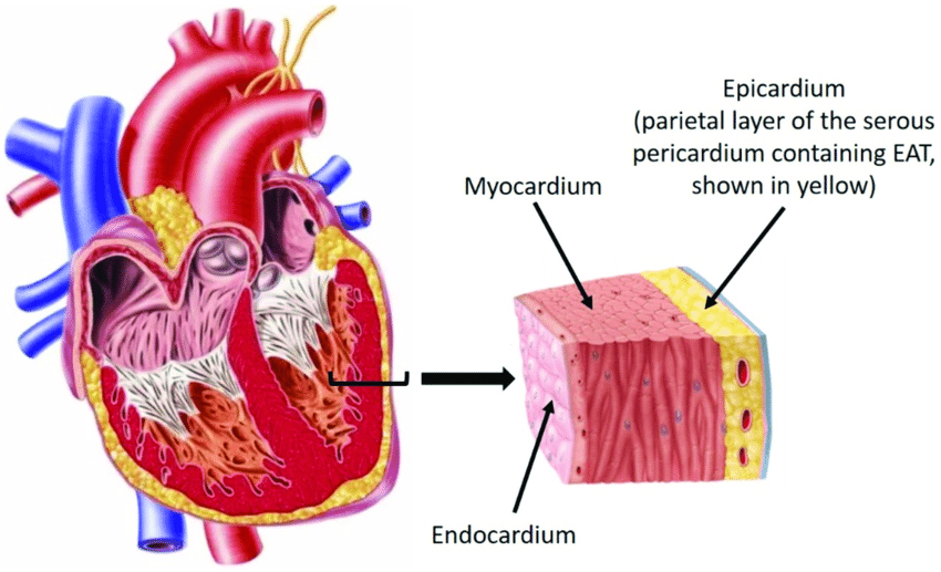
Heart is an organ made up of semi-muscles. Its wall is made up of three types of tissue layers.
The outermost layer of the heart wall is called epicardium or pericardium.
Epicardium or Pericardium.
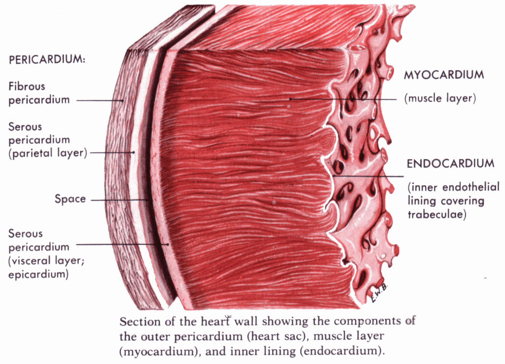
It is thin and transparent and covers the heart from the outside. It is made up of fibrous connective tissue. In which there is a layer of fibrous tissue on the outermost side and a serous membrane on the inner side of the fibrous tissue which is found in a double layer. The outer layer of the serous membrane is known as the parietal layer and the inner layer as the visceral pericardium layer.
The space between the parietal and visceral pericardial layers is called the pericardial space. This space contains fluid called serous fluid or pericardial fluid. Which prevents friction between the two layers.
This layer of outer pericardium protects the heart from the outside and this layer is also seen around the vessels coming out of the heart.
Myocardium..
Myocardium is the middle layer of the heart. It lies below the pericardium. It is made up of a special type of cardiac muscle tissue. The pumping action of the heart is seen due to the contraction of these muscles.
This myocardium layer is thin at the base and thick at the apex. Also, the layer of the wall of the left ventricle is thicker than the wall of the right ventricle.
The contraction of these muscles has an involuntary action that results in the pumping action of the heart and is controlled by the autonomic nervous system and the conducting system in the heart.
endocardium..
It is the innermost layer of the heart wall. It is in contact with layer blood. This layer is made up of epithelium tissue and connective tissue. This layer is smooth and shiny which is important for easy blood flow inside the heart. This layer also covers the valves inside the heart and this layer is continuous in the inner wall of the blood vessels leaving the heart.
Chambers of the Heart.
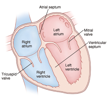
Heart is mainly divided into two parts. Right side and left side.
Between this right and left side lies the septum of the heart. It is again divided into four chambers above the valve and below the valve by the valves located between the right side and the left side of the heart.
Both chambers above the valve are called atrium or auricle. Both chambers below the valve are called ventricles. Thus the heart is divided into a total of four chambers.
The right side of the heart contains deoxygenated blood and the left side of the heart contains oxygenated blood. After birth there is no connection between the right and left sides. Before birth there is an opening between the two atria called the foramen ovale but it closes after birth.
Valve of the heart..

The flaps of tissue inside the heart are called valves. There are two types of valves in the heart.
Atrioventricular valve.
Semilunar valve.
Atrioventricular valve…
These valves are located between the atrium and the ventricle in the heart.The valve between the right atrium and ventricle is called the tricuspid valve. This valve is made up of three tissue flaps.
The valve between the atrium and ventricle on the left side of the heart is called the bicuspid valve or the mitral valve.This valve is made up of two tissue flaps.
Both bicuspid and tricuspid valves have their suspensory ends attached to the inner wall of the ventricles of the heart by chordae tendineae and papillary muscles. This gives strength to the valve and prevents it from opening in the opposite direction.
Semilunar valve…
Since this valve is C-shaped or crescent-shaped, it is called a semilunar valve. These valves are present in the aorta and pulmonary artery. The valve in the aorta is called the aortic valve and the valve in the pulmonary artery is called the pulmonary valve. Which are located at the opening of its vessels. This valve also opens in one direction.
Both the above atrioventricular valves open when the pressure of the atrium increases i.e. during the contraction of the atrium and the blood comes from the atrium to the ventricle and during the contraction of the ventricle the atrioventricular valve closes and the semilunar valve opens and due to this the blood goes into the aorta and pulmonary artery.
Thus, due to the opening and closing of this valve, the circulation of blood can occur in the heart. This valve opens in one direction so that blood cannot flow back in the opposite direction.
Great Vessels Associated with Heart..or
Openings of the Heart..
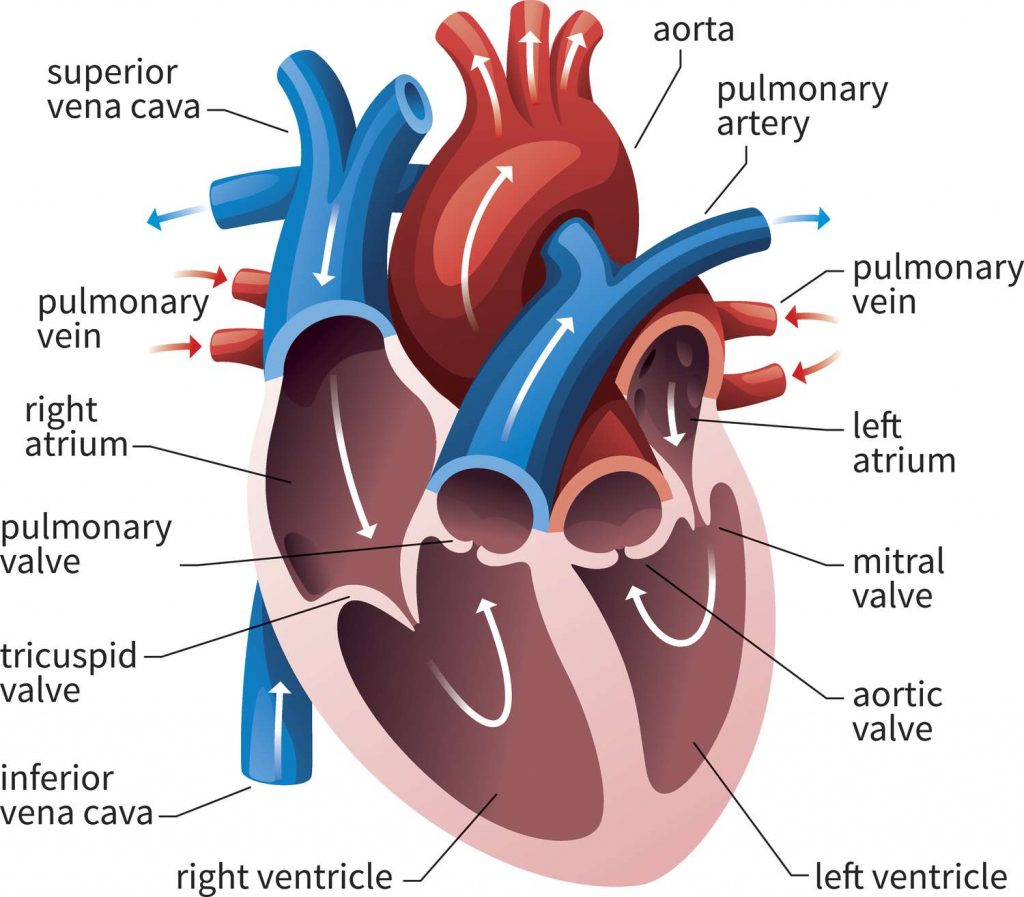
Large blood vessels are mainly connected to the heart. Through which the blood comes from the body to the heart and the heart is pumped and the blood goes out to the body. The vessels and their openings are as follows.
Superior vanacava..
This brings deoxygenated blood from the upper part of the thoracic cavity, head and neck, into the right atrium of the heart. It is in the number of one.
Inferior vanacava..
These blood vessels bring deoxygenated blood from the inferior part of the body and the lower part of the thoracic cavity and open into the right atrium of the heart. It is in the number of one.
Pulmonary artery..
It carries deoxygenated blood from the right ventricle of the heart out of the heart to the lungs. Its number is one.
Pulmonary vein..
Two pulmonary veins from both lungs bring the oxygenated blood to the left atrium of the heart. Its number is four.
Aorta..
The left ventricle of the heart takes oxygenated blood and circulates it throughout the body. Its number is one.
Thus a total of eight blood vessels and their openings are directly connected to the heart. Which is connected with the blood circulation in the heart.
Blood Circulation in the Heart…
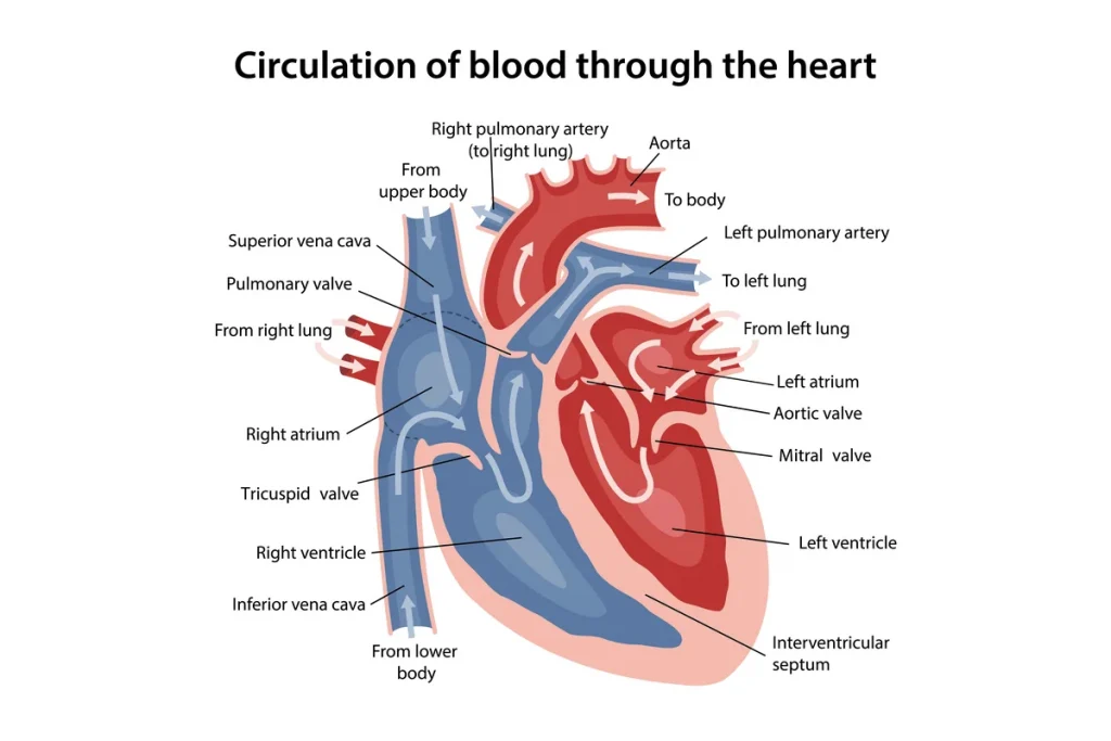
Deoxygenated blood enters the right atrium of the heart through the superior and inferior vena cava from different atria of the heart body. At the same time, the oxygenated blood from the lungs comes to the left atrium of the heart through the four pulmonary veins. Thus both the atriums of the heart are filled with blood at the same time.
Then, due to the contraction of both the atria of the heart, both the atrioventricular valves i.e. the bicuspid valve and the tricuspid valve open, and both the atria are emptied and both the ventricles are filled with blood.
The right ventricle contains deoxygenated blood while the left ventricle contains oxygenated blood. Then due to the contraction of both the ventricles, the blood of the right ventricle is pumped through the pulmonary artery by opening the pulmonary valve and the blood of the left ventricle by opening the aortic valve circulates oxygenated blood throughout the body. Thus both the ventricles become empty.
Then after both the atriums in the heart are filled with blood again, this cycle continues and the circulation of blood takes place.
Blood supply of the heart.
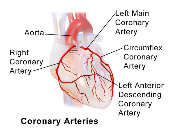
Oxygenated blood supply to the heart to function As soon as the aorta exits the left ventricle of the heart, it branches off into the right and left coronary arteries, which supply oxygenated blood to the heart. These coronary arteries are branches of the ascending aorta.
About five percent of the blood that the heart circulates out of the aorta during a contraction is supplied to the heart through the coronary arteries.
These coronary arteries divide into many branches and supply oxygenated blood to all parts of the heart.
The deoxygenated blood in the heart collects the small coronary veins and drains the deoxygenated blood into the coronary sinuses, and through the coronary sinuses, all the blood drains directly into the right atrium of the heart.
Functions of the Heart…
Heart provides oxygenated blood supply to all organs and tissues of the body.
Heart is an important organ of the cardiovascular system. It functions as a vital organ without which the human body cannot survive.
The heart circulates the blood towards the lungs so that the blood can be oxygenated and purified.
Circulations like pulmonary circulation and systemic circulation are regulated by the heart.
The heart also regulates the heart rate according to the needs of the body and according to the body temperature.
The heart also regulates body temperature as it circulates blood to every part of the body.
As the heart pumps blood to the body’s excretory organs, the blood can be filtered and waste products removed from the blood.
- Conducting System of the Heart…
The heart has a system in its own inner myocardium layer for the transmission of impulses that does not directly require an external nerve supply to function.
In the inner wall of the heart, the myocardium layer contains a special type of neuromuscular cells that have the characteristic of generating impulses and transmitting the impulses further to the myocardium layer. Due to which heart muscle contraction and relaxation takes place. Below is the structure of this conducting system
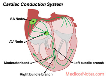
S. a. Node (sino atrial node)..
The SA node is a mass of specialized neuromuscular cells located near the opening of the wall of the right atrium in the myocardium layer. It is also known as a pacemaker as it is the unit that generates the impulses for the contraction of the heart.
Impulses generated by the SA node are transmitted further to the myocardium and contraction of the muscles of the myocardium of the heart occurs.
The S node regulates the heart rate.
a. V. node (atrioventricular node)..
The AV node is a specialized type of neuromuscular cell mass located near the septum of the heart near the atrioventricular valve.
The impulses to contract the myocardium are generated by the SA node, which is received by the AV node and transmitted further to the septum of the ventricle.
Atrioventricular bundle..or
Bundle of His…
The atrioventricular bundle is a network of fibers in the layer of myocardium muscles that starts from the AV node and descends to the septum of the ventricle.
This network of fibers in the wall of the right ventricle is known as the right bundle branch. This network of fibers in the wall of the left ventricle is known as the left bundle branch.
Thus, the network of these fibers is arranged up to the apex of the heart and the impulses from the AV node pass through these fibers and reach the apex of the heart and contraction of the heart muscle takes place.
- Cardiac Cycle of the Heart..
Heart is a continuously pumping organ. The pumping action of the heart is called its cardiac cycle. A healthy person has a cardiac cycle ie pumping action of 68 to 72 times in a minute.One cardiac cycle takes 0.8 seconds to complete. This work runs continuously through the heart of a living human being.
The contraction and relaxation of the heart muscle is done by the impulses generated from the S node of the heart. Contraction is known as systole and relaxation as diastole.
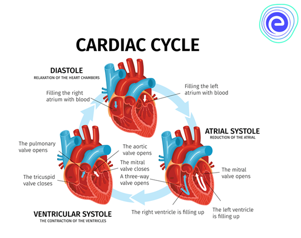
The following events occur in the cardiac cycle.
- Atrial systole..
Atrial systole means simultaneous contraction of both atria which takes 0.1 second.
In this atrial systole, when both atria are filled with blood, impulses are generated by the S node and these impulses reach the AV node. During this time it takes 0.1 second and both atria contract simultaneously both atrioventricular valves open and both atriums empty of blood and both ventricles fill with blood. This phase is called atrial systole.
- Ventricular systole..
Ventricular systole is the simultaneous contraction of both ventricles. It takes 0.3 seconds.
During ventricular systole, when both ventricles are filled with blood, impulses from the AV node reach the bundle of His and Purkinje fibers, i.e., the heart.
During this time it takes 0.3 seconds and both ventricles contract simultaneously. The blood of both the ventricles respectively, the blood of the right ventricle goes to the lung through the pulmonary artery and the blood of the left ventricle circulates throughout the body through the aorta. This phase is called ventricular systole.
- Complete cardiac diastole..
Complete cardiac diastole means simultaneous relaxation of both the atria and the ventricles i.e. the four chambers of the heart. This action takes 0.4 seconds.
During complete cardiac diastole there is no electrical activity in the heart. Myocardium muscles are relaxed. During this time both the atria and both the ventricles dilate i.e. relax. Both atriums are refilled with blood during this time. This relaxation time is 0.4 seconds. This is called complete cardiac diastole.
Thus, it takes 0.8 seconds to complete a complete cardiac cycle and blood circulation occurs due to the contraction and relaxation of the heart.
- Factors affecting heart rate..
Heart rate means the beating action of the heart In a normal healthy person, the heart beats 68 to 72 times per minute. This heartbeat is controlled primarily by the cardiac center located within the medulla in the brain.
Apart from this, the heart rate is also affected by the stimulation of the automatic nervous system and the vagus nerve.
Factors affecting heart rate are as follows.
position.
Changing a person’s position can cause a normal change in heart rate.For example, heart rate may vary in sleeping position, sitting position and standing position.
Circulating chemicals..
Circulating chemicals means different hormones, chemicals, electrolytes etc. secreted in the body. An increase or decrease in the proportion of all these can lead to an increase or decrease in heart rate.
Edge.
Age is an important factor affecting heart rate. Changes in heart rate are seen according to different ages. As the heart rate of a newborn is the highest while an adult person has a different heart rate. Change in heart rate is also seen in old age person.
Emotional status..
Heart rate is affected by emotional changes. Change in heart rate is seen in happy and sad situations.
Fear and Anxiety..
A sudden increase in heart rate is seen during fear or anxiety.
Exercise.
Due to the increase in blood circulation and oxygen demand in the body during exercise, the heart rate also increases.
Gender.
Generally, there is a slight difference in the heart rate of men and women. Which can be due to hormones and metabolic activity.
Temperature..
Due to increase in body temperature, heart rate also increases.
- heart sound..
A sound is produced due to the closure of the valves in the heart which is known as heart sound. This heart sound occurs due to the closure of the atrioventricular valve and the semilunar valve in the heart.
The sound heard in the heart when the atrioventricular valve closes is called the lub sound. Also known as First Sound. A dull sound is heard and it is a long sound.
In the heart, when the semilunar valve closes, a second heart sound is heard, called a dub sound. It has short and sharp type characteristics.
No sound is heard in the heart except for lub and dub. If any additional sound is heard apart from this sound then it is called murmur sound. Which shows any kind of abnormality related to the valves of the heart.
- blood pressure…
The pressure created due to blood flow on the walls of blood vessels is called blood pressure.
Normal blood pressure in a healthy person is found to be 120 / 80 mm/hg.
Measurement of blood pressure is done by applying force on the wall of the artery. The equipment used to measure this is the sphygmomanometer.
Blood pressure is measured in two parts.
- Systolic blood pressure.
This is the highest level of blood pressure. Which is obtained by measuring the blood flow passing through the wall of the artery during the contraction of the heart.Which is usually seen around 120 mmhg.
- Diastolic blood pressure.
It is the lowest blood pressure. When the heart is in the relaxed phase, the blood pressure measured through the arteries is called diastolic blood pressure. Normally it is seen around 80 mmhg.
The difference between systolic blood pressure and diastolic blood pressure is called pulse pressure. A normal pulse pressure is usually around 40 mmhg.
- Electrocardiogram..(ECG)..
The procedure of printing the heart’s electrical activity on a graph of paper is called an electrocardiogram.
From the waves of the electrocardiogram, the normal activity of the heart and any related abnormalities can be known.
A normal electrocardiogram shows five waves P,QRS,T.
In which the P wave shows the contraction of the atrium.
The QRS complex shows the contraction of the ventricle.
The T wave shows dilation of the ventricles.
We can also calculate the heart rate from a normal ECG.
If more than 100 hard beats are recorded in a minute, the condition is called tachycardia. If the pulse is less than 60, the condition is called bradycardia.
- Cardiac output..
The amount of blood flowing from the left ventricle of the heart to the aorta in one minute is called cardiac output.
The volume of blood flowing into the aorta during one contraction of the left ventricle is called the stock volume.
A healthy adult has a storage volume of 70 ml
If a healthy person has a heart rate of 72/min and a tidal volume of 70 ml, his work output is approximately five liters per minute.
Cardiac output = stochastic volume * heart rate