—ENGLISH-ANATOMY UNIT 14 MUSCULAR SYSTEM
MUSCULAR SYSTEM:
INTRODUCTION (Introduction):
- Muscles means contract Fibers that have the power to contract gather together and form strong muscle tissue. Which, when contracted, causes different types of movements.
- The study of the arrangement of muscles, their structure and their function is known as Myology.
- Muscles are made up of fibers, nerves and connective tissue. Which is found in approximately 40% of the human body weight.
Functions of Muscle tissue:
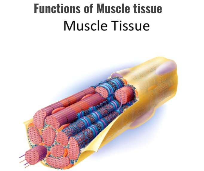
- It helps in different types of movements in the body due to contraction and relaxation.
- It helps in maintaining the body posture.
- It functions in heat production in the body.
- It maintains normal posture by maintaining proper muscle tone through the function of the nervous system.
- Due to the contraction of muscles, lymph circulation is observed in the lymphatic vessels.
- Provides the framework to the body and maintains the body posture.
Classification of Muscle tissue:
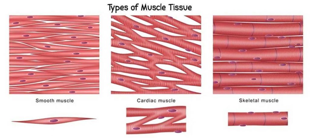
Skeletal muscles:
Structure of skeletal muscles:
- These muscles are also known as Voluntary Muscles because they work according to our will. With their help, the body moves. Therefore, they are called Voluntary Muscles muscles.
- These are straight type muscles.
- The structure of skeletal muscles consists of many bundles of muscle fibers.
- Fiber cells are located between the muscle fibers. They are arranged in a cylindrical shape. It also contains a nucleus.
- Skeletal muscles function for different types of movements.
- Skeletal muscles are found in large numbers in the body and are attached to bones. They are straight muscles of the striped type.
- There are more than 600 muscles in the body in total.
Structure (Structure):
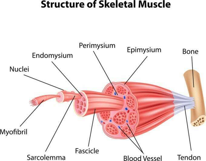
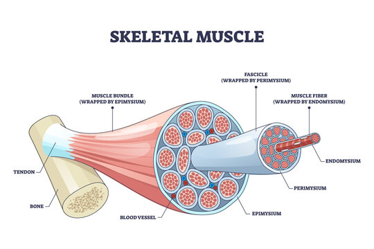
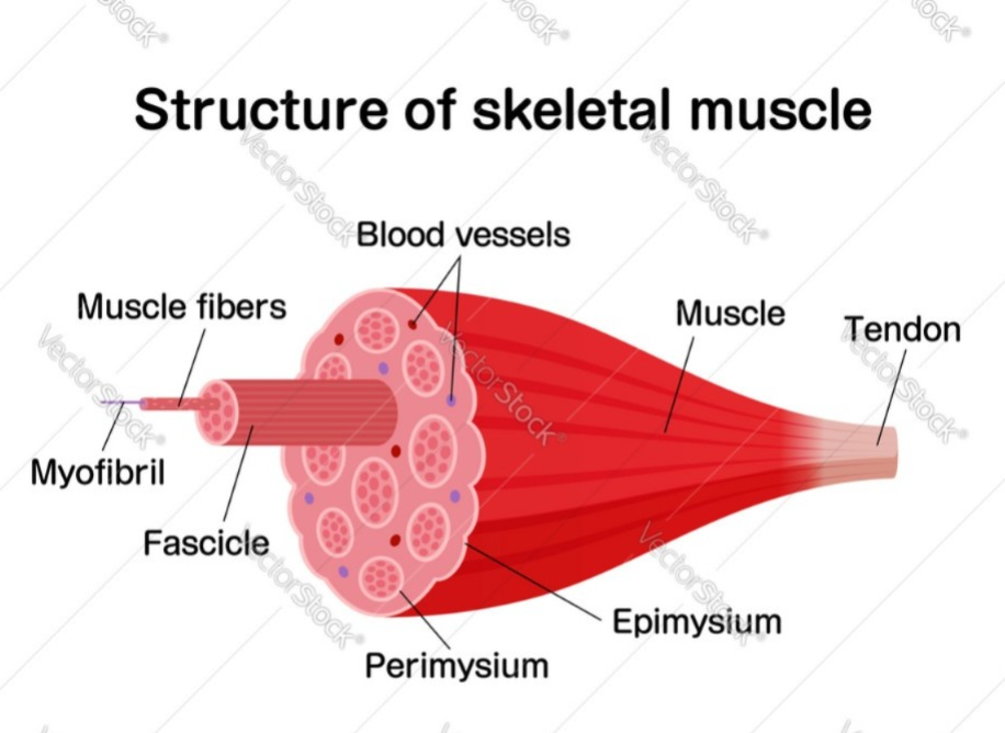
- Muscles are made up of a large number of muscle fibers. Muscles are attached to bones by fascia and tendons. The following structures are found on muscle fibers.
- A layer of Epimysium is present around the muscles as an outer layer. These muscle fibers contain many bundles of fibers, which are called Fascicles, the layer surrounding these bundles of muscle fibers is called Perimysium layer and these fascicles are made up of many single muscle fibers. The layer on these indivision muscle fibers is called Endomysium.
Smooth Muscles:
Structure of smooth muscles
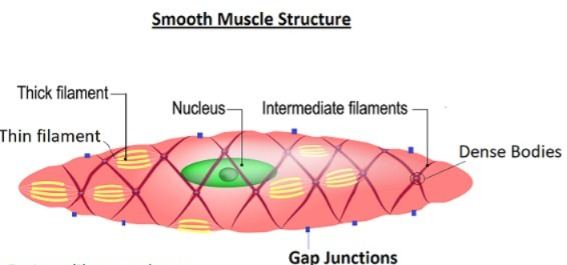
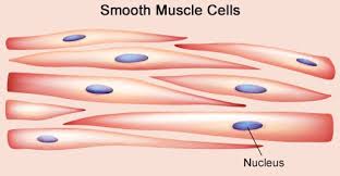
- These muscles are also called Involuntary Muscles because they are not under our voluntary control. These muscles are Unstriped type i.e. circular shape.
- When these muscles are examined under a microscope, cigarette-shaped cells are seen inside them. Nucleus is present in them. These types of muscles are found in the lining of blood vessels, respiratory tract, and alimentary tract.
Cardiac Muscles:
Structure of cardiac muscles
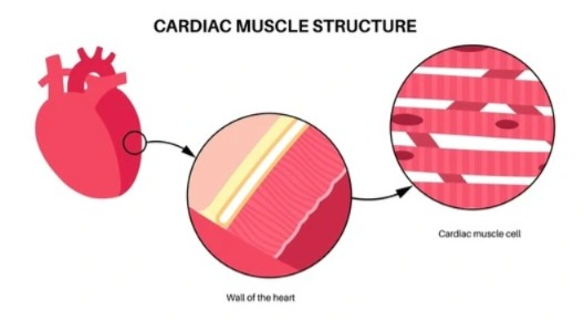
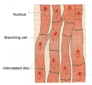
- These muscles are of the Involuntary Muscles type. Their function is also not under our control. These muscles are specially located in the middle layer of the heart Myocardium (Myocardium). They are called Cardiac Muscles.
- These muscles have straight fibers. They are closely connected to each other. The pumping action of the heart is seen due to the contraction and relaxation of these muscles.
The function and structure of each of the above muscles are different and they are associated with different functions in different parts of the body. The muscles of different areas in the body and their functions are as follows.
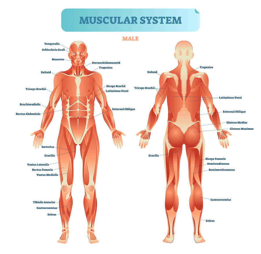
Muscles of the Face :
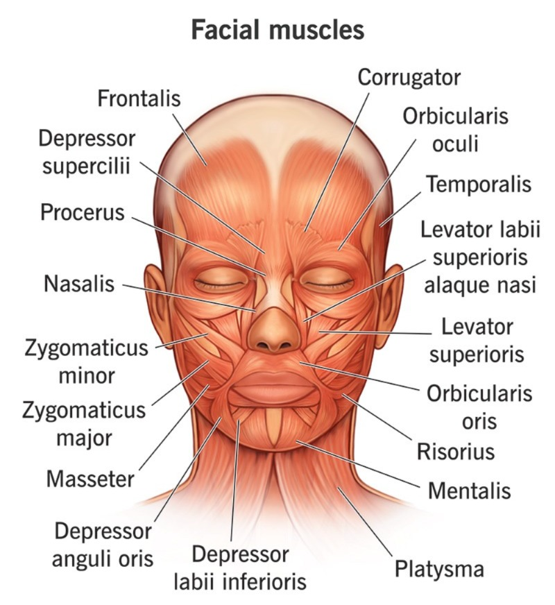
The muscles in the face perform a very important function for different movements of the face, facial expressions, speaking, and chewing. The muscles of the face are as follows.

Occipitalis (Occipitalis):
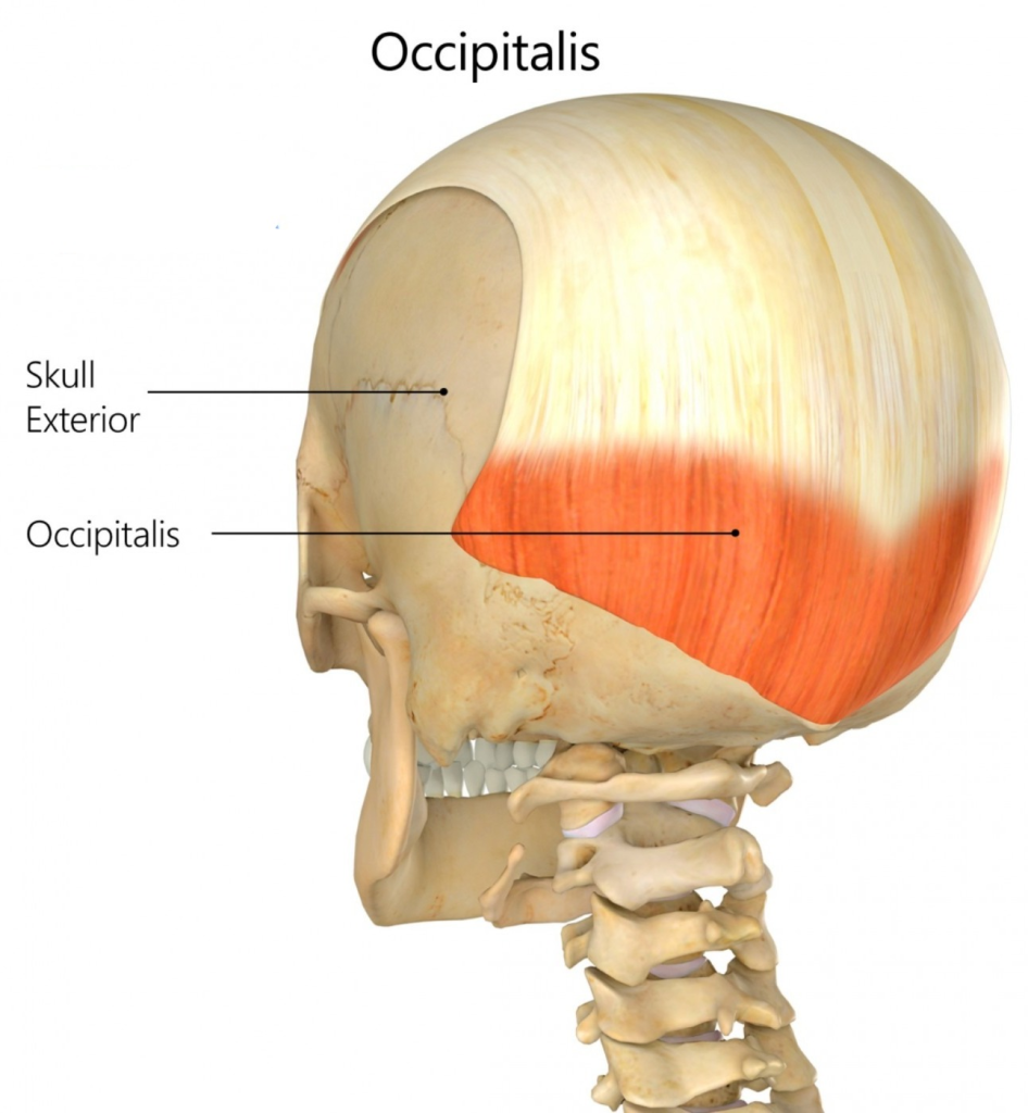
It is a muscle located between the occipital bone and the temporal bone. It is important for moving the scalp backwards.
Levatorpalpebrae superiors:
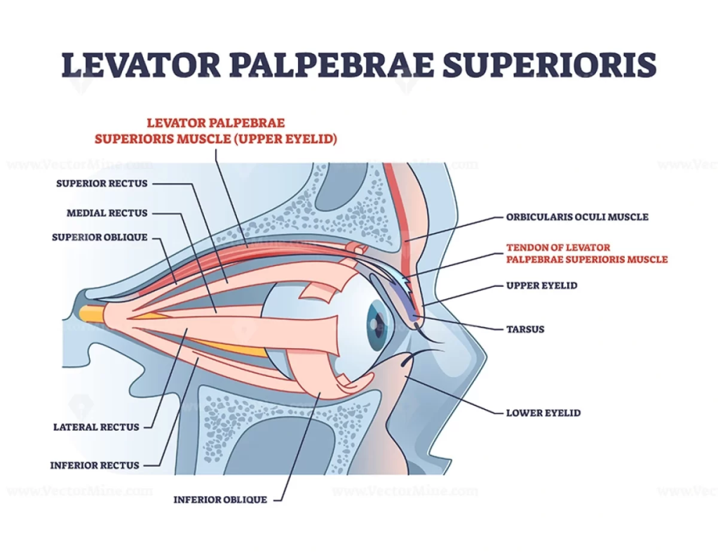
A muscle extending from the orbital cavity to the eyelid. It elevates the upper eyelid.
Orbicularis oculi:
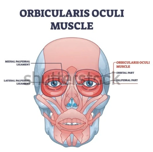
It is a circular arrangement of the orbital cavity. Which helps to close the eye.
Masseter:
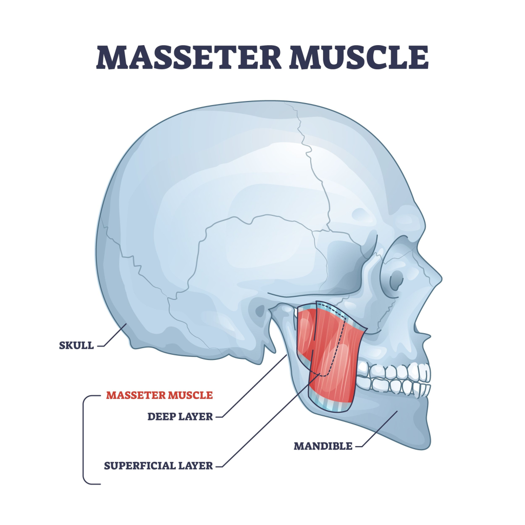
It is a muscle extending from the maxilla to the mandible. It is important in the movement of the mandible.
Temporalis (Temporalis):
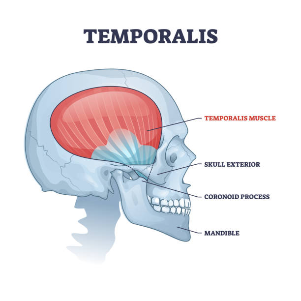
It is a muscle extending from the temporal bone to the mandible bone. It does the movement of the mandible.
Frontalis (Frontalis):
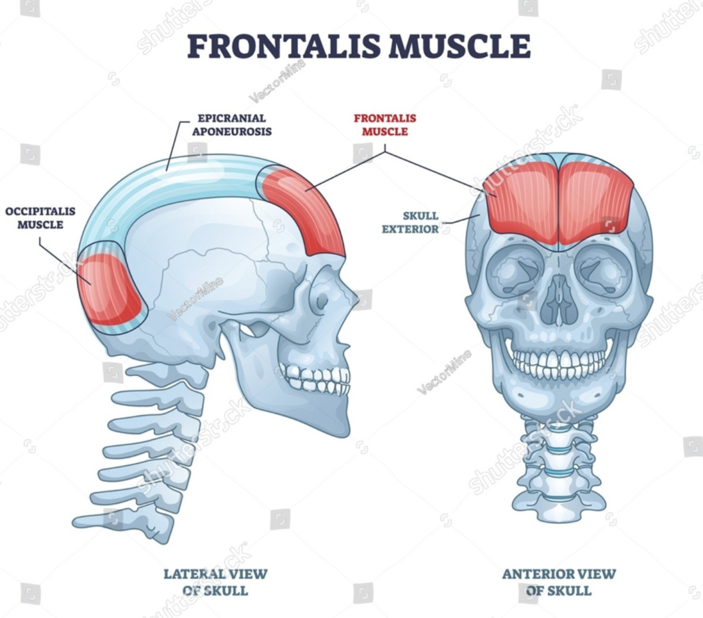
It is a muscle located in the fore head. It moves the scalp, forehead and eyebrows.
Buccinator:
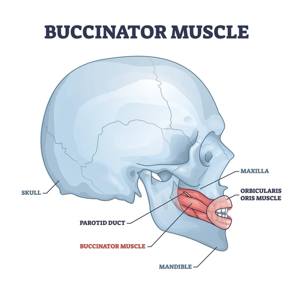
It is a muscle that extends between the maxilla and mandible. It plays an important role in the movement of the cheek.
Orbicularis oris (Orbicularis oris):
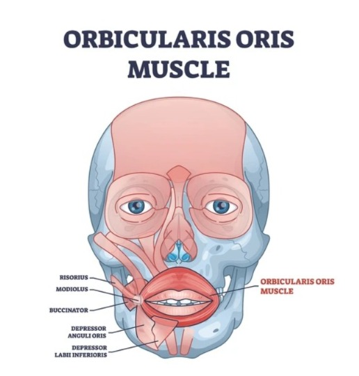
The orbicularis oris is a skeletal muscle that is present around the mouth.
The main function of the orbicularis oris muscle is to move the lips. This muscle forms a ring-like structure around the mouth and is often referred to as the “kissing muscle” because it is responsible for pursed and protruded lips.
Location:
The orbicularis oris is located around the circumference of the mouth and is related to other muscles of the face such as the buccinator, levator labii superioris, and depressor anguli oris.
Functions (Function):
- Close the lips
- Protrude the lips
- Assist in speech articulation
- Assist in keeping food within the oral cavity during eating cavity)
Muscles of Neck:
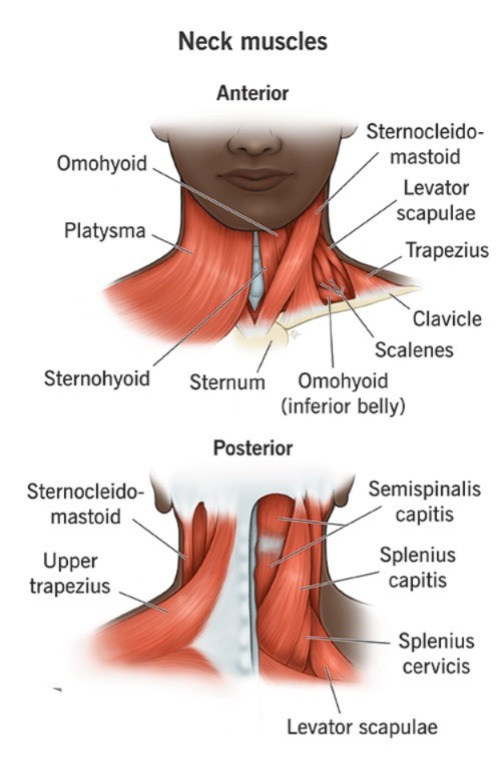
- The muscles in the neck perform different types of movements of the neck and are also responsible for different movements of the head.
- The muscles in the neck region are as follows.
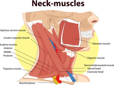
Sternocleidomastoid Muscles:
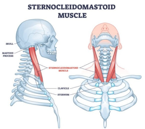
It is a muscle extending from the sternum and clavicle to the mastoid process of the temporal bone. It plays an important role in the movement of the head, neck and chin.
Trapezius Muscles:
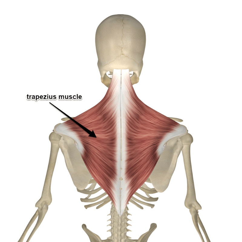
It is a muscle attached to the occipital bone, cervical and thoracic vertebrae, and the scapula bone. It moves the head and shoulder and is also connected to the movement of the scapula bone.
Muscles of Abdomen (Muscles in the abdominal region):
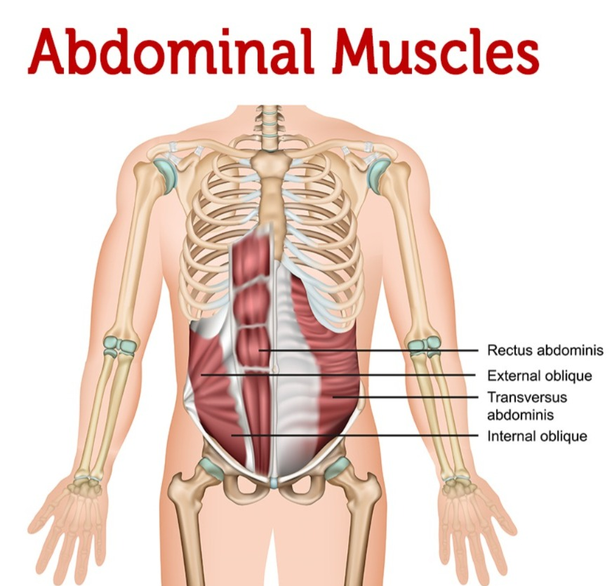
The muscles in the abdominal region are as follows.
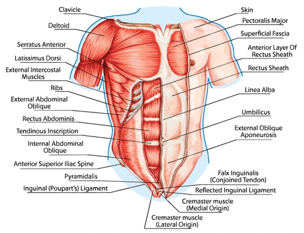
Rectus Abdominis:
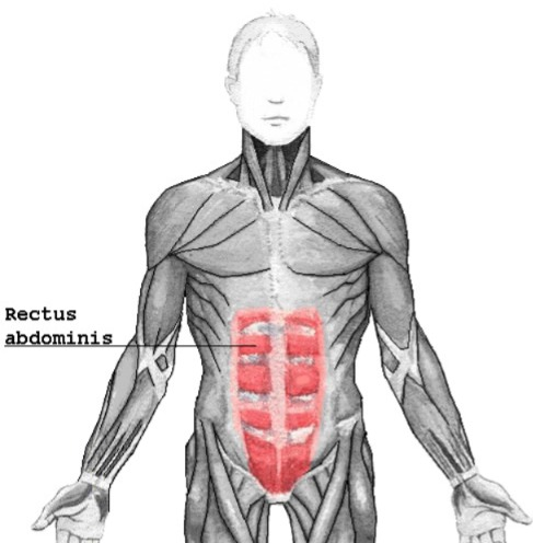
It is pubic bone to rib and It is a muscle extending from the xiphoid process of the sternum. It causes flexion movement of the vertebral column.
Transversus Abdominis:
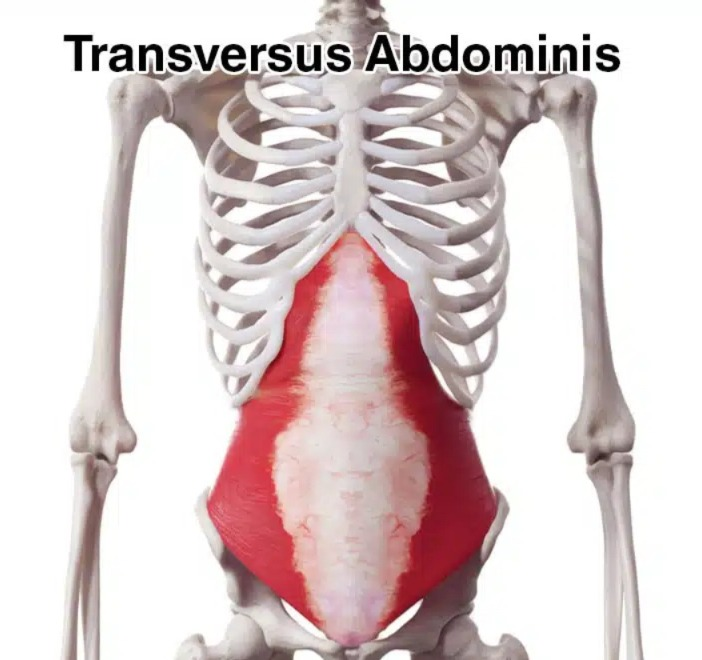
It extends from the iliac crest to the xiphoid process of the rib and sternum. It compresses the abdomen.
External oblique:
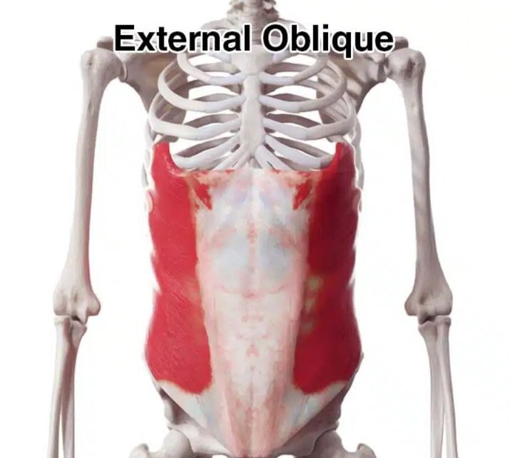
There are muscles extending from the ribs to the iliac crest. It compresses the abdomen and bends the vertebral column.
Internal oblique:
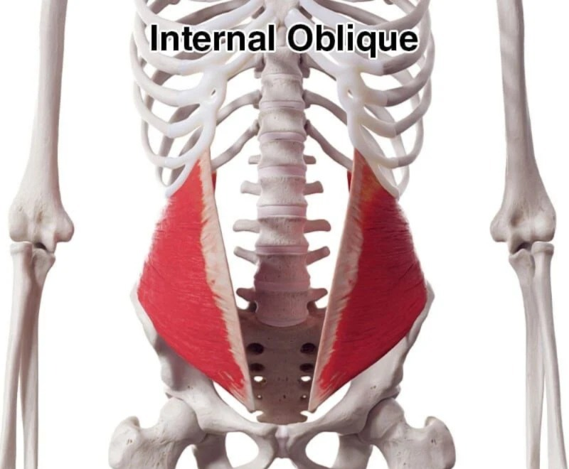
They are muscles attached from the iliac crest to the ribs. It compresses the abdomen and bends the vertebral column.
Psoas muscles (Psoas Muscles):
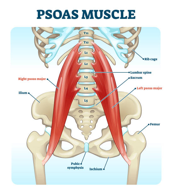
It is a muscle extending from the thoracic and lumbar vertebrae to the ilium and pubic bone. It performs different movements of the thigh.
Quadratus Lumborum:
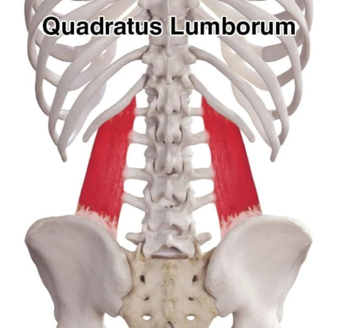
It extends from the iliac crest to the ribs and lumbar vertebrae. It causes lateral movement of the vertebral column.
Muscles of Back (Back muscles):
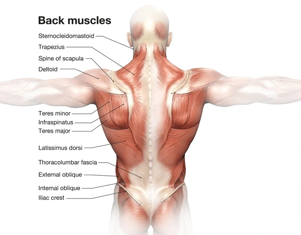
The muscles of the back are as follows.

Psoas muscles (Psoas Muscles):
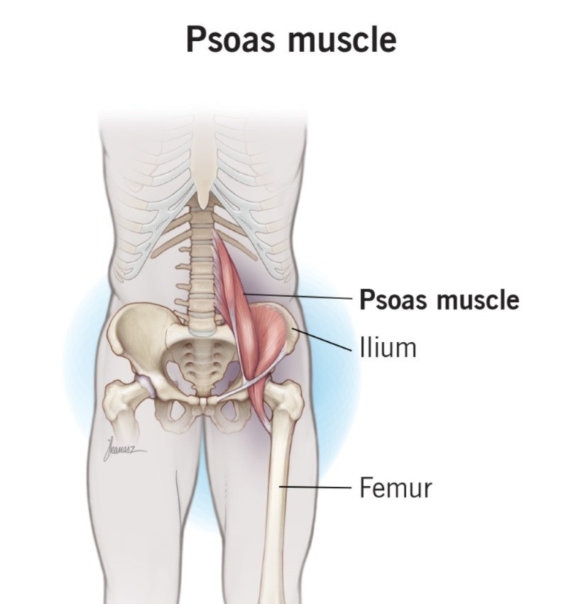
It extends from the thoracic and lumbar vertebrae to the ilium and There are muscles extending to the pubic bone. It performs different movements of the thigh.
Quadratus Lumborum:
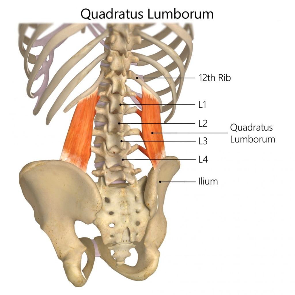
It extends from the iliac crest to the ribs and lumbar vertebrae. It provides lateral movement of the vertebral column.
Sacrospinalis :
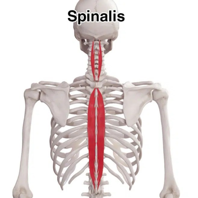
It extends from the iliac crest to the sacrum and lumbar and thoracic vertebrae. It is a group of erector spinae muscles. Which is responsible for different types of movement of the spine.
Latissimus dorsi:
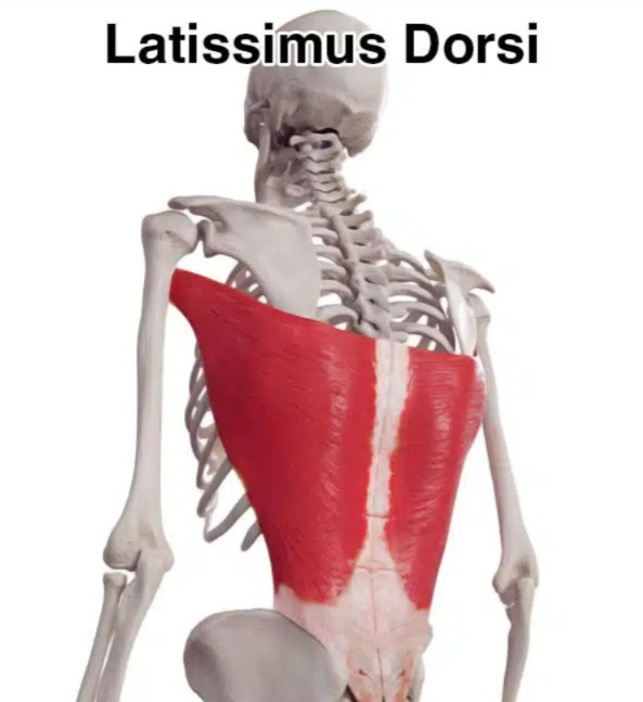
It extends from the iliac crest to the lumbar and thoracic vertebrae, scapula, and humerus bone. It works for different movements of the back, trunk and hands.
Trapezius:
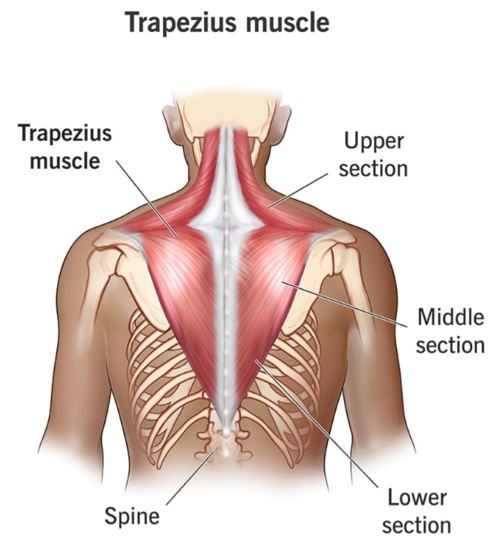
It extends from the occipital bone to the scapula, thoracic vertebrae. It is important for the different movements of the back and scapula and the trunk.
Teres major:
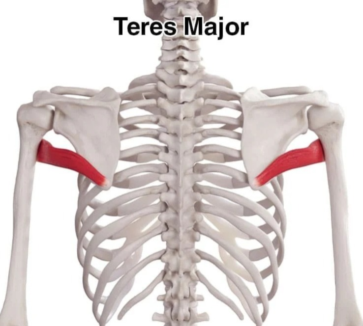
It is a muscle extending from the humerus to the scapula. It is important for the movement of the upper limb.
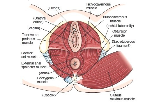
Muscles of pelvic floor:
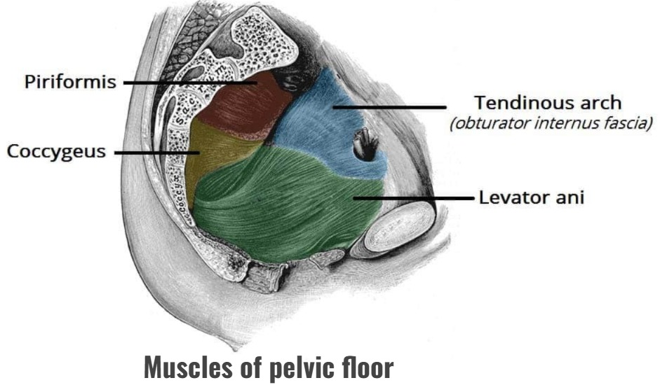
These muscles are important for the structural support of the bowel, bladder, uterus and sexual organs.
The muscles of the pelvic floor are as follows.
Levater Ani:
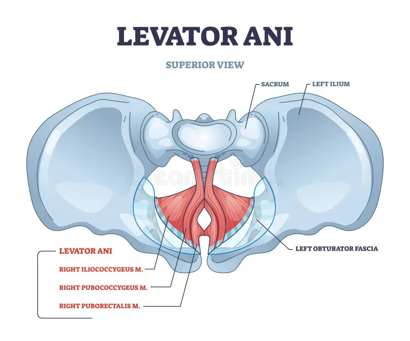
- It forms the floor of the pelvic cavity. It provides support to the pelvic structures. Supports the structures of the anal canal, urethra, and vagina.
- The iliococcygeus and pubococcygeus are also muscles of the levator ani group, they also support the structures of the pelvic floor.
Coccygeus (coccygeus):
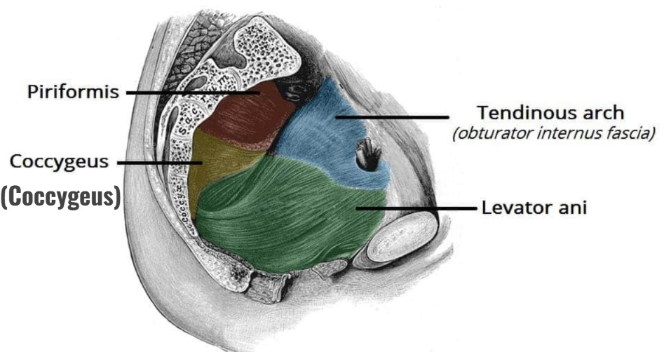
- They are muscles that extend from the ischium to the sacrum and coccyx. The pubococcygeus muscles extend to the pubis. It helps in the process of defecation, and supports the structure of the pelvic floor.
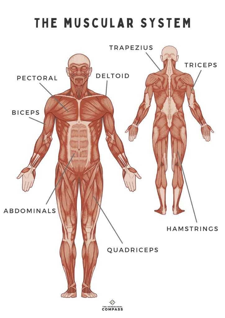
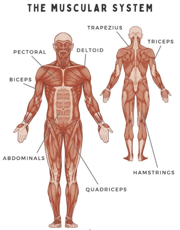
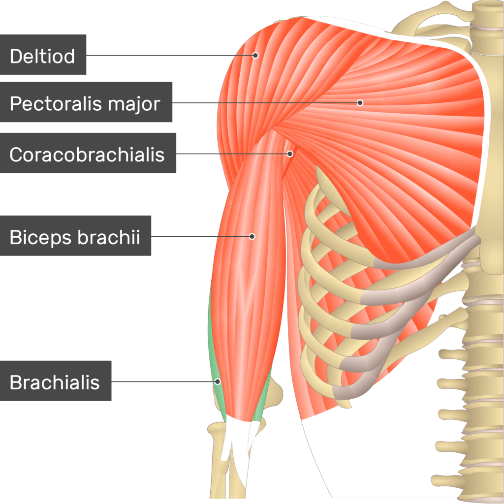
Muscles of the upper limb:
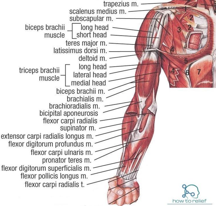
- Deltoid muscles
- Coracobrachialis muscles (coracobrachialis Mussels)
- Pectoralis major
- Biceps
- Triceps
- Brachialis (Brachialis)
- Supinator
- Pronator
- Flexor carpi radialis radialis)
- Flexor carpi ulnaris (Flexor carpi ulnaris)
- Extensor carpi radialis and ulnaris (Extensor carpi radialis and ulnaris)
- Palmaris longus (Palmaris longus) longus)
The above muscles are located in the ankle joint and upper limb. Due to these muscles, different movements of the ankle joint and upper limb are seen.
Upper limb Muscle Origin and its function:
Deltoid:
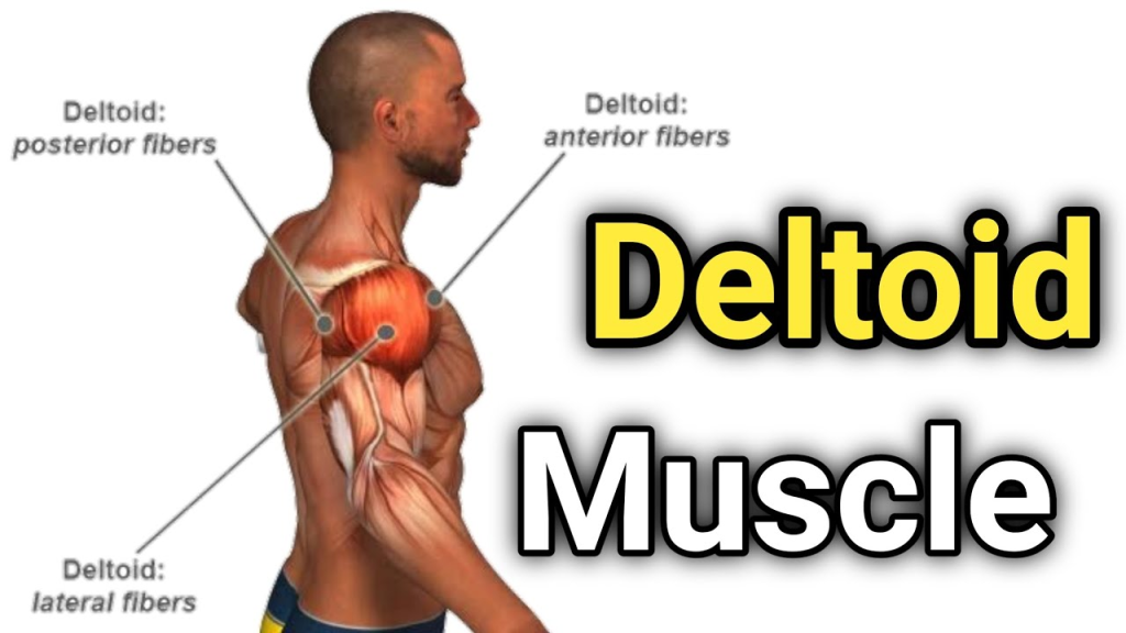
Origin:
- Clavicle
- Acromion
- Spine of Scapula
Function:
- Shoulder abduction
- Flexion and medial rotation by front fibers
- Extension and lateral rotation by back fibers
Biceps Brachii (Biceps Brachii) Brakii):
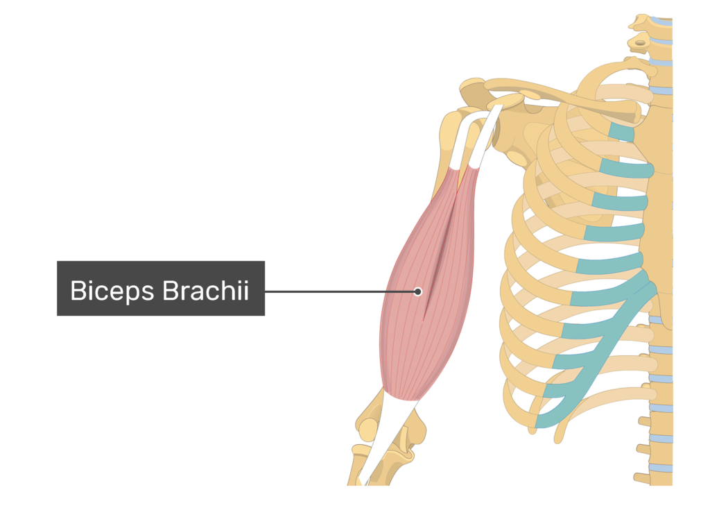
Origin:
- Long Head: Supraglenoid Tubercle
- Short Head: Coracoid Process
Function:
- Flexion of elbow
- Supination of forearm
- Slight flexion of shoulder
Triceps Brachii:
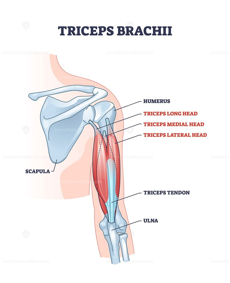
Origin:
- Long Head: Infraglenoid Tubercle
- Lateral head: Above the radial groove on the posterior surface of the humerus
- Medial head: Below the radial groove on the posterior surface of the humerus
Function:
- Extension of elbow
- Long head Extension of the shoulder by
Brachialis (Brachialis):
Origin:
- Lower half of anterior humerus
Function:
- Primary flexor of elbow
Brachioradialis:
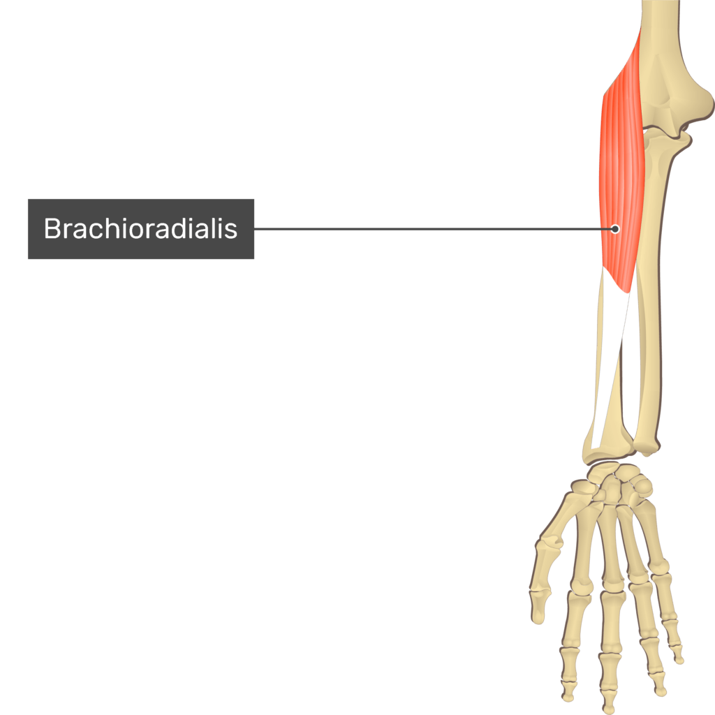
Origin:
- Lateral supracondylar ridge of humerus
Function:
- Forearm flexion in neutral position
Pectoralis Major:
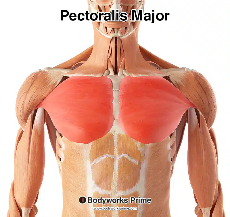
Origin:
- Clavicle, sternum and 1 to 6 costal cartilages
Function:
- Adduction of the Humerus
- Medial Rotation
- Shoulder Flexion
Latissimus Dorsi:
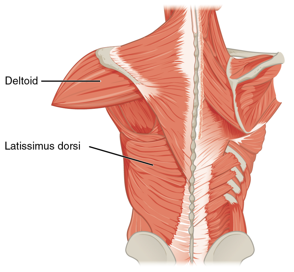
Origin:
- Thoracic vertebrae (T7-T12), lumbar vertebrae, iliac crest
Function:
- Extension of the humerus
- Adduction
- Medial rotation
Teres Major:
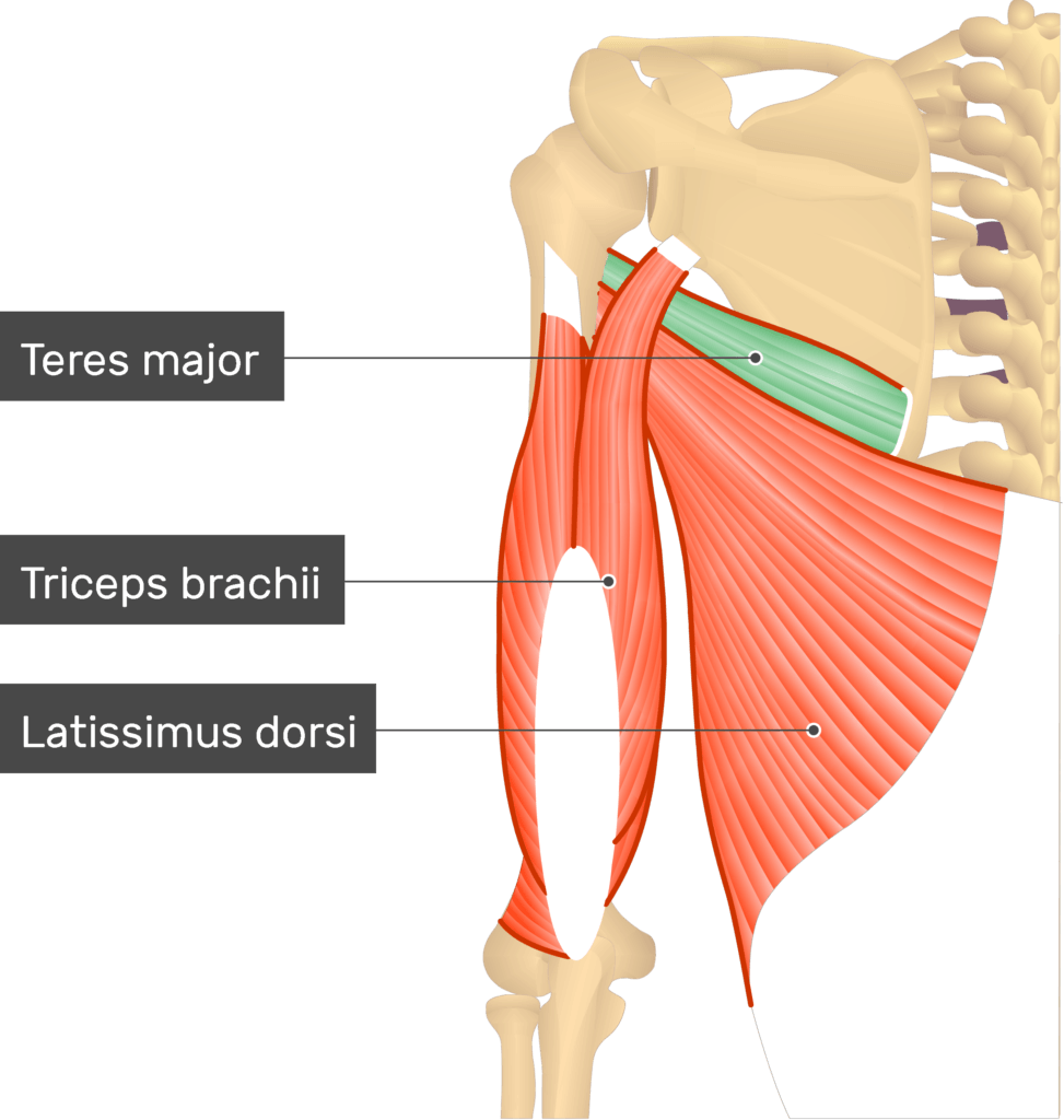
Origin:
- Inferior angle of scapula (Inferior angle of scapula)
Function:
- Adduction of the humerus
- Medial rotation
- Extension
Teres Minor:
Origin:
- Lateral border of scapula
Function:
- Of the humerus Lateral rotation
- One of the four muscles of the rotator cuff
Supraspinatus:
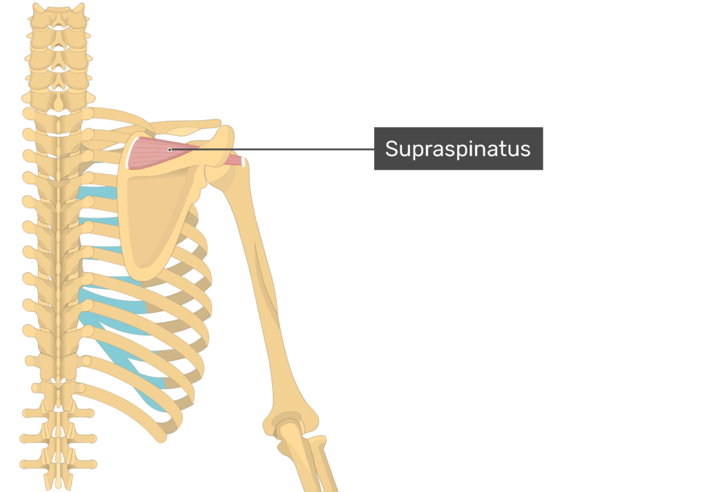
Origin:
- Supraspinous fossa of scapula
Function:
- Early abduction of the shoulder
- Important muscle of the rotator cuff
Muscles of the hip and lower limb:
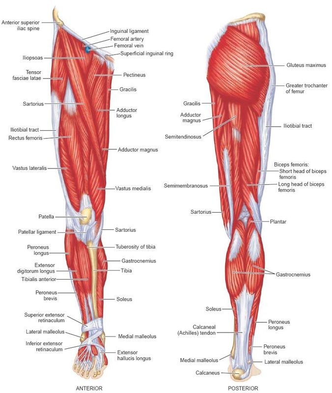
- Psoas
- iliacus (Iliacus)
- Gluteals
- Quadriceps femoris (Quadriceps femoris)
- Sartorius (Sartorius)
- Hamstring
- Obturator
- Compartment group muscles Muscles)
Origine and Functions of Muscles of hip and Lower limb (Origine and Functions of Muscles of Hip and Lower Limb):
Gluteus Maximus (Gluteus Maximus):
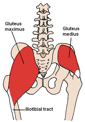
Origin:
- Ilium, Sacrum, Coccyx and Sacrotuberous Ligament
Function:
- Extension of the hip joint
- Lateral rotation (external (rotate)
- Helps with standing and getting up from sitting
Gluteus Medius:
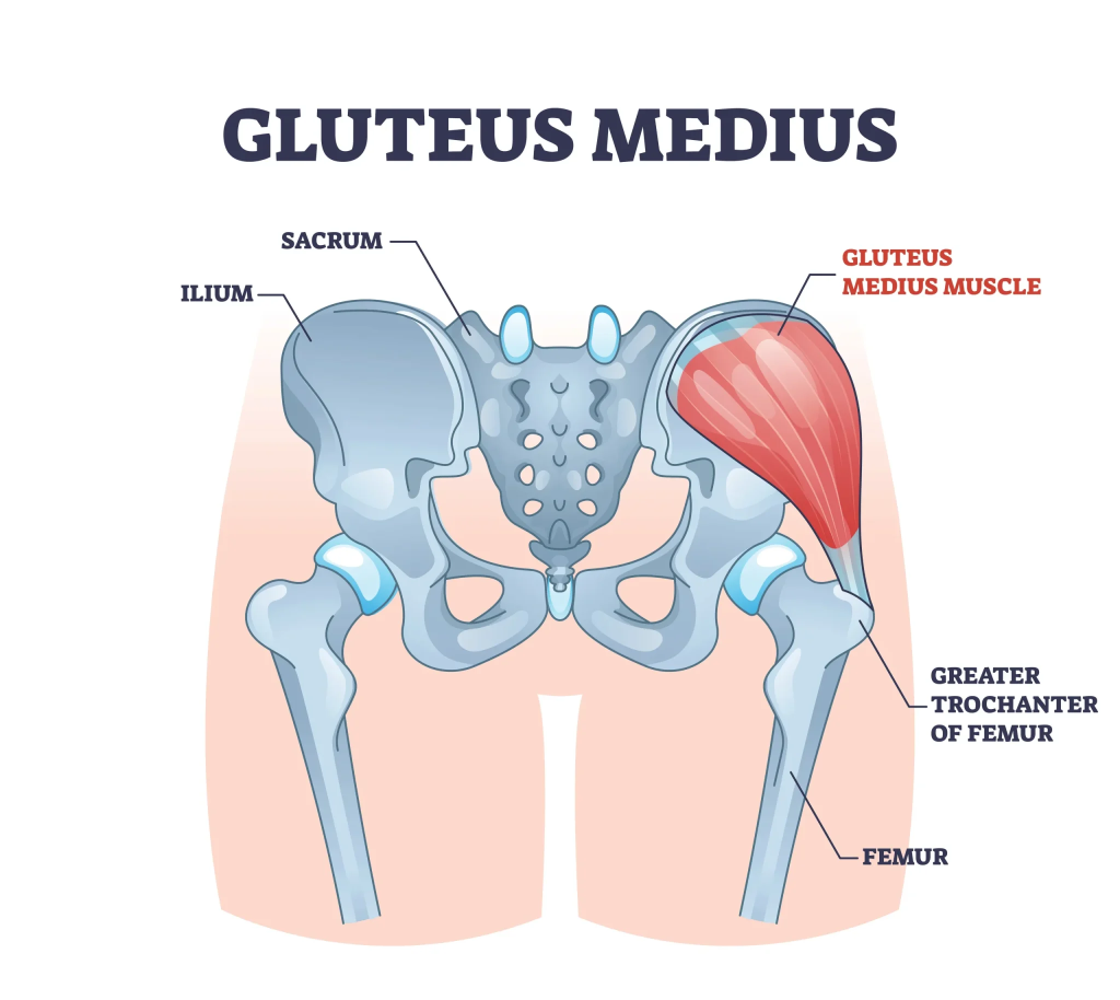
Origin:
- Outer surface of ilium
Function:
- Abduction of hip
- Medial rotation
- Helps in balancing the pelvis while walking
Gluteus Minimus (Gluteus Minimus):
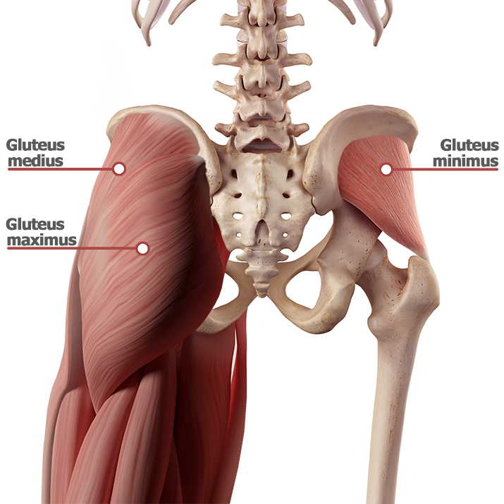
Origin:
- Inferior surface of ilium
Function:
- Hip abduction
- Medial rotation
- Maintaining Gait Balance
Iliopsoas:
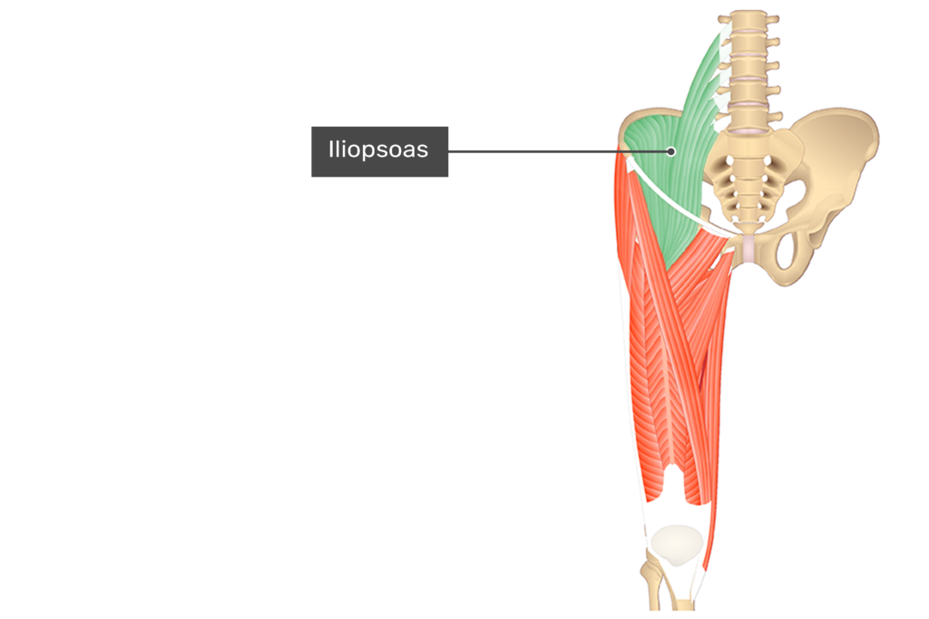
(Combination of Iliacus + Psoas Major)
Origin:
- Iliac fossa (Iliacus)
- Lumbar vertebrae (Lumbar vertebrae – Psoas major)
Function:
- Main hip flexor
- Trunk flexion while sitting
Tensor Fasciae Latae (Tensor Fascia Latte):
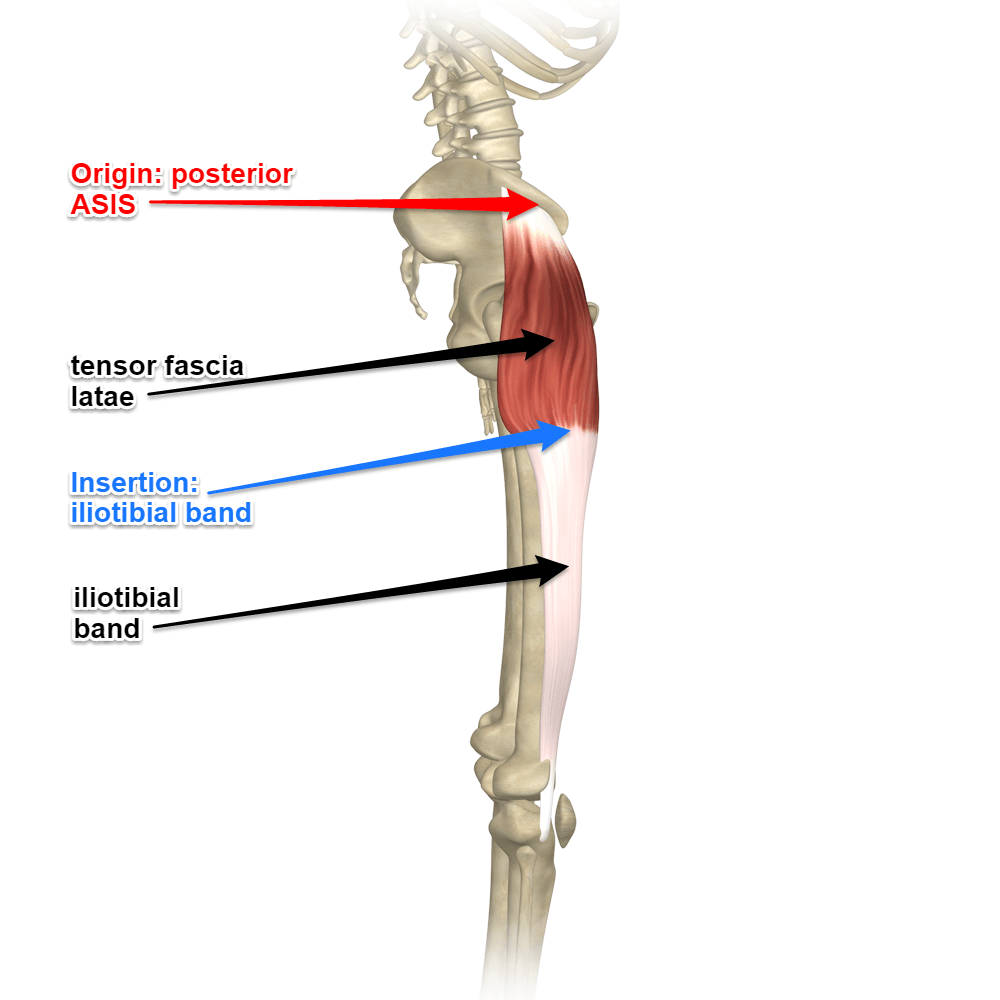
Origin:
- Anterior part of iliac crest
Function:
- Hip abduction and medial rotation
- Supports knee extension (Via iliotibial tract)
Sartorius:
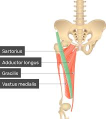
Origin:
- Anterior superior iliac spine (ASIS)
Function:
- Hip Flexion
- Hip Abduction
- Lateral Rotation
- Knee Flexion
(Supports “Cross-legged Sitting” Position)
Rectus Femoris:
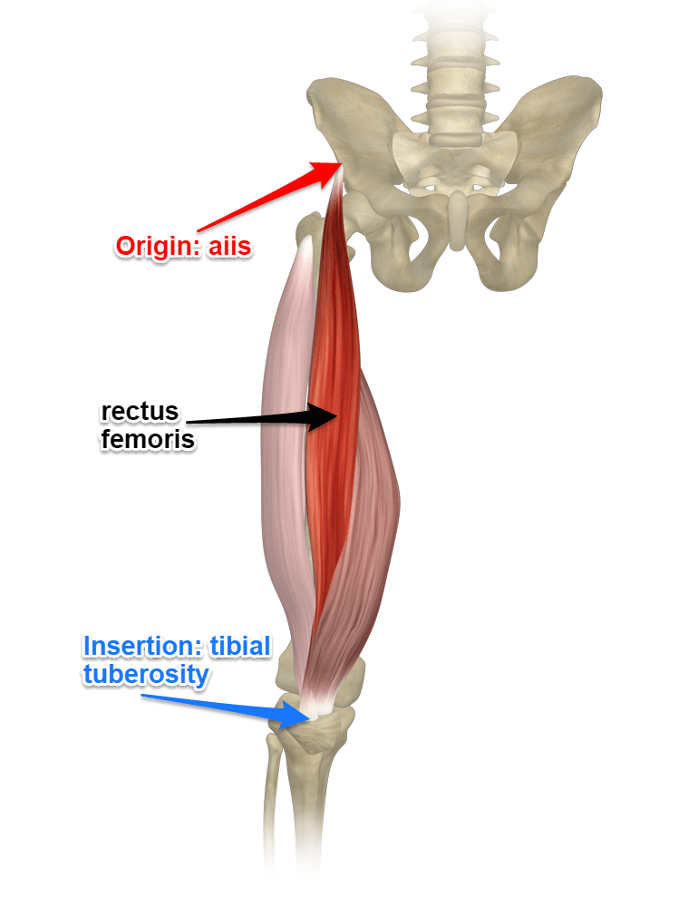
(Part of Quadriceps group)
Origin:
- Anterior inferior iliac spine (AIIS)
Function:
- Knee extension
- Hip flexion
Vastus Lateralis / Medialis / Intermedius:
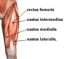
Origin:
- From various surfaces of the femur
Function:
- Of the knee extension
Hamstring Group:
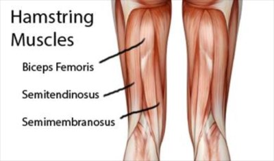
Biceps Femoris, Semitendinosus, Semimembranosus
Origin:
- Ischial tuberosity
Function:
- Hip Extension
- Knee Flexion
- Biceps femoris performs lateral rotation
Adductor Group (Adductor group):
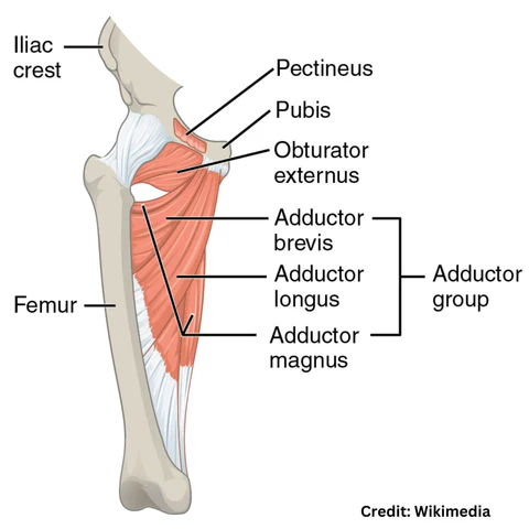
Adductor Longus, Brevis, Magnus
Origin:
- Pubis
Function:
- Hip adduction
- Slight flexion and Medial rotation
Gracilis (Gracilis):
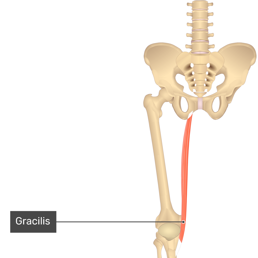
Origin:
- Inferior ramus of pubis
Function:
- Hip Adduction
- Knee Flexion
Tibialis Anterior:
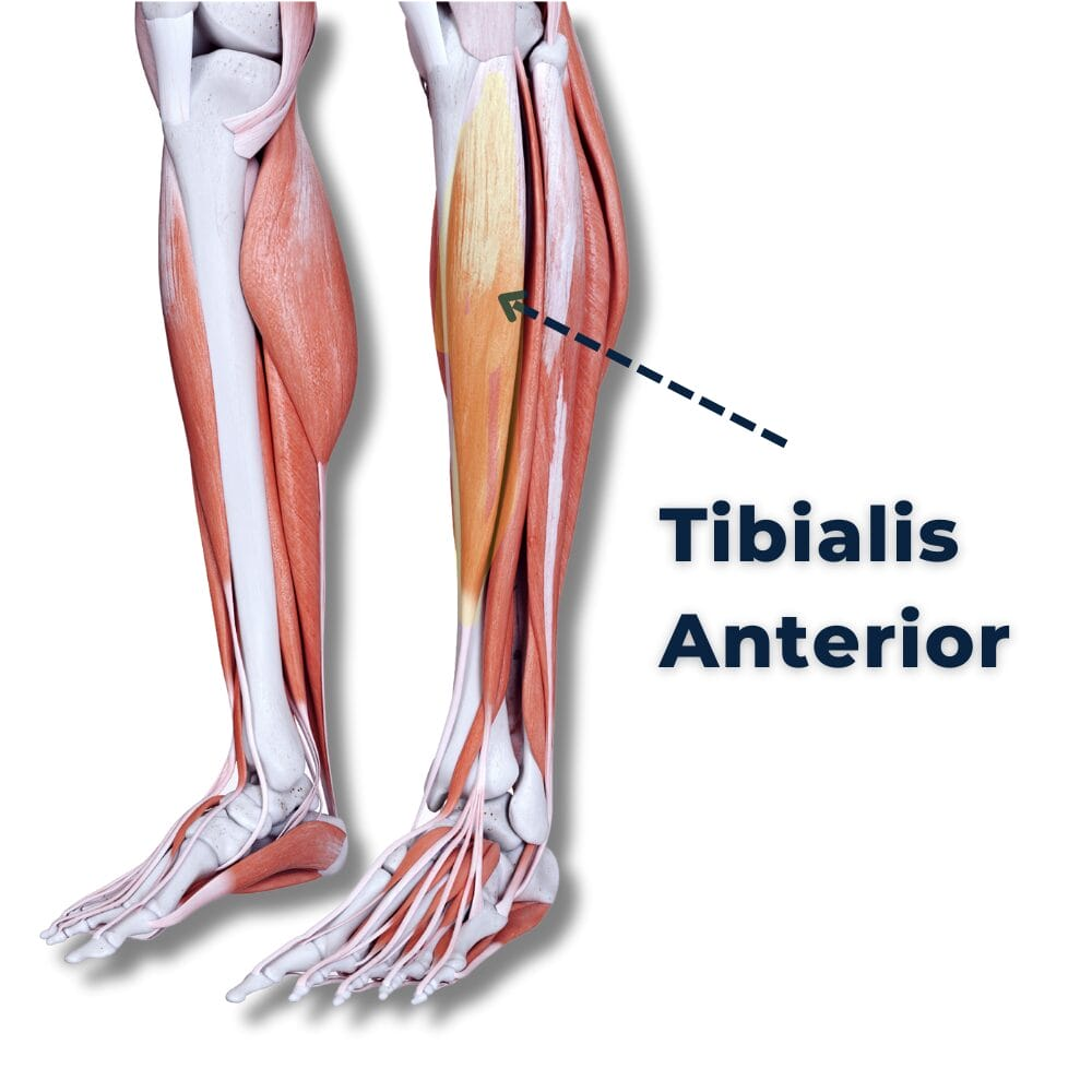
Origin:
- Lateral condyle of tibia and interosseous membrane
Function:
- Ankle dorsiflexion (Dorsiflexion)
- Foot inversion
Gastrocnemius (Gastrocnemius):
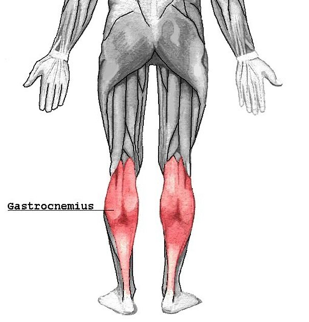
Origin:
- Medial and lateral condyles of the femur
Function:
- Foot plantar flexion
- Knee flexion
Soleus:
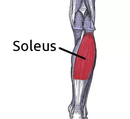
Origin:
- Tibia and fibula
Function:
- Plantar flexion
- To stand Auxiliary
The above muscles can cause different movements of the pelvic region as well as the lower limbs. In which the above muscles are important for walking, running, weight bearing etc. movements.