ENGLISH ANATOMY UNIT 14 MUSCULAR SYSTEM
MUSCULAR SYSTEM
INTRODUCTION
Muscles are bundles of fibers that have the power to contract and form strong muscle tissue. Due to the contraction, different types of movement occur.
The study of the arrangement of muscles, their structure and their function is known as myology.
Muscles are made up of fibers, nerves and connective tissue. Which is estimated to be 40% of the human body weight.
- Functions of muscle tissue. (Functions of Muscle Tissue).
Contraction and relaxation help the body to perform different types of movement.
Helps maintain body posture.
The body performs the function of heat production.
It maintains normal posture by maintaining proper muscle tone through the functioning of the nervous system.
Muscle fatigue occurs due to prolonged muscle contraction.
Due to contraction of muscles, circulation of lymph is seen in lymphatic vessels.
Gives framework to the body and maintains body posture.
- Classification of muscle tissue.
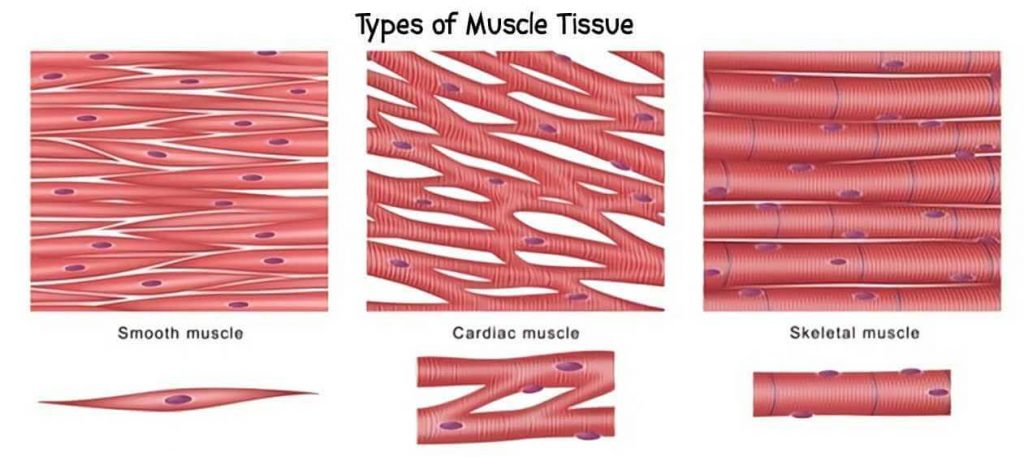
Skeletal muscles. (Skeletal muscles). Write the structure of skeletal muscles.
These muscles are also known as other voluntary muscles because they act according to our will. With its help the body moves. So it is called voluntary muscles.
These are straight type muscles.
Many muscle fiber bundles are present in the structure of skeletal muscles.
Fiber cells are located between muscle fibers. They are arranged in a cylindrical shape. It also contains the nucleus.
Skeletal muscles function for different types of movement.
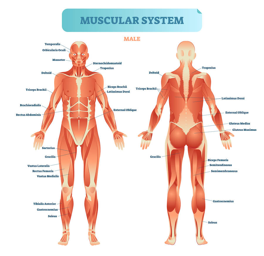
Skeletal muscles are the most abundant in the body and are attached to bones. They are straight muscles of the stripped type.
A total of more than 600 muscles are found in the body.
Structure
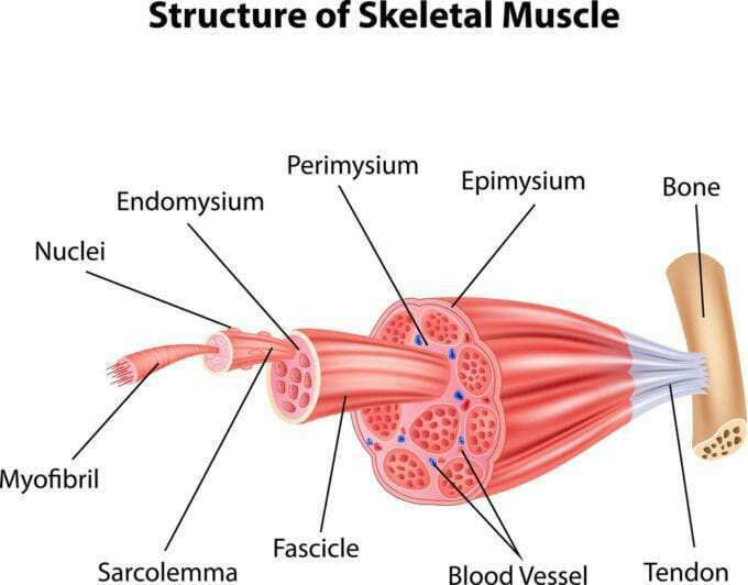
Mussels contain a large number of muscle fibers. Muscles are attached to bones by fascia and tendons. The following structures are found on muscle fibers.
Surrounding the muscles is a layer of epimycium which is divided as outer layer. These muscle fibers consist of bundles of many fibers, which are bundles of muscle fibers called fascicles, the surrounding layer is called perimysium layer and these fascicles are made up of many single muscle fibers. The layer above this indivision muscle fiber is called endomycium.
Smooth Muscles. Write the structure of smooth muscles.
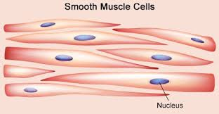
These muscles are also called involuntary muscles because they are not under our voluntary control. These muscles are of unstripped type i.e. circular shape.
Looking at these muscles under a microscope, cigarette-shaped cells are seen inside. A nucleus is present in them. These types of muscles are located in blood vessels, lining of respiratory tract, lining of alimentary tract.
Cardiac Muscles. Write the structure of cardiac muscles.
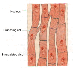
These muscles are voluntary type muscles. Its function is also not under our control. These muscles are especially located in the middle layer of the heart, the myocardium. It is called cardiac muscle.
These muscles have straight fibers. They are closely related to each other. The pumping action of the heart is seen due to the contraction and relaxation of these muscles.
Each of the above muscles has a different function and structure and is associated with different functions in different parts of the body. The muscles of different areas in the body and their functions are as follows.
- Muscles of the Face.
The muscles in the face play a very important role in different movements of the face, facial expression, speaking and chewing. The muscles of the face are as follows.
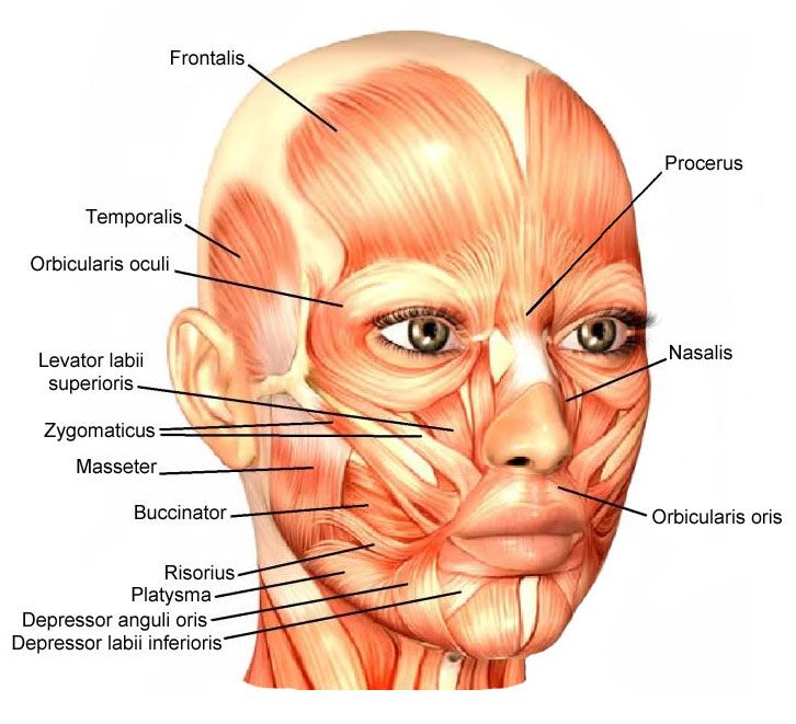
Occipitalis
It is a muscle located between the occipital bone and the temporal bone. It is important to move the scalp backwards.
Levatorpalpebrae superiors.
The orbital muscle extends from the cvt to the eyelid. It elevates the upper eye lid.
Orbicularis oculi.
It is a circular arrangement of orbital KV. Which helps to close the eye.
Masseter.
It is a muscle extending from the maxilla to the mandible. It is important in the movement of the mandible.
Temporalis.
It is a muscle extending from the temporal bone to the mandible bone. It does the movement of the mandible.
Frontalis.
It is a muscle located near the fore head. It does the movement of the scalp, fore head and eyebrows.
Buccinator.
It is a muscle that stretches between the maxilla and the mandible. It plays an important role in the movement of the cheek.
Orbicularis oris.
It is the muscle surrounding the orbital cavity. It plays an important role in closing the eyes.
Muscles of Neck.
The muscles in the neck are responsible for various movements of the neck and also for various movements of the head.
- Muscles in the neck region
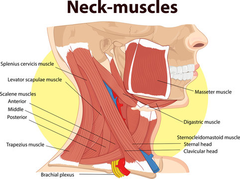
Sternocleidomastoid Muscles.
It is a muscle extending from the sternum and clavicle to the mastoid process of the temporal bone. It plays an important role in the movement of the head, neck and chin.
Trapezius Muscles.
It is a muscle attached to the occipital bone, cervical and thoracic vertebrae as well as the scapula bone. It carries the movement of the head and shoulder and is also associated with the movement of the scapula bone.
- Muscles of Abdomen.
The muscles in the abdominal region are as follows.
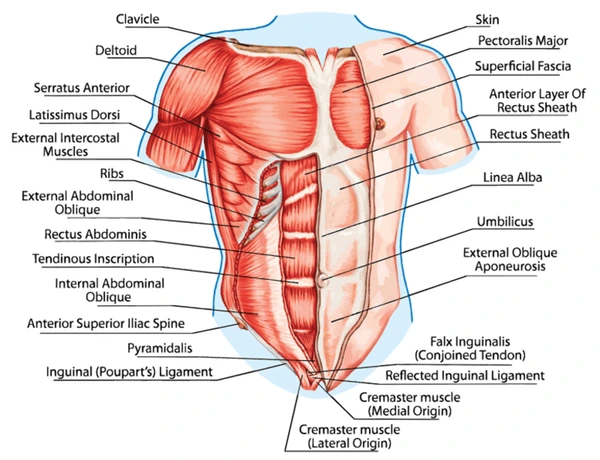
Rectus Abdominis.
It is a muscle extending from the pubic bone to the rib and the xiphoid process of the sternum. It causes flexion movement of the vertebral column.
Transversus Abdominis.
They extend from the iliac crest to the rib and the xiphoid process of the sternum. It compresses the abdomen.
External oblique.
There are muscles extending from the rib to the iliac crest. It compresses the abdomen and bends the vertebral column.
Internal oblique.
They are the muscles attached from the iliac crest to the ribs. It compresses the abdomen and bends the vertebral column.
Psoas muscles.
It is a muscle extending from the thoracic and lumbar vertebrae to the ilium and pubic bone. It makes different movements of the thigh.
Quadratus Lumborum.
It extends from the iliac crest to the ribs and lumbar vertebrae. It causes lateral movement of the vertebral column.
- Muscles of Back.
The muscles of the back are as follows.
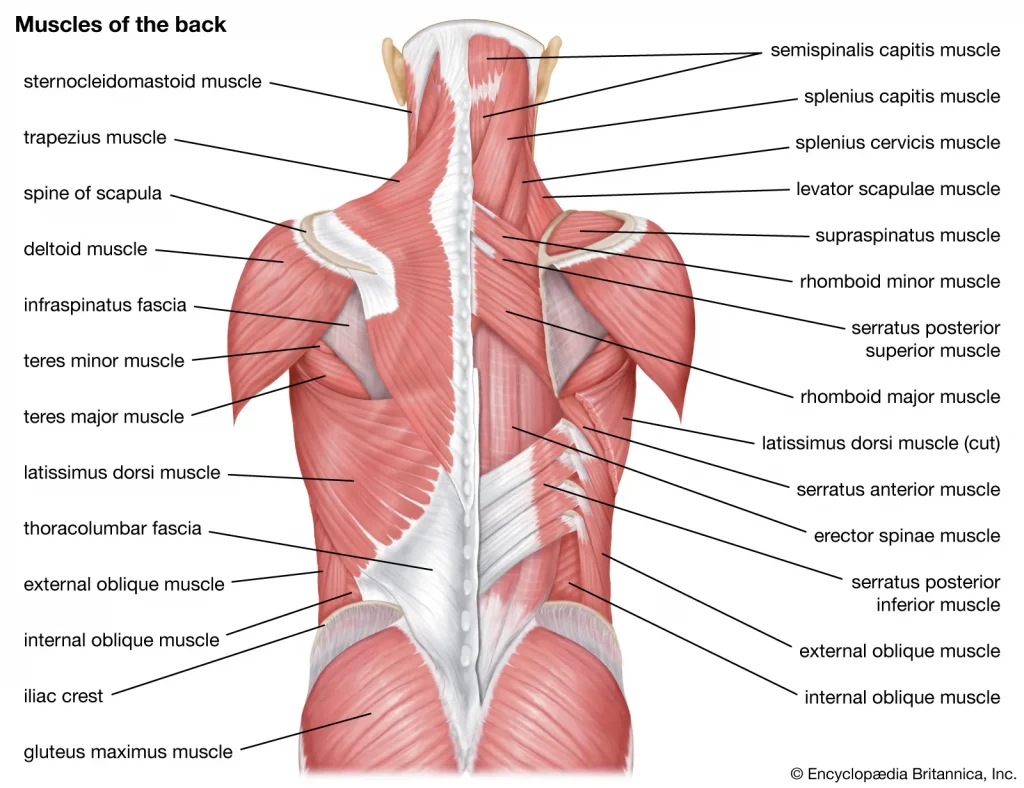
Psoas muscles
It is a muscle extending from the thoracic and lumbar vertebrae to the ilium and pubic bone. It makes different movements of the thigh.
Quadratus Lumborum
It extends from the iliac crest to the ribs and lumbar vertebrae. It causes lateral movement of the vertebral column.
Sacrospinalis.
It extends from the iliac crest to the sacrum and lumbar as well as the thoracic vertebrae. It is a group of erector spinae muscles. Which is responsible for different types of movement of the spine.
Latissimus dorsi.
It extends from the iliac crest to the lumbar as well as the thoracic vertebrae, scapula and humerus bone. It works for different movements of back, trunk and hand.
Trapezius.
It extends from the occipital bone to the scapula, thoracic vertebrae. It is important for different movements of back and scapula and trunk.
Teres major.
It is a muscle extending from the humerus to the scapula. It is important for upper limb movement.
- Muscles of pelvic floor.
These muscles bowel, bladder, uterus and sexual
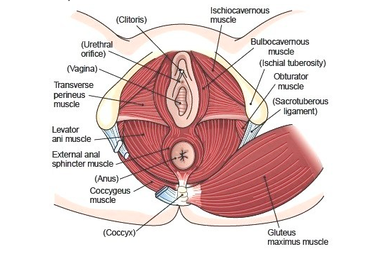
Levator Ani.
This forms the floor of the pelvic cavity. It provides support to the pelvic structure. Supports the structures of the anal canal, urethra and vagina.
The iliococcygeus and pubococcygeus are also muscles of the levator ani group, which also provide support to the structure of the pelvic floor.
Coccygeus.
It is a muscle extending from the ischium to the sacrum and coccyx. The pubococcygeus muscle extends to the pubic area. It helps in the process of defecation, and supports the structure of the pelvic floor.
- Muscles of the upper limb.
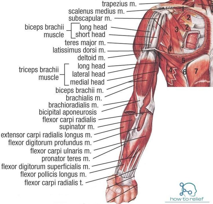
Deltoid muscles
Coracobrachialis muscles
Pectoralis major
Biceps
Triceps
Brachialis
Supinator
Pronator
Flexor carpi radialis
Flexor carpi ulnaris
Extensor carpi radialis and ulnaris
Palmaris longus
The above muscles are located in the solder joint and upper limb. Due to these muscles, different movements of the solder joint and upper limb are seen.
- Muscles of the hip and lower limb.
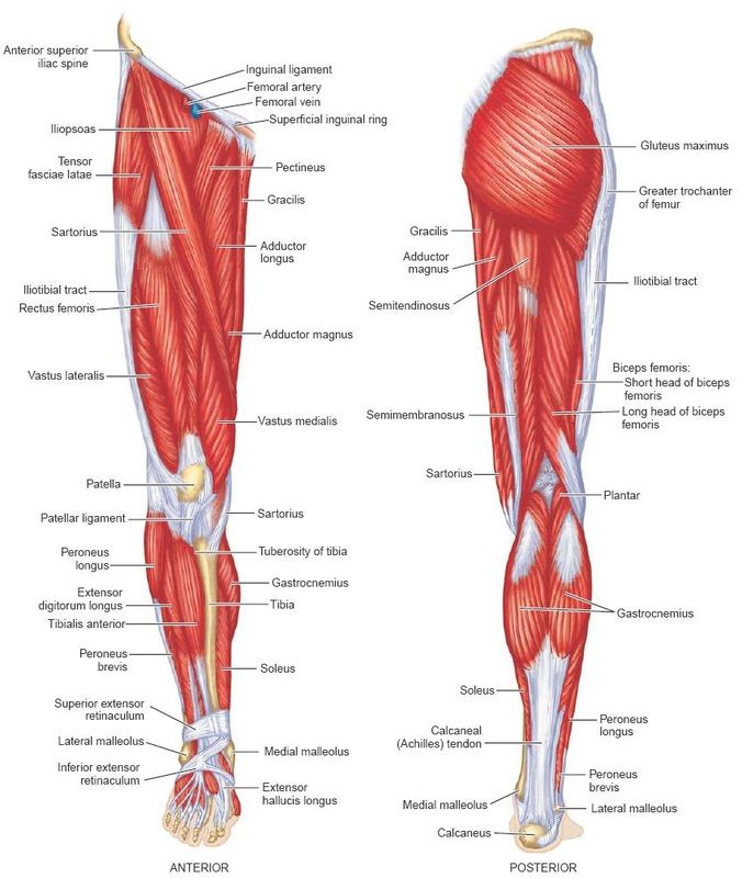
Psoas
iliacus
Gluteals
Quadriceps femoris
Sartorius
Hamstring
Obturator
Compartment group muscles
The above muscles can cause different movements of the pelvic region as well as the lower limb. In which the above muscles are important for walking, running, weight bearing etc. movements.