—ENGLISH-ANATOMY UNIT 13(PART :2) JOINTS
JOINTS:
Joint and Types of joint:
- Two or more bones in the body join together to form a joint. Different movements are seen in the joints. The movement seen in the joints depends on the bone, cartilage, connective tissue and muscular structure that make up the joint.
- These joints are divided into different types based on their mobility. The classification of joints is given as follows based on the movement seen in the joint.
1.Freely movable joint:
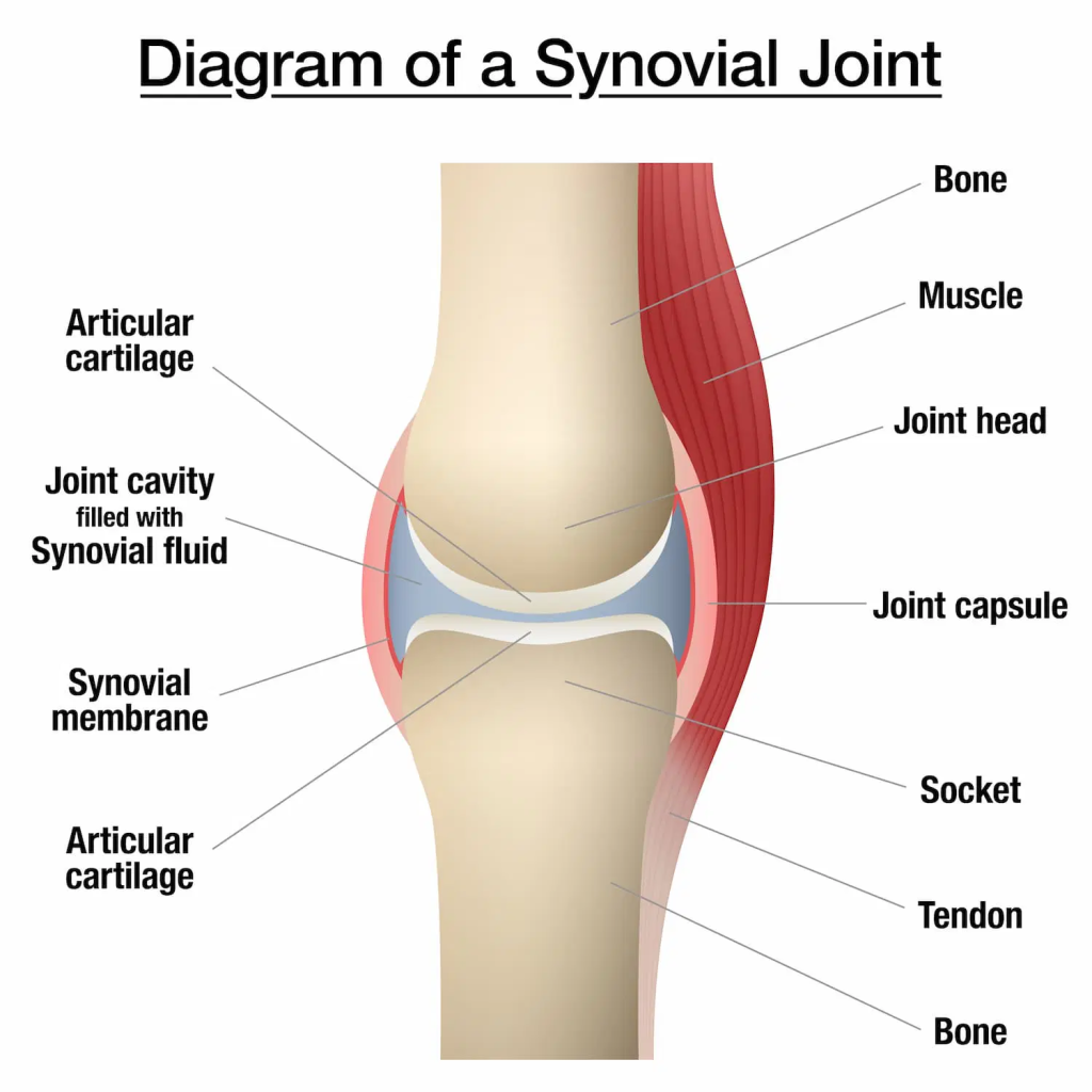
This joint is also called synovial joint. In which two or more bones are connected to each other with the help of articular cartilage to form this type of joint. In this joint, a cavity or space is formed around the joint, which is called the synovial cavity. Maximum movement is seen in this type of joint. Hence it is called a freely movable joint. Synovial joint is also known as another Diarthroses .
Characteristics of Synovial joint. (Characteristics of Synovial Joint):
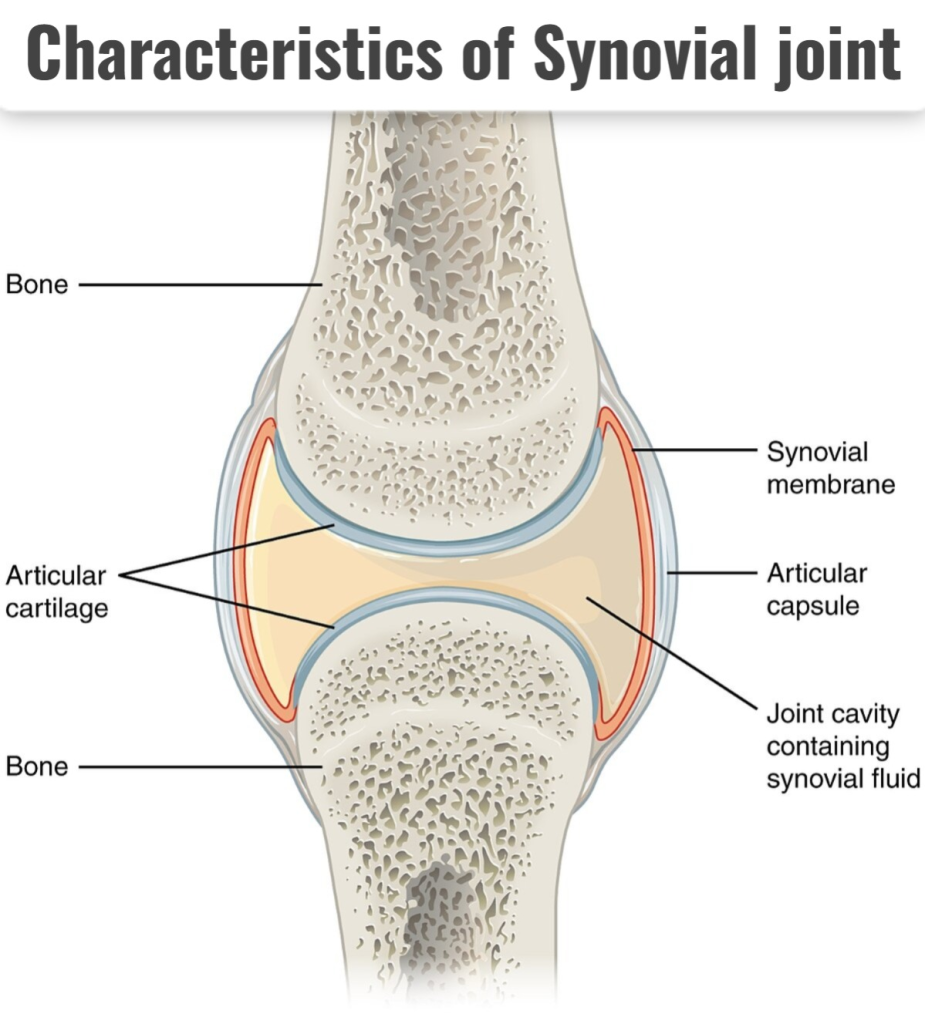
A synovial joint is where two bones form a joint where maximum movement occurs. This type of joint is called a synovial joint or a freely movable joint. Synovial joints have the following characteristics.
HYLIENE CARTILAGE:
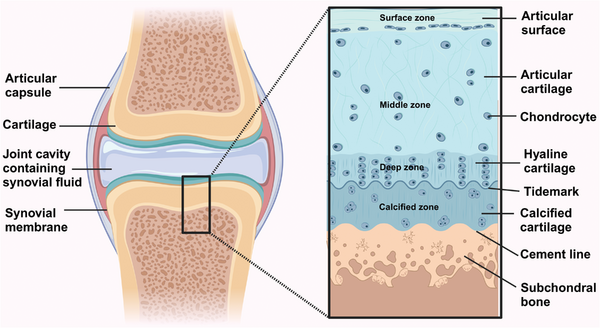
This cartilage is also called articular cartilage. It is located at the end of the bone. This cartilage is found between the two bones where they meet.
This cartilage prevents the ends of the bones from rubbing together. It gives the ability to withstand pressure. Due to this cartilage, the movement between the two bones becomes smooth and painless.
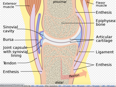
INTRA CAPSULAR STRUCTURE:
A structure is formed around the joint cavity where two bones of a synovial joint meet. The structure inside this capsule is known as the capsular structure or intracapsular structure. This capsule is a double-layered membrane. In which the outer layer is made of fibrous tissue and the inner layer is made of synovial membrane. The structure inside this capsule is called the intracapsular structure. Which includes the following structures.
SYNOVIAL MEMBRANE:
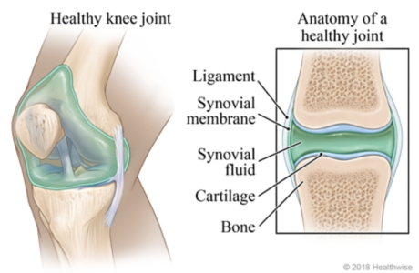
This membrane covers the lining of the capsule and all the membranes surrounding the joint. This membrane is not located in the hyaline cartilage. Apart from this, there is a membrane covering all the structures inside the capsule, which is called the synovial membrane. It is a membrane made of loose connective tissue.
This membrane secretes a fluid called synovial fluid.
SYNOVIAL FLUID:
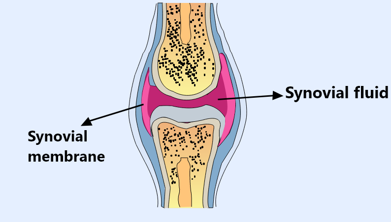
- Synovial fluid is a viscous fluid. It acts as a lubricant in the joint. The cavity where the synovial fluid is located is called the synovial cavity.
- This synovial fluid contains hyaluronic acid. This fluid contains some phagocytic cells which also function to protect the joint by removing microorganisms and some cellular debris present in the joint cavity.
- This fluid functions to nourish the internal structures of the joint by supplying nutrient material. This fluid also performs a wear and tear function on the joint. It is also important for maintaining the stability of the joint.
INTRA CAPSULAR LIGAMENT:
There are bands of connective tissue inside the capsule and at the ends covering the two bones of the joint. These are known as intracapsular ligaments. Ligaments within the capsule provide stability to the joint and help hold the two ends of its bones together.
BURSE:
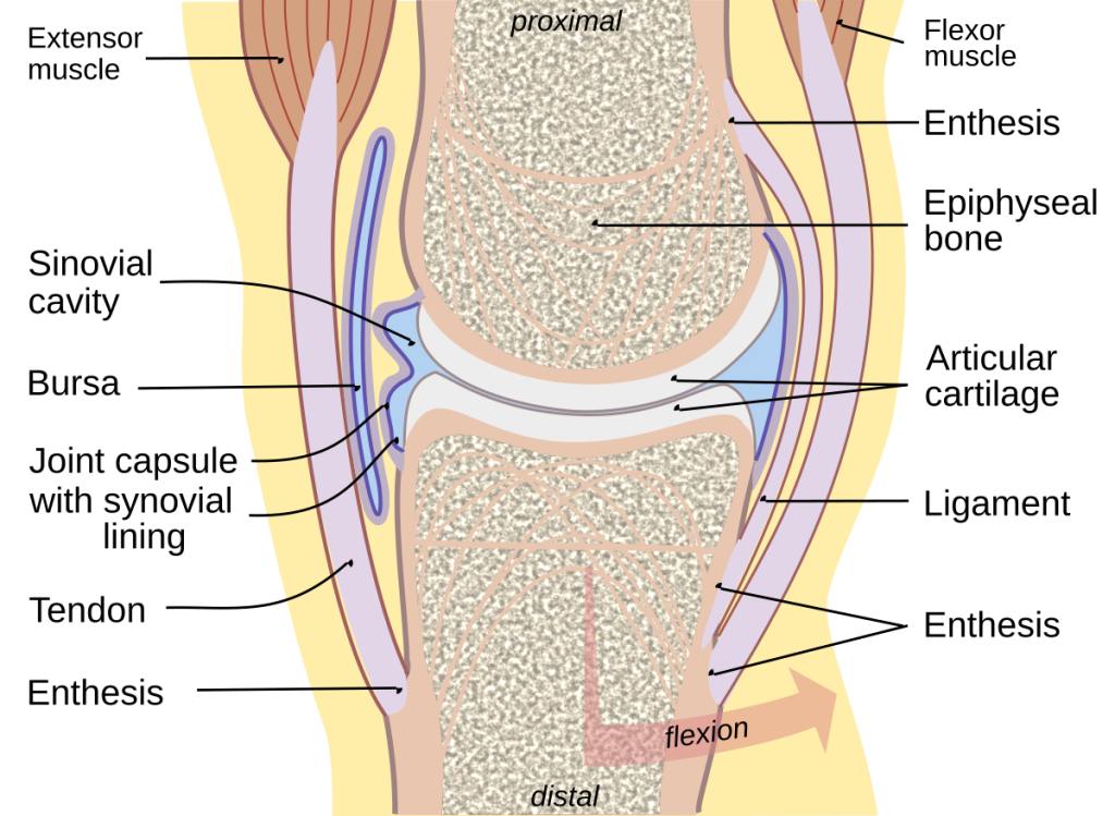
These small sacs are filled with synovial fluid. They are called bursae. These sacs are found around some joints, such as the knee joint. They are not located near the pressure-bearing part, but around it there is a sac that acts as a cushion. Which also plays an important role in preventing friction.
FIBRO CARTILEGINIOUS DISC (Fibro Cartilage Disc):
In some synovial joints, there is a disc made of fibrous cartilage near the articulating surface. This disc acts as a shock absorber for the joint. It gives the joint the ability to withstand additional pressure and stability.
EXTRA CAPSULAR STRUCTURE:
The structure that is located outside the capsule near both ends of the bone is called the extracapsular structure. Which includes the following structures.
LIGAMENT (ligament):
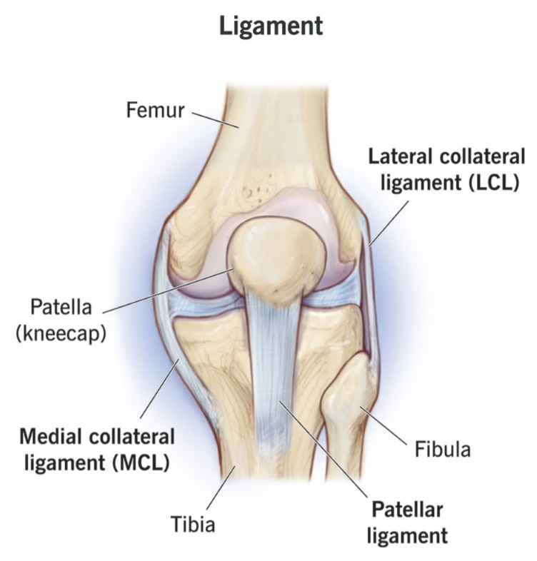
At the ends of the two bones at a joint are structures of fibrous connective tissue called ligaments. These structures pass through each end of the two bones and are connected there for strength and movement. These ligaments maintain the stability and position of the joint.
MUSCLES:
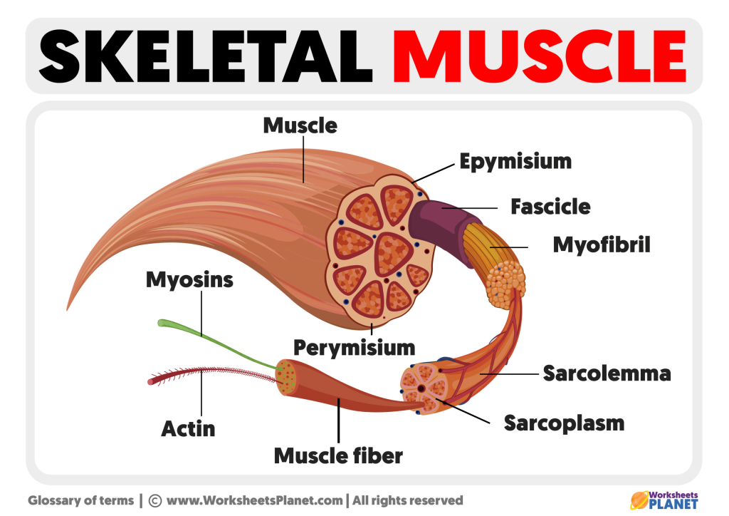
Muscles pass through the structures surrounding the joint. These are skeletal muscles. These muscles are connected to provide support and support to the joint. These muscles are also useful for performing different types of movements at the joint.
TENDON (tendon):
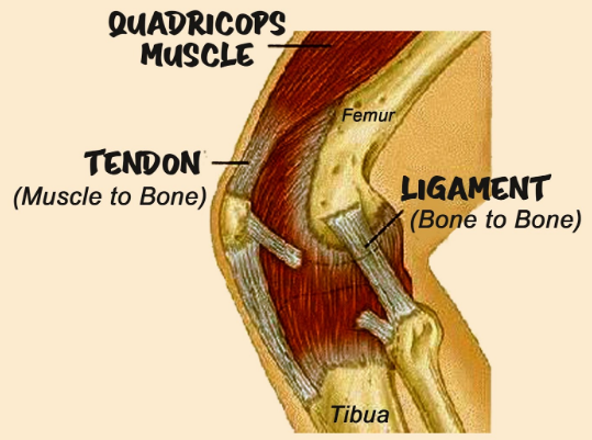
At the joint, the ends of the muscles are attached to the bones by tendons. These tendons are bands of connective tissue. Which attach muscles to bones and allow movement of the joint.
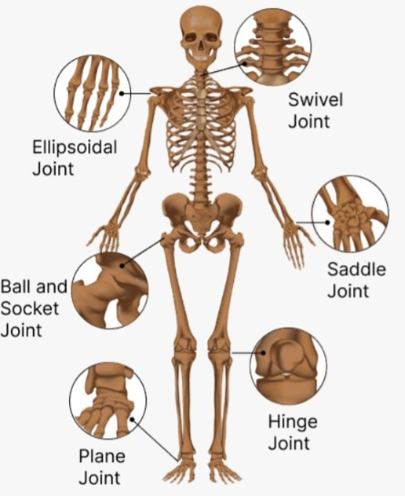
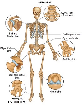
Types of Synovial Joint:
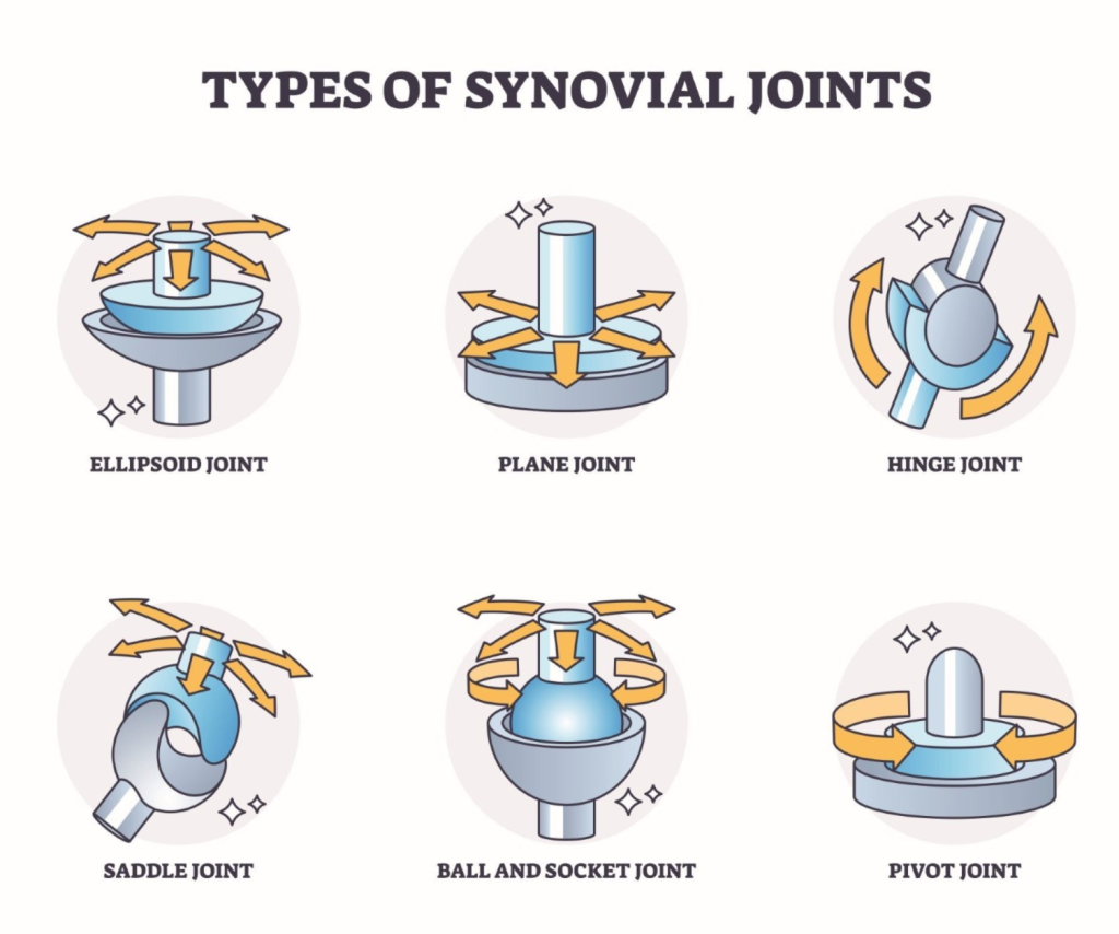
Synovial Joint is one of the softest and most mobile joints found in the body. Such joints have a synovial cavity between two bones, which is filled with synovial fluid. This synovial fluid reduces the friction between the bones and helps the patient for easy and effective movement. Synovial Joints are of different types according to their special structure and work.
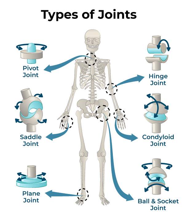
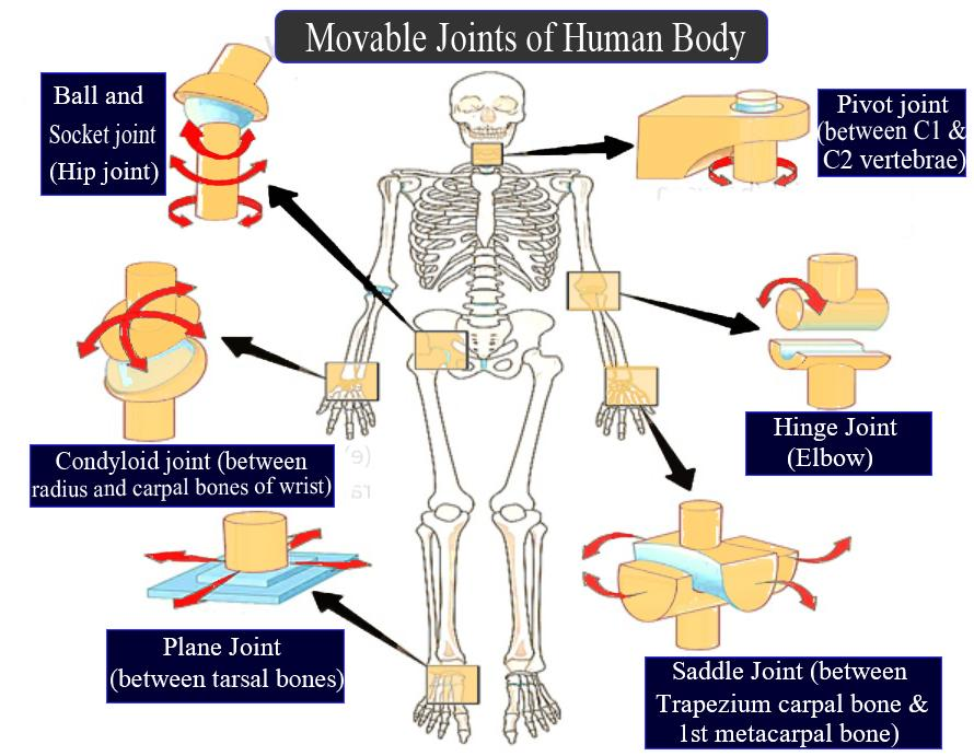
1.Hinge Joint:
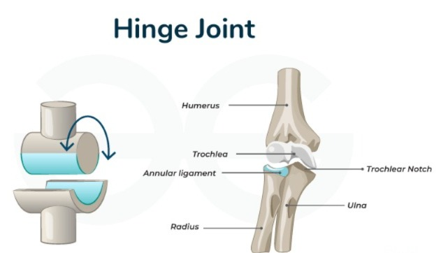
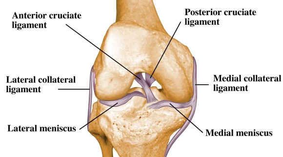
Example: Elbow, Knee, Interphalangeal Joint
This joint works like a hinge – it allows movement in only one direction, forward and backward, such as flexion and extension. This is a uni-axial joint. It is very important for the movement of the hand and foot in the patient’s daily life.
2.Ball and Socket Joint:
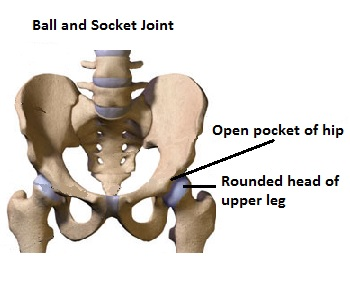
Example: Shoulder, Hip
In this joint, the head of one bone is like a ball and the other bone has a cup-like opening. This is a triaxial joint, in which all directions of movement such as Flexion, Extension, Abduction, Adduction and Rotation are possible. The joint that does the most work for the patient’s body’s major movement is.
3. Pivot Joint:
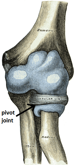
Example: Atlantoaxial Joint
In this joint, one bone fits into another bone like a ring and rotates around it. It is a uni-axial joint. The main joint for the patient to turn his head left and right is the saddle joint.
4. Saddle Joint:
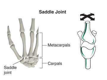
Example: First Carpometacarpal Joint
In this joint, both bones look like a saddle. This is a Biaxial Joint in which movements like Flexion, Extension and Opposition are possible. Specially works for thumb movement.
5.Plane Joint (Plane Joint) :
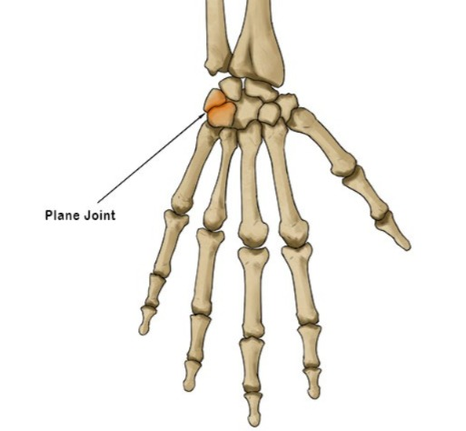
Example: Intercarpal Joint, Intertarsal Joint
In these joints, the bones have flat surfaces and they glide over each other. This allows for limited but important movement. It is necessary for small movements in the hands and feet.
6.Condyloid Joint:
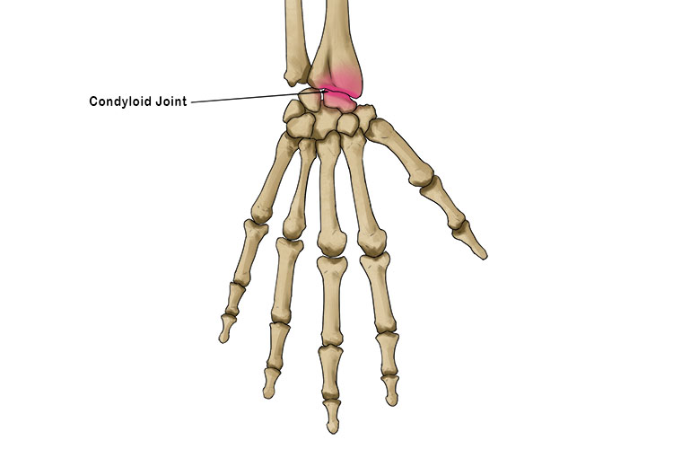
Example: Radiocarpal Joint (Wrist)
This joint is biaxial in which the oval head bone fits into the groove of the other. In this, movements like Flexion, Extension, Abduction and Adduction occur. Wrist Joint works especially for fine movements in the patient’s hand.
General features of Synovial Joint:
- Joint Capsule covers the joint
- Synovial Fluid reduces friction between bones
- Articular Cartilage covers the head of the bone
- Ligaments and Muscles provide stability to the joint
- Synovial Joint plays a major role in the movement of a person’s body. Each type of joint has its own specific structure and function. In cases of orthopedic conditions, movement disorders and body injuries, it is very important to have deep knowledge about Synovial Joint.
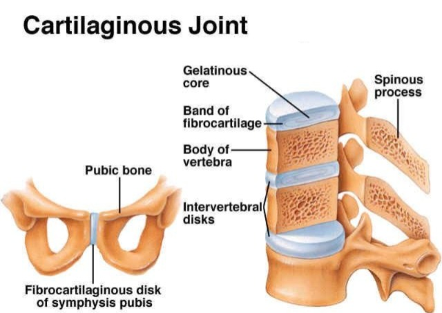
2.Slightly movable joints:
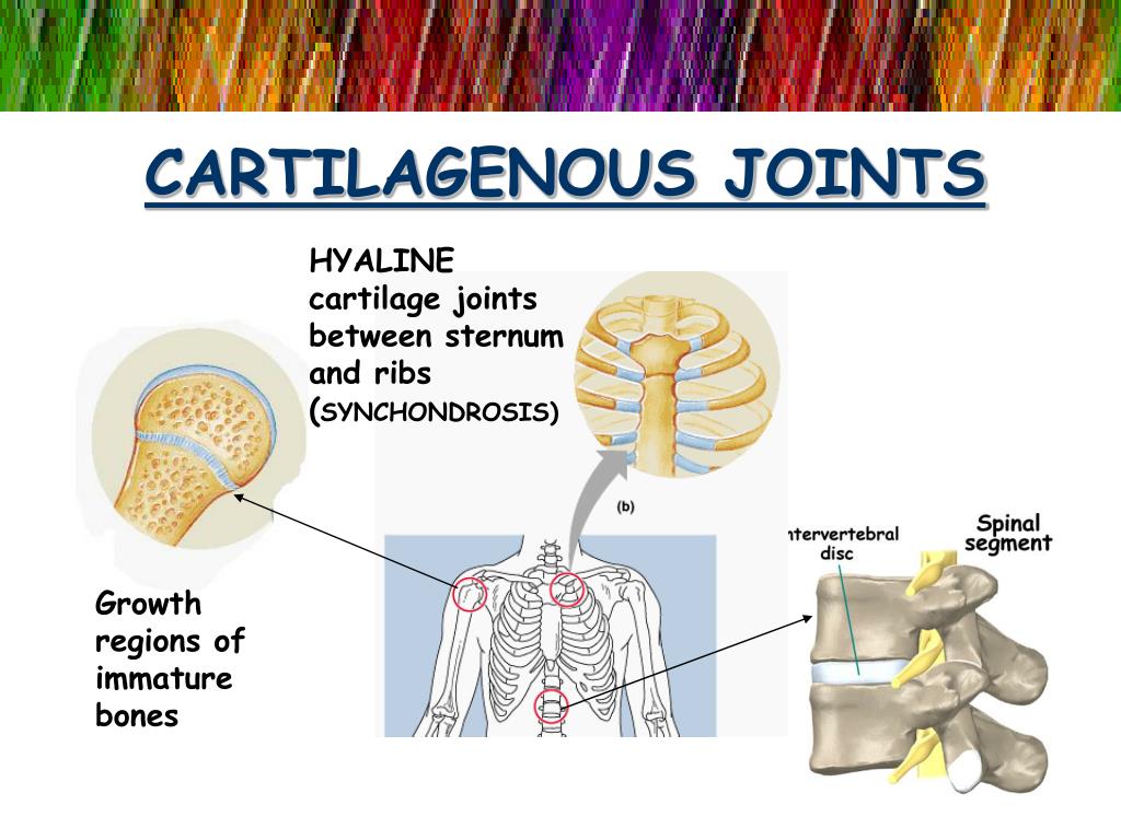
- This joint is also known as another Cartilagenous Joint. In which the bones are connected to each other with the help of cartilage. Due to this, some movement is seen in this joint. It can also be known as another Amphiarthroses.
- The joint formed between the bodies of two vertebrae, the joint formed between the sternum bone and the rib are such joints. Where some movement is seen in the joint area.
3.Fixed joint:

This type of joint is also known as a fibrous joint. In which the bones are closely connected to each other and no movement is seen in the joint. It is also known as
Synarthrosis.
The joints formed between the cranial bones of the skull are of this type. These joints form sutures such as the coronal suture.
Types of movements at various joints:
Joint movement is the movement that occurs through a joint between two or more bones. There are different types of joints in the bones of the body such as Synovial Joints, Cartilaginous Joints and Fibrous Joints, but especially Synovial Joints are responsible for the most movement.
1) Flexion:
Reducing the angles between the circulating bones or decreasing the angle between two bones in flexion.
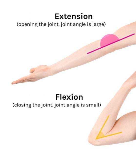
- Bending the head towards the chest.
- Movement of the palm towards the inside of the forearm.
- Making a fist.
- Bending the toes downwards.
2) Extension:
To straighten the joints. To increase the angles between the circulating bones. To restore a part of the body to its anatomical position after flexion or to increase the angle between the bones in extension.
- To provide an erect (straight) position to the head.
- Straighten the toes.
3) Hyperextension:
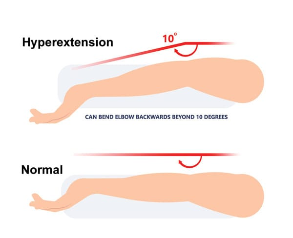
The normal movement of a part of a joint, extension or extension beyond the anatomical position of the condition is called hyperextension. When a joint of the body is extended beyond its normal range, it is called hyperextension.
4) Abduction:
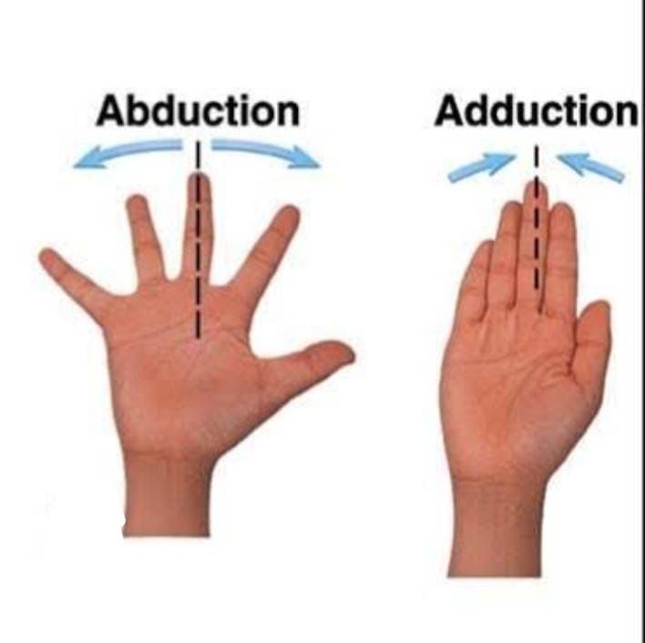
In abduction, the joint or extremities are moved away from the midline.
- The palm is moved laterally (from the side) at the joint of the palm )Move.
- Move the femur laterally in the hip joint.
5) Adduction:
In adduction, the joint or extremities are moved towards the midline.
- Fingers together to bring.
6) Circumduction:
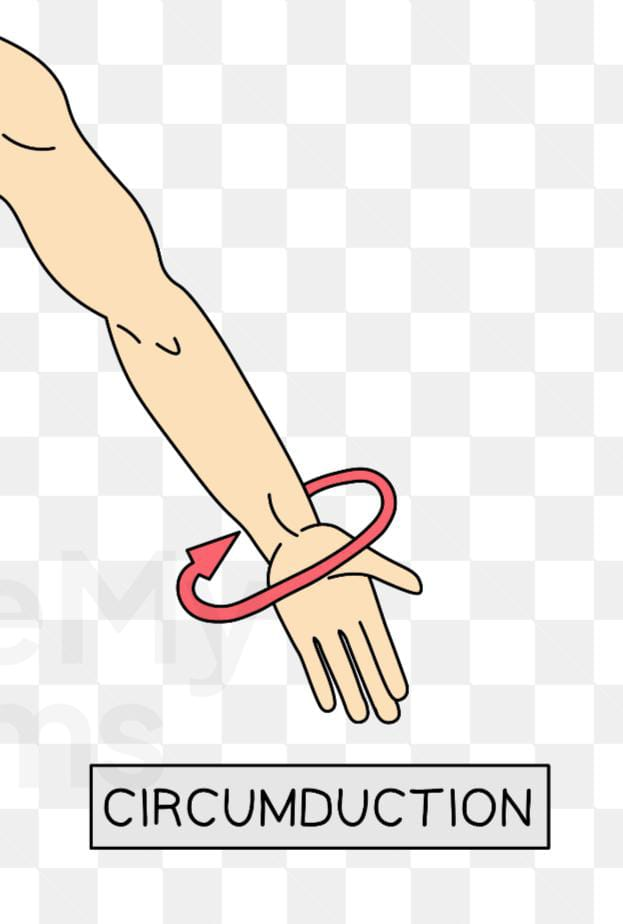
To move a part of the body inwards in a widening circle. It results from a continuous sequence of flexion, abduction, extension, and adduction. Eg.
- Moving the humerus in a circle of the solder joint.
7) Rotation:
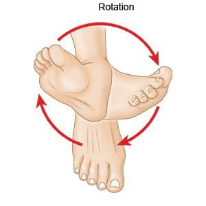
In rotation, the bone rotates around its longitudinal axis.
- Rotating the head from side to side
Two Types:
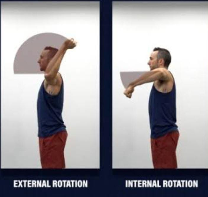
I) Internal rotation:
In internal rotation, the axis of the joint or The extremities are turned towards the midline of one’s body.
- Turning the foot or leg towards the other leg.
II) External rotation:
In external rotation, the axis joint or extremities are moved away from the midline of one’s body.
- Remove the foot and leg from the other leg.
8) Supination:
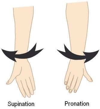
In it, the body parts are turned upward.
9) Pronation:
In it, the body parts are turned downward.
10) Inversion:
Feet Turn inward, so that the toes point towards the midline.
11) Eversion:
Turn the foot outward so that the toes point away from the midline or move the sole laterally at the intertarsal joint so that the soles are away from each other.
12) Dorsi Flexion:
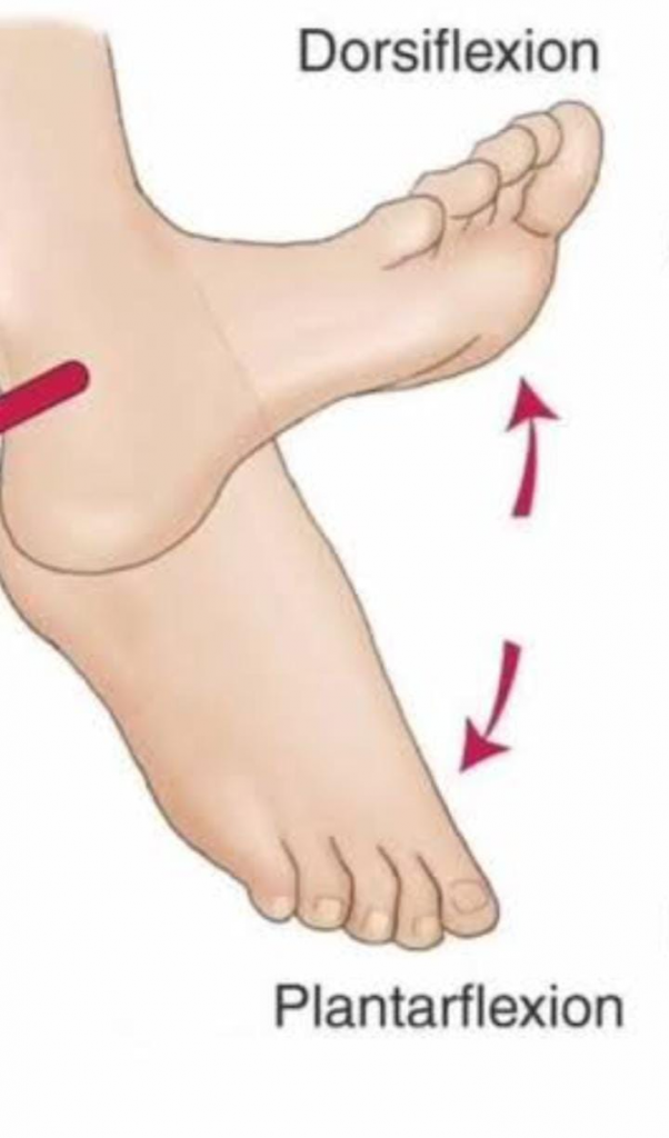
Bending of the foot in the direction of the dorsum (upper surface) at the ankle. E.g. Standing on heels.
13) Palmer Flexion:
Bending of the foot at the ankle joint in the plantar direction (inferior surface) e.g. standing on tiptoes.
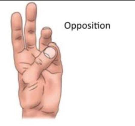
14) Opposition:
Apposition is the movement of the thumb across the palm to touch the fingertips of the same hand.
15) Lateral flexion:
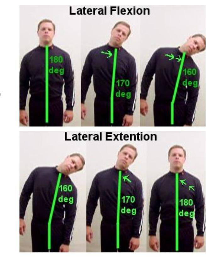
Lateral flexion occurs in the frontal plane. Movement of the trunk occurs in the frontal plane. It involves the involvement of intervertebral joints, e.g. Tilt the head as far as possible towards each solder.