ENGLISH ANATOMY UNIT 12. SENSE ORGANS. EYE
- Eye ball ..
The eye is an important organ for seeing which works like a camera. its shape is spiracle. its diameter is 2.5 cm it carries the nerve supply to the brain through the optic nerve and the function of seeing is done.
The eyeball is located in the orbital cavity in the skull. In the wall of this cavity there is a layer of adipose tissue to protect the eye. The wall around the eyeball is made of bone and this adipose tissue works to protect the eyeball. .
Structure…
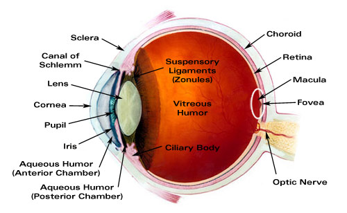
There are three layers in the structure of the eyeball.
- Outer layer consists of fibrous tissue which is the support layer containing sclera and cornea.
- The middle layer is the vascular layer containing the choroid, ciliary body and iris.
- The inner layer is made up of nervous tissue containing the retina.
Sclera…
It is the white colored part of the eyeball. The outer layer is composed of fibrous tissue. It is continuous anteriorly and forms the corneoscleral junction where it joins the transparent cornea. It functions to protect the delicate structures of the eye and maintain the shape of the eyeball. works. It provides attachment for attachment of extrinsic muscles.
Cornea…
It forms the anterior portion of the eye and forms the coloring part of the eye. It is convex from the front and transparent through which the rays of light enter and fall on the retina.
Choroid…
It is the middle layer of the eye and contains blood vessels so it is the vacuolar layer. It has blue, brown, gray colored pigmentation and is divided anteriorly into three structures, ciliary body, suspensory suspensory ligament and iris.
Ciliary body…
It is the thick part of the uveal track that is continuous anteriorly with the iris and posteriorly continuous with the choroid. At its front is the lens cavity which helps in accommodation of the lens for near vision. The ciliary body contains ciliary muscles of the unstripped type that provide attachment to the suspensory ligament. These ciliary muscles are important in maintaining the shape of the lens during near and distance vision. Thus, due to the change in the shape of the lens, the light rays can be focused directly on the retina. Nerve supply to these muscles is through the parasympathetic nerves.
Iris…
It forms the front colored part of the eye It is long and flat It is arranged vertically between the cornea and the lens It divides the eyeball into anterior and posterior chambers and these two chambers contain a fluid called atreus humor. It consists of circular and radial smooth muscle fibers and is connected by the ciliary body. The iris has an opening in the middle called the pupil. Fluctuations in the size of the pupil control how much light is allowed into the eye. Nerve supply by sympathetic and parasympathetic sympathetic dilates the pupil and parasympathetic constricts the pupil.
Lens…
The lens is the transparent portion of the eye. It has bicon cave structure. It is located behind the posterior chamber of the eye and is about 1 cm in diameter. The contraction and relaxation of the ciliary muscles causes changes in the thickness of the lens.If the object is close, the lens becomes thicker and allows light rays to focus more easily on the retina.
Retina…
It is the innermost layer of the eyeball. It is composed of nervous tissue. This layer of retina is continuously attached to the optic nerve at the back, next to it is a circular area known as the optic disc. Retina contains photoreceptors, rod cells and cone cells. Rod cells help in dim light vision and cone cells help in bright light and color vision.
At the back of the retina is a depression called the macula lutea, in the middle of this is called the central fovea or fovea centralis where the rays are focused and help to see. From there, the impulses of vision go to the brain through the optic nerve and the interpretation of vision takes place. There is a blind spot near the optic nerve, there are no light sensitive cells.
Interior of the Eye…
Eye ball is of spherical shape. Inside it lies a very large space known as KVT, these KVTs are called anterior and posterior KVTs.
Anterior CVD is again divided into two parts namely anterior chamber and posterior chamber. The anterior chamber is the front chamber of the iris and forms the back part of the cornea. The posterior chamber forms at the back of the iris. Both anterior and posterior chambers contain fluid called aqueous humor.
The posterior cvt is the cvt at the back of the lens and is larger than the anterior cvt. The fluid in this cavity is called vitreous humor. This fluid is high in water, contains some salts and proteins and helps maintain intraocular pressure. Normal intraocular pressure is 16 to 20 mmhg, if this pressure increases, the disease condition is called glaucoma.
- Physiology of site…
After returning from an object, the light rays pass through the conjunctiva and enter the eye through the cornea, then it reaches the anterior chamber and is focused in the lens and reaches the vitreous body, i.e. the posterior chamber of the eye. Concentrated at one place on the layer and through the optic nerve located there, the visual image of these light rays goes to the brain and a clear image is interpreted.
Size of the pupil…
Bright light pupil constriction
Dim light pupil dilate
Dilation of the sympathetic nervous system pupil
The parasitic nervous system causes constriction of the pupil.
- Accommodation of eye to light…
The eye works like a camera. Focusing the eye on any object and seeing it clearly is called accommodation. There are four stages in total.
- Refraction of light raises ..
Refraction takes place through the cornea and lens in which light rays coming from outside are refracted and reflected due to coming from one medium to another medium and it will be focused on a part of the retina through this band so that a clear image can be seen.
- Accommodation of the lens..
The ciliary muscles attached to the lens, the iris, help the lens adjust to see near or far objects. Changing the shape of the lens to see a closer object is called accommodation. To see a distant object, the ciliary muscles relax and the lens becomes flat.
- Construction of the Pupil..
The size of the pupil controls the amount of light entering it.
- Convergence..
Clear vision occurs when light rays falling on the retina can be focused to a single point.
Muscles of the eye…
Two types of muscles are attached to the eye
- Extrinsic…
It is also called as skeletal muscles It is attached to the wall of the orbital cavity on the outside and attached to the sclera part of the eyeball on the inside which is important for movement of the eyeball in different directions These extrinsic muscles include four straight muscles and two oblique muscles. which are as follows.
medial rectus
Lateral rectus
Superior rectus
Inferior rectus
Superior Oblique and Inferior Oblique Muscles
Extrinsic muscles are voluntary muscles that we can move the eyeball in different directions by our will.
Intrinsic muscles…
Intrinsic muscles are those on the inside of the eyeball, including the iris and ciliary muscles that are not under voluntary control. As light from outside enters the eye, its size and shape change. This is controlled by the autonomic nervous system.
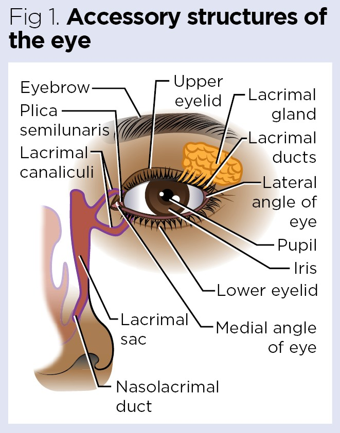
- Accessory Organs of the Eye…
The eye is a very delicate organ and is protected by some accessory structures around it.
- Eye Leads and Eye Laces
- Eyebrows
- Lacrimal apparatus
- Eye Leads and Eye Laces
The eye lid consists of an upper and a lower pair that both serve to protect the eyeball during sleep. The upper eye lid is a moving layer in front of the eye ball that protects the eyeball from any foreign body or object. This eye lead is also called the palpibra. The space between the upper and lower eye lids is called the palpebral fissure. The part where these two eyelids join is called the canthus, which is named medial and lateral according to the side of the body.
Along the margin of the eye lid, there are hair-like processes called eye lasis that protect the eyeball from bright light and prevent it from foreign bodies. At the base of the eye laces in the margin of the eye lead, there is a gland which keeps these hairs lubricated. When this hair follicle becomes infected, it is called a stye. This gland is known as tarsal or Meibomian gland.
Eyebrows…
Eyebrows are the hairs on the supraorbital margin, the upper margin of the orbital cavity. It prevents sweat from entering the eye area and works to protect the eyeball from bright light and injury. These eyebrows are also known for cosmetic purposes.
Conjuctiva…
The protective mucus membrane inside this eye lead is called the conjunctiva. It is a membrane composed of columnar epithelium cells. It contains goblet cells that close the eye when we close it and protect the eyeball.
Lacrimal apparatus…
The lacrimal apparatus consists of the following structures that surround the eyeball and secrete tears that shed on the surface of the eyeball. And the eyeball remains moist The structure of the lacrimal apparatus is as follows.
- Lacrimal gland and its duct…1
- Lacrimal canali culi…2
- Lacrimal sac …1
- Nasolacrimal duct…1
Lacrimal glands are two in number located one on the lateral side of the superior eyelid. It is almond shaped. The fluid it secretes is called tears. This fluid drains through the lacrimal duct and spreads over the surface of the conjunctiva and the surface of the eyeball and keeps the eyeball wet.
Composition of Tears.
Water
Mineral salt
Antibodies and lysosomes that contain bactericidal enzymes
The secretion of this lacrimal gland collects in the lacrimal sac through the superior and inferior canaliculi leading to the middle canthus of the eyeball and from there opens into the nasal cavity through the nasolacrimal duct.
Functions of the tear…
Provides nourishment to the cornea
Works to flush out irritating substances and dust that fall on the eyeball
Having bactericidal property, it prevents infection by micro-organisms
Being oily acts as a lubricant
It prevents the eyes from drying out