ENGLISH ANATOMY REPRODUCTIVE SYSTEM FEMALE
REPRODUCTIVE SYSTEM
- INTRODUCTION
An ability by which an individual can produce a new individual or give birth to its offspring. This process is called reproduction.
Because of reproduction that species maintains its existence.
Almost all the systems of the body start at the time of birth, but the reproductive system is such a system, which starts (active) at the time of puberty.
There are some types of germ cells inside the human being. It is called gametes.
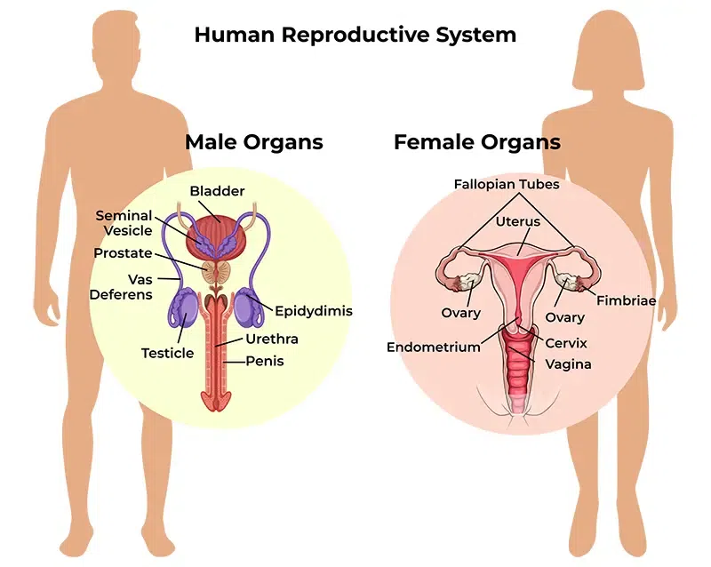
Primary sex organ of female is ovary and male has testes.
Ovary produces female egg and testes produces male sperm.
All other reproductive organs work as accessory organs and supportive organs.
Male and female gametes, when fused, form a zygote, which develops into a fetus.
Human reproductive system is as follows :-
1) FEMALE REPRODUCTIVE SYSTEM
2) MALE REPRODUCTIVE SYSTEM
1) FEMALE REPRODUCTIVE SYSTEM
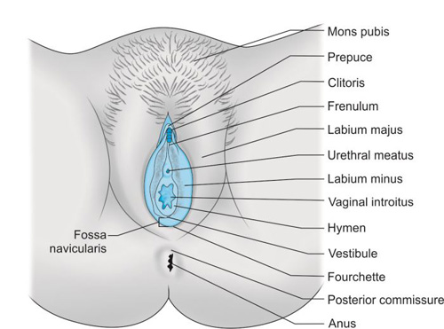
A. External genitalia of female / VULVA / PUEDENDAM
Mons pubis
Labia majora
Labia minora
Clitoris
Perineum
Vestibule
Mammary gland
MONS PUBIS
The mons pubis is a cushion-like structure. Which is made up of fat and skin. which lies in front of the symphysis pubis.
During puberty it becomes covered with hair and forms a horizontal margin.
LABIA MAJORA
It has two thick folds on the front side. which forms the brim of the vulva. (vulva means external reproductive organ is also called vulva).
Labia Majora is composed of skin, fat, areolar tissue, and smooth muscle. Hairs are located on the upper surface. There are sebaceous glands located inside it.
The round ligament ends at the labia majora.
The labia majora meets anteriorly near the mons pubis and posteriorly near the skin of the perineum. As males have testes, females have labia majora.
LABIA MINORA
Labia minora are two small skin folds (which do not contain fat, hairs, but many shibaceous glands) that lie inside the labia majora.
Its upper part joins the clitoris and forms the prepuce (prepus – like the foreskin of the penis in the female), and the lower part forms the floor of the clitoris (frenulum).
CLITORIS
It is of cylindrical shape and triangular shape. It is made up of erectile tissue.
The female has the clitoris as the male has the penis.
Sensory nerve endings are located inside it. Acts as a stimulant during sexual intercourse.
PERINEUM
This is an area which extends from the base of Libya Majora to the anal canal. It is roughly triangular in shape.
It is made up of connective tissue, fat, and muscle. It provides attachment to the pelvic floor muscles.
VESTIBULE
Vestibular gland i.e. (Bartholin gland) is 2 and located on both sides of the vaginal opening. Its size is like a pea. It has ducts that open into the vagina and secrete mucous. This keeps moisture in the vulva and keeps the vaginal cavity wet.
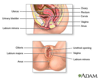
- INTERNAL REPRODUCTIVE ORGANS.
These organs reside within the pelvic cavity. Which includes the following organs.
Internal reproductive organs are as follows.
Vagina
Uterus
Uterine tube or Fallopian tube or Salpinges
Ovaries.
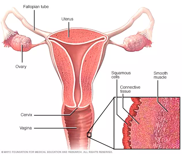
- VAGINA
Vagina is a fibro muscular tube. The lining of which is composed of stratified squamous epithelium cells. Which is the structure connecting the internal and external reproductive organs.
It has urinary bladder in front and rectum behind. Its front wall is 7.5 cm and back wall
Structure of Vagina
There are three layers of vajaina
1.Outer layer
- Middle layer
- Inner layer
1.Outer layer :- The outer layer is made up of areolar tissue
- Middle layer :- The middle layer is made of smooth muscles.
- Inner layer :- It is made up of stratified squamous epithelium tissue. which form rugae (ruga-like projections).
It has no secretions. Due to the secretion of cervical (cervical) it remains moist.
Between puberty and menopause, a bacteria called lactobacillus acidophilus makes the pH of the vagina acidic. Which is 4.9 to 3.5. Because of the acidic pH, infectious microorganisms do not enter the vagina.
Blood Supply:-
through the uterine artery and the vaginal artery which is a branch of the internal iliac artery.
The venous plexus is located in the wall of the muscles and drains into the internal iliac vein.
NERVE SUPPLY
There are parasympathetic nerves, sympathetic nerves and somatic nerve supply
FUNCTIONS
Vajaina allows entry of the penis during intercourse.
Holds the sperm until it moves to enter the uterus.
There is an elastic structural part that helps the baby to come out during birth.
The sensory nerve endings in the vagina provide the filling of sexual pleasure.
Acts as the outer opening of the urinary tract.
Women have monthly menstrual bleeding so blood from the uterus comes out through the vagina.
The acidic pH of the vaginal cavity inhibits the growth of micro-organisms.
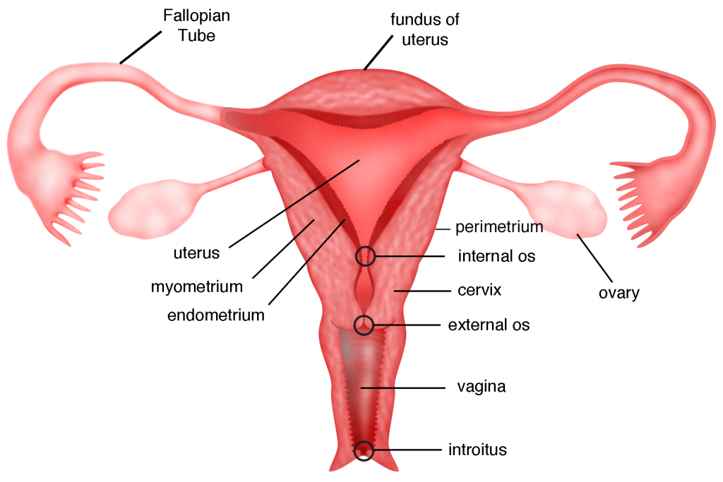
- UTERUS
Uterus is a hollow (polu) and muscular inverted pear (peru) shaped organ. It is an organ located inside the pelvic cavity. Which is located between the urinary bladder and the rectum.
In most of women, the uterus is anteflexed and anteversion and at a 90°-right angle.
Its anterior wall lies above the bladder and forms the vesicouterine pouch (the pouch of peritoneum between the bladder and the uterus is called the vesicouterine pouch).
When the body is in upright position, the uterus is 3 inches in length, 2 inches in diameter and 1 inch in thickness. Its weight is 30 to 40 grams.
The parts of uterus are as follows:-
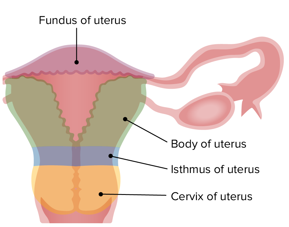
Fundus:– It is dome shaped. The fallopian tube opens from its top.
Body:- Body is the main part. The uterus becomes narrower as it descends. Forms the internal os (mouth). Which continues to form the cervix.
Cervix:- It is called neck of uterus. Anterior part of vagina opens or protrudes. Its opening is called external os.
STRUCTURE
Uterus has 3 layers.
perimetrium
Myometrium
Endometrium
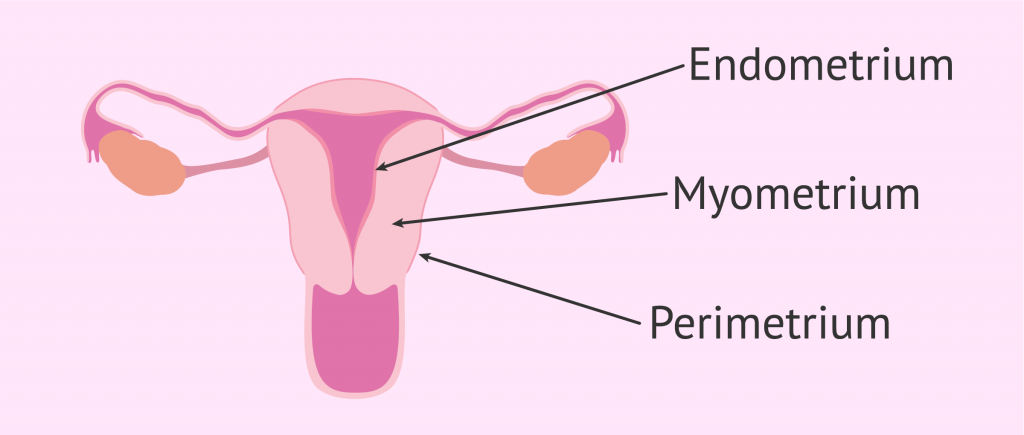
Perimetrium
This is a layer of peritoneum. Which is spread around the uterus. Anteriorly there is a fold of peritoneum over the fundus and body and superiorly over the urinary bladder.
Anteriorly the pouch of peritoneum between the uterus and bladder is called the vesico uterine pouch while posteriorly the peritoneum extends from the fundus to the cervix. The pouch that forms between the posterior rectum and the uterus is called the pouch of Douglas or the rectouterine pouch.
Laterally it is double folded. A layer of peritoneum forms the lateral broad ligament and the round ligament, which join the uterus to the pelvis.
MYOMETRIUM
This is the thickest layer of the uterus. It contains a mass of smooth muscle and is accompanied by areolar tissue, blood vessels and nerves.
ENDOMETRIUM
It is composed of columnar epithelium tissue and tubular cells that secrete mucus are located in the glands.
It has two layers in total.
Functional layer
Basal layer
1.Functional layer (functional layer)
It is the top layer. It is filled with blood vessels for the first 15 days or half of the cycle and if the ovum (egg) is not fertilized, this layer falls off and the menstrual cycle (menstruation) begins.
If fertilized, this remains for 9 months (pregnancy) and then sheds and during 9 months it is called decidua.
2.Basal layer
It lies in front of the myometrium. which does not come out during the menstrual cycle. This is the layer, which creates a new and fresh layer.
2/3rd layer of endometrium is made up of mucous membrane and the bottom part is made up of stratified squamous epithelium up to vagina.
BLOOD SUPPLY
Arterial supply :- Blood is supplied by the uterine artery and it is a branch of the internal iliac artery.
Venous drainage :- Vein is similar to artery but blood drains into iliac vein.
Nerve Supply
The parasympathetic nerve supply originates from the sacrum and the sympathetic nerve supply originates from the lumbar region.
Supporting Structure
The uterus is supported by surrounding organs. Which is in the pelvic cavity and where the ligaments are located. which supports the uterus. Like :-
Broad ligaments
round ligaments
Uterosacral ligaments
Transverse cervical ligaments
Pubo cervical fascia
Due to the above structure, the uterus holds its shape and provides structural support to it.
FUNCTIONS OF UTERUS
Uterus helps in fertilization of ovum and sperm.
After fertilization, it helps the zygote to implant in the inner wall of the uterus and maintain the pregnancy.
In pregnancy, the content inside the uterus increases, the size of the uterus also increases so that the pregnancy can continue.
It functions to provide nutrition to the fetus in the uterus during pregnancy.
The muscles of the uterus contract to help the baby come out during delivery.
The endometrium, the inner wall of the uterus, breaks down during the menstrual cycle. As this cycle continues every 26 to 30 days, the chance of infection is reduced due to influx of wbc.
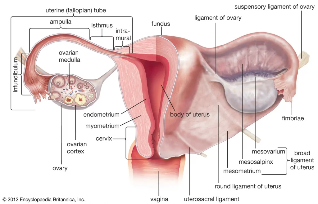
- FALLOPIAN TUBE / SALPHINGES / MULERIAN DUCT / UTERINE TUBE
It is in the number of 2. It is attached to the fundus and body and one on both sides of the fundus. It is 10cm long.
Its main parts are as follows.
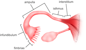
Interstitial:- This part is attached to the uterus. It is part of the opening after the fundus of the uterus.
Isthmus:- This is narrow and about 2.5 cm.
Ampulla:- Ampulla is the most prominent part of the tube. Here the ovum and sperm are fertilized.
Infundibulum:- This is the last part of the fallopian tube and is shaped like a jug.
At the end of this, a finger like projection is formed. It is called fimbriae. Fimbriae receive egg cells. It has three layers
Outer layer which is made up of peritoneum.
The middle layer is made up of muscles.
The inner layer is composed of mucus membrane and ciliated epithelium tissue.
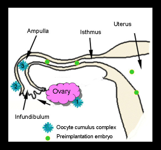
Functions of FALLOPIAN TUBE
It causes peristalsis movement of the egg (ovum) and reaches the ampulla.
The mucus secreted from it provides an ideal environment for the ova and sperm.
Egg and sperm are fertilized here and a zygote is formed.
Fertilize transports the zygote to the uterus.
- Ovaries
The ovary is the female gonad. It lies on either side of the uterus in the ovarian fossa. It lies behind the broad ligament.
It is in number of 2 (pair of overs). It is homologous to testes.
It is attached to the pelvic wall by ovarian wall ligaments.
Location :- It surrounds the ovarian fossa (fossa is a pit-like structure inside which any organ is located) which is behind the ureters (behind the ureters), obliterated umbilical artery (an artery that forms shortly after birth is eliminated) in front of it
and is supported below by obturated interosseous muscles, vessels and nerves.
Shape and Size :– Ovary is of oval shape. It is 3 cm long, 1.5 cm high and 1 cm thick.
Surface and Color :- Ovaries are pink in color and smooth inside young adults. In older women, it is rough, irregular and gray in color because of frequent ovulation.
Attachment :- Both ovaries are attached to the uterus superiorly by the ovarian ligament and posteriorly by the broad ligament. It is called mesovarium, blood vessels and nerves pass through mesovarium.
Blood Supply
It originates from the ovarian artery and is a branch of the abdominal aorta.
The vein is called the pampiniform plexus. It emerges from the ovary itself.
The right vein drains into the inferior vena cava and the left vein drains into the left renal vein.
Nerve Supply
Sympathetic fibers that originate from the T10 and T11 spinal ligaments.
The parasympathetic nerve originates from the vagus nerve.
- Histology Of Ovary
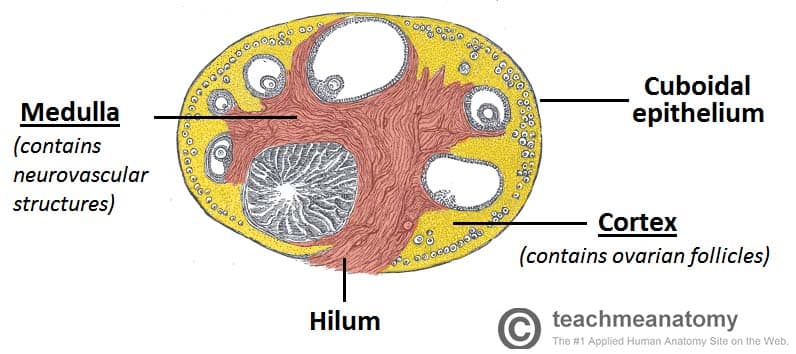
The ovary is oval in shape.
Inside it is a single layer composed of cuboidal epithelium cells. It is called germinal epithelium.
Dense tissue is present in it. It is called tunica albinia and is located inside the germinal epithelium.
It has total 2 parts.
Cortex:- It consists of stoma and ovary follicles.
Medulla :- Made up of connective tissue and connected by a network of blood vessels and made up of elastic fibers.
- Oogenesis.
This is a process of making female gametes. The process by which ova develop from primary germ cells is called oogenesis.
About 6 million oogonia are present when the intrauterine period is 6 months. Oogonia undergo meiotic division and convert to primary oocyte and arrest in prophase.
The 1st meiotic division arrests at puberty. Menstruation begins when puberty begins. The first menstrual cycle is called menarche.
At the beginning of the first menstrual cycle, ovulation occurs and the egg is released and ovulation occurs, the first egg is released.
Follicles form when ovulation occurs. It occurs with the help of FSH and then it is seen due to estrogen. Those follicles mature and become Graafian follicles.
A Graafian follicle develops into a mature ovum. The ovum is released on the 14th day of the menstrual cycle and if it is fertilized it becomes fitus and if it is not fertilized then menstruation occurs.
- Menstruation Cycle
Menstruation cycle occurs after puberty phase in females.In which changes are seen in the function of ovaries and uterus.
A menstrual cycle occurs every 26 to 30 days. This is seen due to changes in blood hormone levels.
The onset of the menstruation cycle is known as menarche.
Females find this cycle continuous after the age of puberty. Which stops temporarily during pregnancy and stops completely after the period of menopause.
The onset of menstruation is due to the degeneration of the corpus luteum layer in the uterus and bleeding occurs through the vaginal cavity.
Menstruation cycle has the following phases.
- Menstrual phase..
This phase occurs every 28 days and lasts for about four days. When fertilization of the egg does not take place in the female, the hormones estrogen and progesterone that support the uterine wall decrease and the hormone oxytocin increases. So the stimulation of contraction of the uterus increases and degeneration of the corpus luteum layer of the wall of the uterus starts and blood drains from the uterus through vaginal discharge. This phase lasts from 1 to 4 days.
This menstrual flow contains endometrial glands, endometrial cells, blood and unfertilized ovum. Approximately 100 to 200 ml of blood is shed during the 3 to 5 days of this phase which is called the menstrual phase.
- Proliferative phase..
The menstrual phase ends on the 5th day. After that, the proliferative phase starts from day 6 and lasts for 14 days.
In this phase follicle stimulating hormone stimulates the ovarian follicles and hence increases estrogen production. This estrogen stimulates the proliferation of the endometrium.
The endometrium of the uterus begins to develop from the sixth day.Its cells multiply and due to this increase in mucus secreting glands and blood capillaries. Thus the endometrium of the uterus becomes bulky and vascular.
At the end of this phase, the inner wall of the uterus is ready for implantation of the fertilized egg. This phase ends with ovulation. Towards the end of this phase, there is a decrease in estrogen levels.
- Secretary phase.
After the completion of the proliferation phase, the secretory phase begins. The secretory phase is seen starting from the 15th day of the menstrual cycle to the 28th day.
As progesterone hormone is important in this phase, this phase is also called progesterone phase.
When the mature egg is released by the ovary due to ovulation, the amount of estrogen and progesterone hormones decreases, but the corpus luteum, the wall of the uterus, maintains the pregnancy by secreting progesterone.
As this mature ovum is not fertilized by a sperm, the corpus luteum decreases progesterone and due to the decrease in progesterone hormone, there is an increase in the amount of oxytocin hormone and the uterine muscles begin to contract.
The next cycle begins at the end of this phase due to corpus luteum not receiving a fertilized ovum and increased uterine contractions.