ENGLISH – ANATOMY & PHYSIOLOGY-2019 PAPER 1
GUJARAT NURSING COUNCIL PAPER 2019.
QUE 1.🔸 (a) Explain the gross structure of the heart 03 marks
Heart…
Heart is an important organ of the circulatory system. Heart beats continuously during human life. Due to its pulsation, the blood circulates continuously in the blood vessels.
The heart is an organ made up of blood and muscles. It weighs approximately 310 grams in males and approximately 250 grams in females. The heart beats approximately one lakh times during the day.
Location of the heart..
The heart lies above the diaphragm in the mediastinum space between the two lungs in the thoracic cavity.
The heart is a rough cone shape. In it, its upper broad part is known as the base and the lower angled part is known as the projection.
The heart is arranged slightly to the left between the two lungs in the thoracic cavity.
Structure of the Heart..
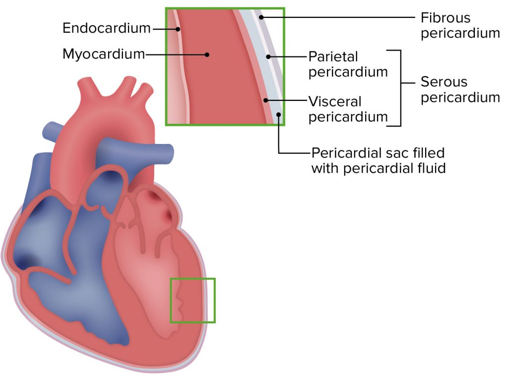
Heart is an organ made up of semi-muscles. Its wall is made up of three types of tissue layers.
The outermost layer of the heart wall is called epicardium or pericardium.
Epicardium or Pericardium.
It is thin and transparent and covers the heart from the outside. It is made up of fibrous connective tissue. In which there is a layer of fibrous tissue on the outermost side and a serous membrane on the inner side of the fibrous tissue which is found in a double layer. The outer layer of the serous membrane is known as the parietal layer and the inner layer as the visceral pericardium layer.
The space between the parietal and visceral pericardial layers is called the pericardial space. This space contains fluid called serous fluid or pericardial fluid. Which prevents friction between the two layers.
This layer of outer pericardium protects the heart from the outside and this layer is also seen around the vessels coming out of the heart.
Myocardium..
Myocardium is the middle layer of the heart. It lies below the pericardium. It is made up of a special type of cardiac muscle tissue. The pumping action of the heart is seen due to the contraction of these muscles.
This myocardium layer is thin at the base and thick at the apex. Also, the layer of the wall of the left ventricle is thicker than that of the wall of the right ventricle.
The contraction of these muscles has an involuntary action that results in the pumping action of the heart and is controlled by the autonomic nervous system and the conducting system in the heart.
endocardium..
It is the innermost layer of the heart wall. It is in contact with layer blood. This layer is made up of epithelium tissue and connective tissue. This layer is smooth and shiny which is important for the smooth flow of blood inside the heart. This layer also covers the valves inside the heart and this layer is continuous in the inner wall of the blood vessels leaving the heart.
(b) Write the Functions of the heart.. 04 marks
Heart provides oxygenated blood supply to all organs and tissues of the body.
Heart is an important organ of the cardiovascular system. It functions as a vital organ without which the human body cannot survive.
The heart circulates the blood towards the lungs so that the blood can be oxygenated and purified.
Circulations like pulmonary circulation and systemic circulation are regulated by the heart.
The heart also regulates the heart rate according to the needs of the body and according to the body temperature.
The heart also regulates body temperature as it circulates blood to every part of the body.
As the heart pumps blood to the body’s excretory organs, the blood can be filtered and waste products removed from the blood.
(c) Explain the pulmonary circulation. 05 marks
Pulmonary circulation started from the right ventricle and the blood goes to the lungs and from there returns to the left atrium, so the circulation from the right ventricle to the left atrium is called pulmonary circulation.
In the pulmonary circulation, deoxygenated blood in the right ventricle exits the right ventricle through the pulmonary artery. As it exits, the pulmonary artery divides into a right and a left pulmonary artery and both enter the lung. In which two branches in the left lung and three branches in the right lung enter the pulmonary artery which is according to each lobe of the lung.
Gas exchange takes place between the blood in the lungs and the tissues of the lungs and two pulmonary veins carry oxygenated blood from each lobe and enter the left atrium of the heart from the lungs on both sides.
Pulmonary circulation converts deoxygenated blood in the heart to oxygenated blood via the lungs. This blood enters the left ventricle and supplies oxygenated blood to the whole body through the systemic circulation.
Circulation from right ventricle to left atrium is called pulmonary circulation.
or
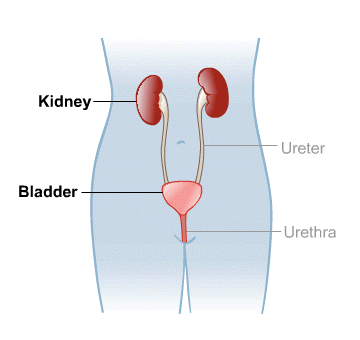
Que 1🔸(a) List out the organs of the urinary system ..03 marks
The urinary system is the excretory system of the body. Which removes waste products from the body. It has the following organs.
Kidney (Right and Left) 2
Ureter (right and left) 2
Urinary Bladder 1
Urethra 1
🔸(b) Describe the gross structure of the Kidney.. 04 marks
There are two kidneys in the human body. They are located one on both sides of the vertebral column on the poster side of the body on the right and left sides of the abdominal cavity.
Kidney is a shapeless organ. It lies from the level of the twelfth thoracic vertebra to the level of the third lumbar vertebra.
Kidney is 11 cm long by 5 to 6 cm wide. Its weight is approximately 150 grams. The right kidney is positioned slightly lower than the left kidney because the liver occupies a larger portion on the right side.
Veins around the kidney.
The kidney is an organ located in the abdominal cavity. One is located on both the right and left sides. Abdominal cavity organs like liver, small intestine, adrenal glands, stomach, spleen, pancreas etc. are located around both kidneys.
Structure of the Kidney..
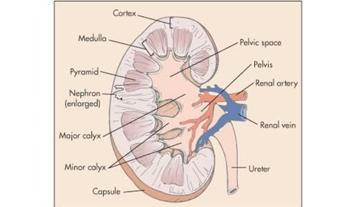
Kidney is a shapeless organ. A groove in the middle is called hilum or renal hilum. Through which the structures of renal artery, renal vein, nerves, lymph vessels and ureter enter and exit.
The inner border or hilum of the kidney is found on the side of the vertebral column. Its outer border is convex. The kidney is a hanging organ on both sides of the abdominal cavity. To hold it in position, it is surrounded by a network of fatty tissue and fibroelastic connective tissue called the renal fascia. With the help of this kidney can maintain its position and it also gets protection.
When the kidney is viewed in a longitudinal section, it is seen to be distributed into three kidney structures.
- Fibrous capsule.
It is part of the fibrous tissue that surrounds the kidney. This membrane is arranged around the kidney. Which acts as a layer to protect and maintain the shape of the kidney.
- Cortex.
It is redis brown in color made up of tissue. Which is located under the kidney capsule.
- Medulla.
In the kidney, the inner part from the cortex is called the medulla. It also has redish brown color. The triangular shaped pyramidal structure is called renal pyramid. The base part of this renal pyramid is towards the cortex and the pointed part of the pyramid i.e. the part of the renal papilla is arranged inwards towards the hilum.
The renal papilla forms a cup-like structure anteriorly called the calyx. The part with large space is called major calyx and the part with small space is called minor calyx. The minor calyx opens into the major calyx. Beyond this calyx is the wide funnel-shaped portion called the renal pelvis.
The urine filtered by the kidney falls into the wide part of the calyx, the funnel shape, i.e. the renal pelvis. Urine collects here and then passes anteriorly from the renal pelvis through a narrow structure called the ureter that exits the kidney and reaches the urinary bladder.
Urine filtered by the kidney passes from the minor calyx to the major calyx and from the major calyx to the renal pelvis. It then reaches the urinary bladder through the ureter. This action is not controlled by any kind of nervous system. In the wall of the renal pelvis there are special muscles and pacemaker cells due to the contraction of which this urine flows forward.
🔸(c)Write functions of Kidney Write.. 05 marks
Kidney is mainly responsible for urine formation.
Kidneys filter the blood and remove the waste products through urine.
The function of the kidney is to maintain the normal balance of electrolytes.
It works to maintain blood pH.
The body functions to remove waste products accumulated at the end of metabolism from the body.
Kidneys secrete a hormone called erythropoietin which plays a very important role in the production of RBCs.
Kidneys secrete a hormone called renin which plays a very important role in maintaining blood pressure.
Kidneys are responsible for maintaining water balance in the body.
Kidney prevents the elements that are needed in the body from leaving the body.
Que 2🔸(a) Explain the gross structure and functions of the Liver ..O8 marks
Among all the glands in the body, the liver is the largest gland. which lies below the diaphragm in the right quadrant of the abdominal cavity. It weighs approximately 1.4 kilograms inside an adult. It is located below the ribs of the chest. Ribs protect it. A part of it is also located in the region of the left abdominal cavity.
The upper surface of the liver is smooth. This portion is attached to the diaphragm and has an irregular surface and margin on the back and underside of the liver.
Organs around the liver.
Organs such as the diaphragm, anterior abdominal wall muscles, stomach, duodenum, kidney, inferior vena ceva, gallbladder etc. are arranged around the liver.
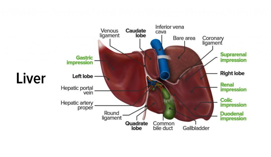
Liver is mainly divided into two lobes, right lobe and left lobe.
The right lobe is larger than the left lobe and both lobes are separated by the falciform ligament.
The quadrate lobe is found on the posterior side of the liver and the quadrate lobe is found on its inferior side.
Thus four lobes of liver can be seen anatomically.
Portal fissure…
The groove on the posterior surface of the liver is called the portal fissure. From this fissure some structures enter the liver and some structures exit the liver viz.
The portal vein carries deoxygenated and nutritious blood from the intestine and enters the liver through this fissure.
The hepatic artery carries oxygenated blood and enters the liver through this fissure.
Nerve fibers of the sympathetic and parasympathetic nerves enter the liver through this fissure.
Left and right hepatic ducts exit this fissure to carry bile from the liver to the gall bladder.
The hepatic vein exits this fissure to carry deoxygenated blood from the liver.
Structure of the Liver…
The liver is an organ located in the right quadrant of the abdominal cavity. It is mainly composed of 2 lobes. These lobes are made up of many lobules. These lobules are made up of special type of epithelium cells and cells called hepatocytes. These cells are found in a hexagonal pattern in the liver.
After entering the hepatic artery and portal vein within the liver, a capillary network of arteries and veins is formed. This network of capillaries is called sinusoids. Kupffer cells are found in the wall of these sinusoids, which Kupffer cells act to protect the liver from bacteria, foreign material or toxins and perform a protective function in the liver.
Hepatocytes cells within the liver secrete bile. This bile enters the bile canaliculi. These bile canaliculi carry the bile into small bile ducts. These small bile ducts join to form the right and left hepatic ducts. which drains bile from the liver through the common hepatic duct. The common hepatic duct joins with the cystic duct from the anterior gallbladder to form the common bile duct. Bile is drained into the small interstitium and plays an important role in the digestion of fat.
Blood supply of the liver.
The liver is supplied with blood by the hepatic artery and the portal vein carries nutritious blood and enters the liver.
The hepatic vein drains deoxygenated blood from the liver and joins the inferior vena cava.
Functions of the Liver..
Liver is a very important organ of our body. It is associated with many important functions. These functions are as follows.
The liver is responsible for the metabolism of carbohydrates.
The liver functions to maintain normal carbohydrate ie blood glucose levels. When blood glucose levels fall, glucose is produced from glycogen through the action of glycogenolysis and blood glucose levels are maintained.
When the amount of glucose in the blood increases, glucose is converted into glycogen by the action of glycogenesis.
The liver converts fat into fatty acids so that it can be used in the body, this is called desaturation of fat, thus it helps in lipid metabolism.
The liver helps in protein metabolism so that amino acids are synthesized in the body.
The liver converts ammonia and makes urea so that this waste product can be excreted through urine.
The death of red blood cells releases bilirubin, which is modified by the liver to help remove excess bilirubin from the body.
Liver helps to detoxify the toxic substances, alcohol, drugs etc. introduced in the body and remove them from the body.
The liver synthesizes bile salts, which are essential for emulsification of fats and hence absorption of lipids and cholesterol.
Helps to maintain the level of vitamin D in the body.
Since Kupffer cells are located inside the liver, they perform a protective function by protecting the liver from harmful substances and performing phagocytosis.
The liver acts as a storage of vitamins and minerals and releases these vitamins and minerals into the body when needed.
Liver is responsible for heat production in the body.
🔸(b) Describe Menstrual cycle.. 03 marks
Menstruation cycle occurs after puberty phase in females.In which changes are seen in the function of ovaries and uterus.
A menstrual cycle occurs every 26 to 30 days. This is seen due to changes in the level of hormones in the blood.
The onset of the menstruation cycle is known as menarche.
Females find this cycle continuous after the age of puberty. Which stops temporarily during pregnancy and stops completely after the period of menopause.
The onset of menstruation is due to the degeneration of the corpus luteum layer in the uterus and bleeding occurs through the vaginal cavity.
Menstruation cycle has the following phases.
- Menstrual phase..
This phase occurs every 28 days and lasts for about four days. When fertilization of the egg does not take place in the female, the hormones estrogen and progesterone that support the uterine wall decrease and the hormone oxytocin increases. So the stimulation of contraction of the uterus increases and degeneration of the corpus luteum layer of the wall of the uterus starts and blood drains from the uterus through vaginal discharge. This phase lasts from 1 to 4 days.
This menstrual flow contains endometrial glands, endometrial cells, blood and unfertilized ovum. Approximately 100 to 200 ml of blood is shed during the 3 to 5 days of this phase which is called the menstrual phase.
- Proliferative phase..
The menstrual phase ends on the 5th day. After that, the proliferative phase starts from day 6 and lasts for 14 days.
In this phase follicle stimulating hormone stimulates the ovarian follicles and hence increases estrogen production. This estrogen stimulates the proliferation of the endometrium.
The endometrium of the uterus begins to develop from the sixth day.Its cells multiply and due to this increase in mucus secreting glands and blood capillaries. Thus the endometrium of the uterus becomes bulky and vascular.
At the end of this phase, the inner wall of the uterus is ready for implantation of the fertilized egg. This phase ends with ovulation. Towards the end of this phase, there is a decrease in estrogen levels.
- Secretary phase.
After the proliferation phase is completed, the secretory phase begins. The secretory phase is seen starting from the 15th day of the menstrual cycle to the 28th day.
As progesterone hormone is important in this phase, this phase is also called progesterone phase.
When the mature egg is released by the ovary due to ovulation, the amount of estrogen and progesterone hormones decreases, but the corpus luteum of the uterine wall maintains the pregnancy by secreting progesterone.
As this mature ovum is not fertilized by a sperm, the corpus luteum decreases progesterone and due to the decrease in progesterone hormone, there is an increase in oxytocin hormone and the uterine muscles begin to contract.
The next cycle begins at the end of this phase due to corpus luteum not receiving a fertilized ovum and increased uterine contractions.
or
(a) Write the functions of the following (Any two) .. 08 marks
write the functions blood..
Blood is a major liquid in the body. Which performs the following functions.
- Blood A blood mainly performs functions related to transportation which includes the following activity functions.
Blood transfers oxygen from the lungs to the body tissues and carbon dioxide from the body tissues to the lungs.
Blood transports nutrients absorbed from the alimentary canal throughout the body.
Blood carries hormones from endocrine glands to their target cells.
Transports the waste produced at the end of metabolism to the body’s excretory organs.
Helps to transport the hits generated in the body throughout the body.
- Blood acts for the regulation of certain functions in the body which are as follows.
Blood maintains the pH of both acids and bases and blood acts as a buffer solution.
Body maintains water and electrolyte balance in the blood.
Blood regulates body temperature.
- Blood also performs some protective functions which are as follows.
Due to the specific characteristics of the white blood cells present in the blood, they protect the body by providing protection against micro-organisms.
Protects the body from some toxic substances.
Due to the property of blood to clot in the cells, blood clotting mechanism prevents excess blood loss from the body.
- Write the functions of uterus.
Uterus helps in fertilization of ovum and sperm.
After fertilization, it helps the zygote to implant in the inner wall of the uterus and maintain the pregnancy.
In pregnancy, the content inside the uterus increases, the size of the uterus also increases so that the pregnancy can continue.
It functions to provide nutrition to the fetus in the uterus during pregnancy.
The muscles of the uterus contract to help the baby come out during delivery.
The endometrium, the inner wall of the uterus, breaks down during the menstrual cycle. As this cycle continues every 26 to 30 days, the chance of infection is reduced due to influx of wbc.
- write the functions of Pituitary gland…
The pituitary gland acts as the body’s master gland.
Due to which many other glands help to maintain normal function.
The growth hormone of the pituitary gland maintains the normal growth of the body.
The pituitary gland regulates steroid hormones in the body.
The prolactin hormone of the pituitary gland plays an important role in milk production.
Hormones of the pituitary gland play an important role in maintaining fertility.
Hormones of the pituitary gland play an important role in maintaining normal delivery, breast development and breast feeding.
Hormones from the pituitary gland play an important role in maintaining body water balance and blood pressure.
(b) What Precautions you will take while collecting blood sample for culture test?
Aseptic technique should be followed while taking blood culture sample
A culture specimen should be obtained before initiation of anti-microBL therapy
Because it may result in a negative report
The container for taking the specimen should be suitable
Appropriate culture media should be used. Care should be taken to avoid air contact when taking specimens for culture of anaerobic organisms.
The culture temperature and condition should be maintained for proper identification of bacteria
5 to 10 ml of venous blood is taken and placed in a culture bottle through the hole in the stopper of the container. The bottle is mixed as required and kept at 37 degree temperature.
Do not forget to mention Name Age Gender Ward No. in marriage and do not forget to write the date and time of collection and mention it on samples of hepatitis patients Systematically send the sample to the laboratory and store it at proper temperature.
Que 3 Write short answer (Any two) 2×6 =12 marks
🔸 1. Describe the mechanism of hearing
Mechanism of hearing i.e. physiology of hearing means act of hearing. The wavelength for hearing is 20 to 20,000 Hz. The human ear is capable of frequencies between 500 and 5,000 hz. The frequency at which the sound waves vibrate is known as the pitch, as the vibration increases, the pitch increases.
Every sound produces sound waves and they strike the outer part of the auricle and from there enter through the external auditory canal, these sound waves vibrate the tympanic membrane i.e. the ear drum which is the junction between the external ear and the middle ear.
The sound waves are connected to the tympanic membrane by the malleus bone to the incus and incus to the stapes and the stapes bone is further connected to the oval window. goes and from there goes to the endolymph and the round window vibrates and the vestibule goes to the cerebrum through the cochlear nerve and the sound is recognized.
b. Write the role of nurse in Bio-Medical Waste management..
Nurses have a very important role in the management of bio medical waste
Disinfect the biomedical waste generated in the ward as early as possible so that it does not become a source of infection.
To reduce as much as possible the transportation and storage of waste
According to the medical waste policy, it should be put in separate bags or containers
To discount disposable items so that they are not reused
Infectious plastic waste that can be recycled should be done only after disposal.
Sharp instruments should be kept in a puncture proof white container
Sharp instruments should be treated for dish effect before transport
A chemical like sodium hydrochloride should be used
If there is any mistake in segregation of bio medical waste, it should be corrected by the nursing staff
Bio medical waste should be surveyed in case of infection
Should be clean and used in dusting
Careful handling should be done to prevent
Bio medical waste should be segregated according to the color coding of bio medical waste
Education should be provided for buy medical waste
Proper records and reports of bio medical waste should be maintained
c. Describe health hazards of Bio-Medical Waste..
Bio medical waste containing harmful micro-organisms which can harm hospitalized patients, health workers working in hospitals and general public
Bio medical waste contains sharp instruments, syringes, spectacles, etc. which may cause injury.
Toxic products of pharmaceutical products, especially antibiotics, are released into the environment, mercury and boxing also cause harm.
Chemical burns can occur during disinfection of waste or waste treatment activities
The incineration process causes air pollution
Infection occurs by using needle etc
Due to lack of safe injection practices, infectious diseases like HIV, hepatitis B, hepatitis B etc. are caused by medical waste.
All these diseases can also be caused by needle stick injury
People handling bio medical waste can get special injuries and other diseases
🔸D. List out cavities of the human body and explain about the Thoracic cavity.
Vacancies in Body are called cavities. Important organs are arranged in this cavity. These organs are supported and protected by cavity. Due to cavity each of these organs are arranged separately from each other.
The following major cavities are located in the body.
Cranial cavity
Thoracic cavity
Abdominal cavity
Pelvic cavity.
There are also some small cavities in the body like..
Orbital cavity
Nasal cavity
Oral cavity etc
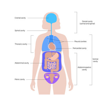
Thoracic cavity..
Thoracic cavity is also called chest cavity. This cavity is located in the upper part of the chest. This cavity is formed by the ribs, sternum bone, clavicle bone and thoracic vertebrae.
Important organs like lungs, heart, trachea, bronchi, esophagus etc. are located inside this cavity.
A layer of tissue is arranged around this cavity called the pleura.
Thoracic cavity also contains smaller cavities like pleural cavity, mediastinum cavity and pericardial cavity.
Que. 4 Write short notes (Any three) 3×4=12 marks
🔸(a) Cross Infection-
An infection that occurs between two different hosts is called cross infection
Through direct body contact :-
Due to skin-to-skin contact, body-to-body contact of the person discharging the organism from the patient to another person who has a cut or abrasion, the organism can easily enter that person.
E.g. In tracks, gonorrhea etc. is spread by direct body contact.
By infected hand :-
Human hands come in contact with another person or environment, they come in contact with faecal matter. Harmless staphylococcus and streptococcus are permanently on hand. But many times Pathogen also comes to hand. D. T. While handling the host’s urine, body secretions or fecal matter, a person acquires a pathogen in his own body or in another person’s body. Intestinal and respiratory tract diseases are spread by infection hands. Therefore, direct handling of infection materials should be avoided, thus also cross infection
By Droplet :-
A person also acquires the infection by inhaling the droplets released from coughing, laughing or sneezing from the speaking of an infected person. The pathogen of Diphtheria, influenza, T.B., Whooping cough, Measles, Mumps is in nasal discharge or saliva. By Contaminated Foamite :-
Diphtheria, trachoma, small pox, measles, pneumonia etc. are spread through eating or drinking utensils, handkerchiefs, towels, clothes, sputum cups etc. Respiratory, digestive and skin infections also spread from beddings, clothes etc. Through transgenic infection :- Through the equipment used to perform the procedure by the hospital
(b) Reflex action
Spinal cord acts as a connection between the brain and body parts. The length of the spinal cord is 45 cm.
It carries sensory impulses to the brain and motor impulses from the spinal cord to different parts of the body.
•Spinal code individuates certain activities which are not required for the brain to function and the brain becomes aware of them after the function is completed.
which action is accomplished by spinal reflexes. It is called reflex action.
•Sensory and motor neurons in the spinal cord are connected by connecting neurons found at different levels in the cord for the action of spinal reflexes.
A reflex arch is formed by the spinal cord which results in a quick action which reduces the workload of the brain.
(c) right lung
Lung is an important organ of respiratory system. They are located on either side of the mediastinum space in the thoracic cavity in total number of two.
The lungs take in oxygen from the atmosphere and expel carbon dioxide from the body.
Structure of the Lung..
Lungs are located in the thoracic cavity in number of two. They are conical in shape.
The lungs are separated from the heart and the thoracic cavity by the mediastinum space.
The lungs are made of spongy tissue within which many air field cavities are located. Its color is brown or grey.
The weight of the right lung is approximately 625 grams and the weight of the left lung is approximately 575 grams. The right lung is heavier in weight and larger in structure than the left lung.
Lungs are divided into lobes. The right lung has three lobes, namely the superior lobe, middle lobe and inferior lobe, while the left lung has two lobes, the superior lobe and the inferior lobe. These lobes are separated by a fissure. There are two fissures in the right lung. A fissure is seen in the left lung.
Lungs are classified into the following parts.
- Apex..
The upper triangular and round part of lunge is called Apex.Which is seen up to the level of the clavicle bone.
- Base.
The lower broad part of the lung is called the base. The base portion is attached to the diaphragm at the bottom. This part is of concave shape.
- Anterior border..
It is thin. It is shorter than the posterior border. It has a cardiac notch. In which the heart part is arranged.
- Posterior border..
It is thick. It is found from the 7th cervical vertebra to the 10th thoracic vertebra.
- Inferior border..
It is located at the bottom of the lung. It separates the costal surface and the medial surface. The costal surface is large and convex. It is in contact with the costal pleura. It is attached to the ribs and intercostal muscles by costal cartilage.
- Medial surface
It is concave. There is a groove in the middle of it which is called the hilum. The hilum lies at the level of the fifth, sixth and seventh thoracic vertebrae. Through this hilum, bronchi, pulmonary blood vessels, lymphatic vessels and nerves enter and exit each lobe of the lung.
In the middle of the medial surface lies the mediastinum space. which separates the two lungs. In this space there are structures like heart, great vessels, trachea, bronchi, esophagus etc. which separate both the lungs.
Structure of the Lobe of the Lung..
The lobes of the lungs are made up of many lobules. One lobe is separated from the other lobe by a fissure. In the center of the lung is a groove called the hilum. From this hilum the following structures are found in each lobe.
Bronchi enter each lobe of the lung. After entering, it divides and transforms into secondary bronchus, tertiary bronchus, terminal bronchioles, alveolar shakes and small grape-like alveoli. Thus, this structure is seen in a tree-like structure in the lobes of the lung, which is called bronchial tree or respiratory tree.
Surrounding these alveoli is a network of capillaries of the pulmonary artery and pulmonary vein. Gas exchange takes place here between oxygen in the alveoli and carbon dioxide in the blood capillaries through inspiration. This is known as external respiration.
Thus, in each lobe of the lung there is a network of bronchial tree, capillaries of pulmonary vessels, lymph capillaries, nerves and parenchymal tissue of the lung.
pleura..
The pleura is the serous membrane surrounding both lungs. Which is found in double layer. The outer layer is known as the parietal pleura and the inner layer is known as the visceral pleura.
Between the parietal pleura and the visceral pleura lies a cavity called the pleural cavity. There is serous fluid which is also called pleural fluid.
Due to the pleural fluid in the pleural cavity, the two layers do not rub against each other and due to this, the lungs get enough space for expansion. The pleural fluid in this cavity is viscous and also acts as a lubricant.
The visceral pleura is the layer adjacent to and adjacent to the lungs. Whereas the parietal pleura is the layer attached to the ribs and muscles.
🔸(d) Pasteurization of milk-
Discovered by Louis Pashwar in 1860.
It was first used in wine.
But currently it is used for milk.
Pasteurization is a method of sterilizing milk. In which only harmful bacteria are destroyed. But lactic acid and necessary organisms are not destroyed. There are few changes in the protein and sugar in the milk. There are three methods of pasteurization
(1) Holder Method or Wet Method
In this milk is heated at 62 C for 30 min. It is then rapidly cooled to less than 5°C.
T.B. and for Typhoid organisms is heated at 121’c for 15 to 20 seconds. It is called flash method.
(2) H.T.S.T. (“Temp, ↓ Time)
Milk is rapidly heated to 72°C and cooled to less than 5°C.
(3) Ultra high temperature
Milk as high temp as 121’c to 150’c. It is heated and then cooled very quickly. Phosphatic method is used to check the pasteurization of milk. Phosphate is present in raw milk. which is destroyed during pasteurization.
🔸(e) Culture Media –
Examination of micro-organism shows only the morphology of bacteria.
But to know bacteriological characteristics it is necessary to know the culture of organisms.
Many factors are responsible for the growth of microbes
Like moisture, Nutrients, Absence of toxin substance, Osmosis, PH Value, 02 etc.
Certain culture media are used to cultivate bacteria. Micro-organisms grow very fast in this media.
Required for antigen making and vaccine preparation.
Type of growth media
1) Liquid Media :-
Which is done in test tube, flask or bottle. It is also known as “Broth”. Use for large number of bacteria
(2) Solid media:- This is made by adding Agar-Agar to the liquid. It is made from red algae.
Concentration 1.5 to 2.5 %
3) Semi-solid media:– In this, Agar-Agar is added to the liquid. This is done to know micro-aerophilic bacteria or bacterial motility.
(4) Basal media:- water, agar, peptone are used. Ex staphylococcus
(5) Enrich media :- Blood is used as media. When the blood is heated at law temperature, the color of hemoglobin in it becomes dark brown, which is used as media, which is known as chocolate media.
(6) Cell culture media :- This is used for cultivation of Virus and Rickettsia. Tissue cell culture technique is used.
(7) Chick Embryo Media:-This is also used for culture of Virus and Rickettsia. Eggs are kept in incubator for 10-12 days after which fertile egg is used as media.
Que 5. Define following (Any six) 6*2=12 marks
Microbiology: “micro” means micro and “bio” means life and “logy” means study. Thus, microbiology is the study of microscopic organisms that cannot be seen by the naked eye.
Histology is a branch of anatomy that deals with the scientific microscopic study of cells and tissues of the body.
Carrier: A person or an animal that harbors certain disease-producing micro-organisms that can spread or carry the disease but does not show any signs or symptoms of the disease is called a carrier.
E.g. Typhoid
Pathogen: Any harmful living micro-organism that produces disease or has the ability to produce disease is called a pathogen.
Da. T. virus
Epidemic: If many cases of the same disease occur at the same time in a specific geographical area, it is called an epidemic.
D. T. Dengue
Tidal volume (tidal volume): Tidal volume is the amount of air going in and air coming out. The volume of air used by a person in inhalation and exhalation in normal situation is called tidal volume. In a normal person this is 500 ml.
Aseptic technique: Aseptic technique is a technique used to perform a procedure free from pathogenic micro-organisms using only sterile materials.
Antigen – Antigen is any toxic substance or foreign substance that produces an immune response, especially the formation of anti-bodies against it.
ex. Microorganisms.
Que 6 🔸(A) Fill in the blank 05 marks
1.Tears are produced______ by gland. Tears (tears) are produced from the gland. (lacrimal gland)
2._ is the longest bone in body. …… is the longest bone in the body. (Femur)
3. ______is located in the right side under the diaphragm. At the bottom right of the diaphragm is ……….. (liver)
4. Normal PH value of blood is_______ H. Value is …………….. (7.35 to 7.45)
5. Bile is secreted by _ organ. Bile …. . . .. is secreted from the organ. (liver)
🔸(B) Connect the joints. 05 marks
A B
1) Sternum bone 1) Patella bone
2) Hip bone Hip bone 2) Blood Blood
3) Clavicle bone-clavicle bone 3) Breast bone Breast bone
4) Fluid connective tissue 4) Innominate bone
5) Sesamoid bone Ceramoid bone 5) Sacrum bone Rom bone
6) Collar bone
Answer :-
1-3
2-4
3-6
4-2
5-1
(c) State whether the following statements are true or false. 05 marks
Anti-Diuretic Hormone is secreted by posterior lobe of pituitary gland. (correct)
There are three main lymphatic ducts in the body. . (false)
Optic nerve is a sensory nerve.. (correct)
Fertilization of ovum occurs in fallopian tube.. (correct)
The dermis is the outermost layer of the skin. (false)