ENG – Midwifery – First Year-BIO SCIENCES-26-03-2021-PAPER SOLUTION UPLOAD NO.8
First Year-BIO SCIENCES-26-03-202-PAPER SOLUTION UPLOAD NO.8
Date:- 26/03/2021
Q-1
a. List out bones of skull. 03
The skull is found in the upper part of the body above the vertebral column.
Which has the following 2 parts.
- cranium
- face
The cranium in the skull is made up of many different flat and irregular bones. There are a total of 8 bones in the cranium, which are as follows:
- Frontal bone 1
- Parietal bone 2
- Temporal bone 2
- Occipital bone 1
- Sphenoid bone 1
- Ethmoid bone 1
The bones in the face are called facial bones. They are 14 in total. These bones are found as follows.
- Maxilla bone. 02
- Zygomatic bone. 02
- Mandible bone. 01
- Nasal Bone. 02
- Lacrimal bones. 02
- Vomer bone. 01
- Palatine bones. 02
- Inferior conchae 02
b. Write down functions of Vertebral column.
The skull is located at the top of the vertebral column and supports the skull.
Between the bodies of the two vertebrae is an intervertebral disc that absorbs shock during movement and protects the brain from jerks.
A small canal-like strong structure is formed in the middle of the vertebral column through which the spinal cord passes. This serves to protect the spinal cord.
The transverse processes or pedicles of the vertebrae form a foramen on both sides. Nerves, blood vessels and lymph vessels pass through these foramen.
Provides attachment for other bones to connect. Provides a framework for the body. So that many moments are possible.
It provides a place for the ribs to connect. Thus, the thoracic cage is formed and also provides attachment for the formation of the solder joint and the pelvic joint.
It connects the axial skeleton with the appendicular skeleton.
Due to this, the posture of the body is maintained in different movements, sitting and walking.
c. Write down structure of cell. 05
Cell is the basic functional and structural unit of the human body. It is the main working unit.
Cell is known as a mass of protoplasm. Inside the cell there are organelles which are covered by plasma membrane.
In the human body, the fusion of ovum and sperm results in the formation of zygote. The growth of this zygote and cell division results in the formation of the human body.
The liquid part inside the cell is known as cytoplasm. In which there are many organelles. The structure of the cell is as follows.
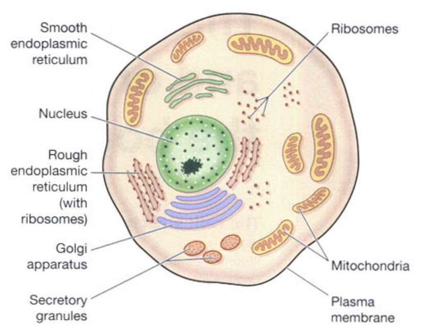
(Plasma membrane)
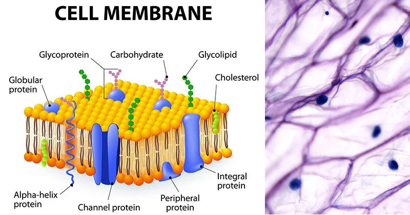
The membrane surrounding the cell is called the plasma membrane. This membrane has selective permeability (movement of only selected substances). Due to which some substances can enter the cell and some substances can go out of the cell. Thus, the cell maintains the structure of its cytoplasm through this membrane.
Plasma membrane is a double layer membrane made of phospholipids. It works to protect the organelles of the cell and to give the shape of the cell.
Nucleus.
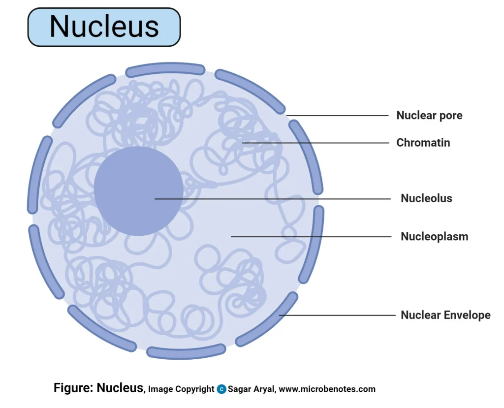
The nucleus is located in the center of the cell. It contains a liquid called protoplasm. The nucleus is surrounded by a nuclear membrane. This membrane also has selective permeability. The nuclear membrane partially separates the cytoplasm and protoplasm. The nucleus controls all the activities inside the cell and only with its help can the cell remain alive.
Inside the nucleus are thread-like proteins called chromatin. This chromatin is converted into chromosomes during cell division and performs the function of cell division.
These chromosomes inside the nucleus are responsible for the hereditary characteristics of the individual. Chromosomes are found in 23 pairs in the cells of the human body. Of these chromosomes, 22 pairs are called ordinary chromosomes while 1 pair is called sex chromosomes.
Mitochondria.
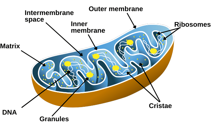
Mitochondria are rod-shaped structures located in the cytoplasm of the cell. They are surrounded by a double membrane, the structure of which is similar to the plasma membrane. The outer layer of this membrane is a smooth layer and the inner layer is folded. The series of these folds are called cristae.
Inside these cristae are enzymes that release ATP. That is why mitochondria are called the powerhouse of the cell.
Ribosomes.

They are small granules found in the cytoplasm. They are made up of proteins and RNA. They carry out the function of protein synthesis from amino acids. Some ribosomes are found free in the cytoplasm and some are attached to the surface of the endoplasmic reticulum.
(Endoplasmic Reticulum)
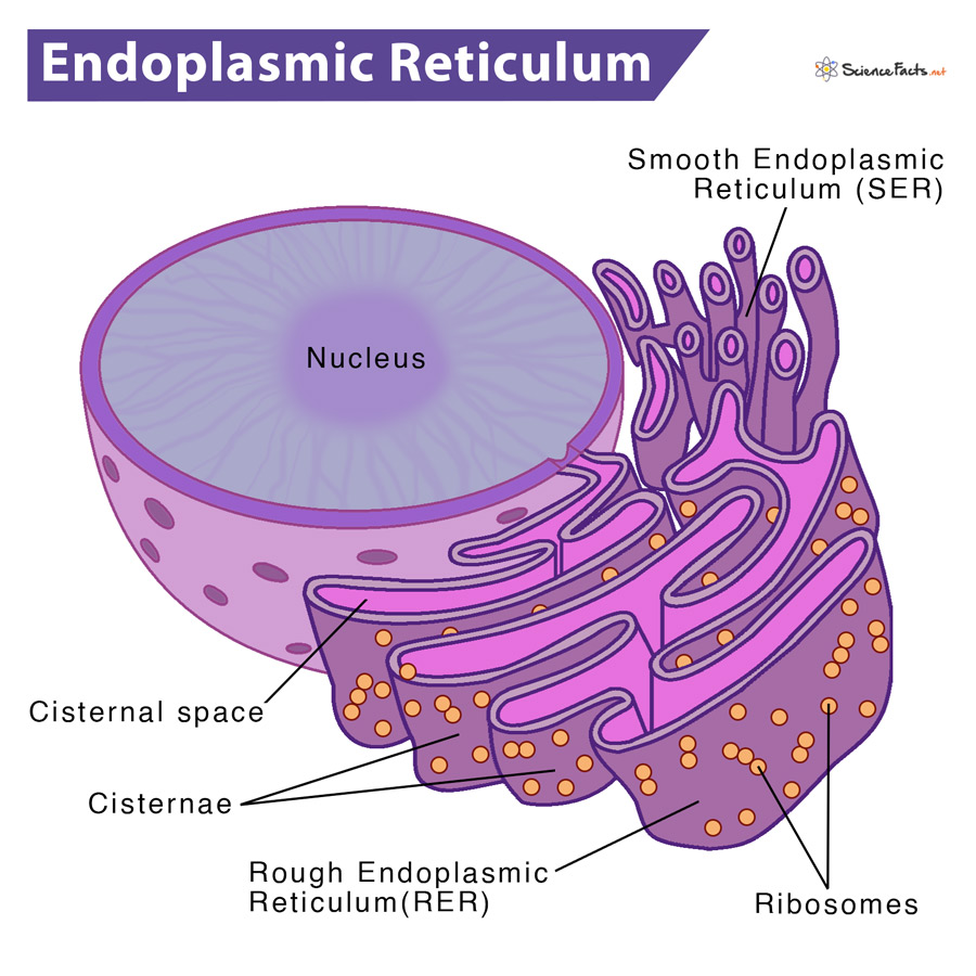
It is a series of interconnecting membranes or canal-like structures that connect one structure of the cytoplasm to another. Two types of which are found.
- Smooth endoplasmic reticulum..
Its surface is smooth. It functions in steroid hormone and lipid synthesis. It also helps in detoxifying some drugs.
- Rough endoplasmic reticulum..
Its surface is rough. Ribosomes are present on its surface. These ribosomes perform the function of protein synthesis.
Endoplasmic reticulum also helps in transporting substances from one place to another in the cytoplasm of the cell.
(Golgi Apparatus)
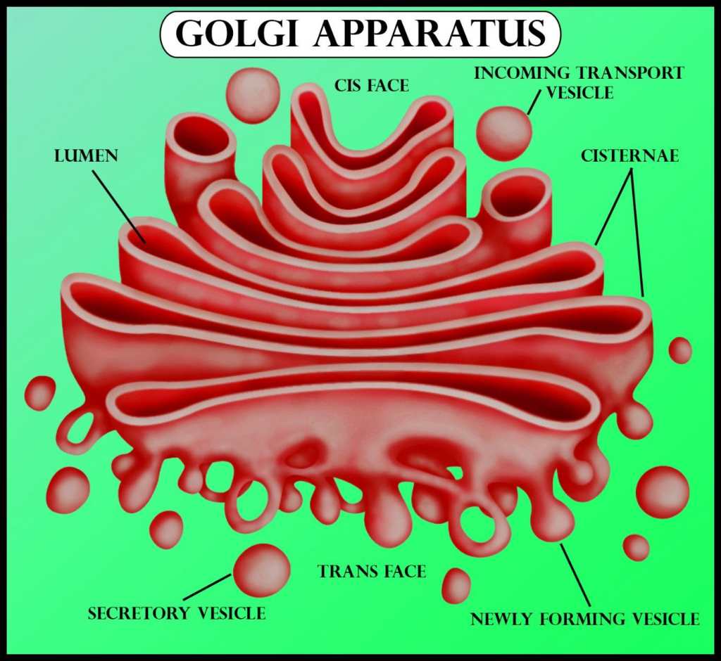
The Golgi apparatus is a bag-like structure with four to eight folds. These folds are stacked on top of each other. The ends of this structure form a pouch-like structure called a cisterna. Proteins synthesized by ribosomes are collected and stored in the ends of these cisterna in the form of secretory vesicles. When needed, these secretory vesicles release the proteins into the cytoplasm. The Golgi apparatus is a structure located near the nucleus.
(Lysosome)
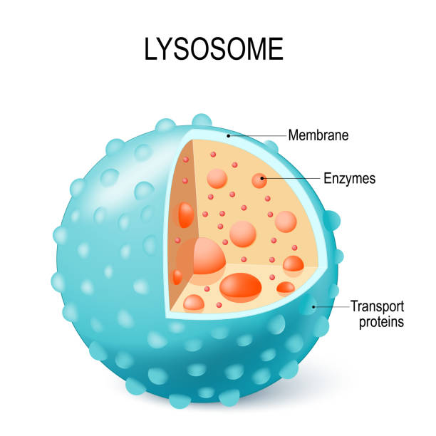
Lysosomes are a type of secretory vesicles that are secreted by the membrane of the Golgi apparatus. These lysosomes contain certain enzymes that work to break down certain large molecules present in the cytoplasm of the cell. They work to protect the cell from foreign material and microorganisms. These lysosomes also work to remove waste material accumulated inside the cell.
The cytoplasm of the cell also contains a sun-shaped centrosome which plays an important role in cell division.
In addition, the cytosol of the cell also contains a network of microfilaments and microtubules that function to maintain the shape of the cell and to protect and support the structure of the cell.
d. Write down gross structure of the Heart 08
The heart is an important organ of the circulatory system. During human life, the heart beats continuously. Due to its beating, blood circulates continuously in the blood vessels.
The heart is an organ made of cartilage and muscles. Its weight in men is approximately 310 grams and in women its weight is approximately 250 grams. The heart beats approximately one lakh times during a day.
Location of the Heart..
The heart is located in the thoracic cavity between the two lungs in the mediastinum space above the diaphragm.
The heart is roughly cone-shaped. Its upper flat part is known as the base and the lower angled part is known as the apex.
The size of the heart is about the size of a human clenched fist. Its length is 12 cm, width is 9 cm and thickness is 6 cm.
The heart is located in the thoracic cavity between the two lungs, slightly tilted to the left.
The heart is found in the middle of the thoracic cavity between the second and fifth intercostal ribs.
Organs surrounding the heart..
The heart is located in the thoracic cavity. There are two lungs on both the left and right sides of it.
On the lower side, there is the diaphragm and the central tendon.
On the upper side of the heart, there are the vena cava and aorta and the pulmonary artery and pulmonary vein.
On the back side of the heart, there are the esophagus, trachea, bronchi and bronchioles, and the descending aorta and thoracic vertebrae.
The sternum bone and ribs and intercostal muscles are located on the front side of the heart
Structure of the Heart..
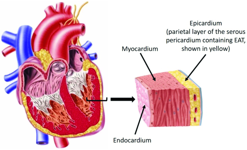
The heart is a semi-muscular organ. Its wall is made up of three types of tissue layers.
The outermost layer of the heart wall is called the epicardium or pericardium.
Epicardium or pericardium.
It is thin and transparent and covers the heart from the outside. It is made up of fibrous connective tissue. In which the outermost layer of fibrous tissue is present and on the inner side of the fibrous tissue is a serous membrane which is found in a double layer. The outer layer of the serous membrane is known as the parietal and the inner layer is known as the visceral pericardium layer.
The space between the parietal and visceral pericardial layers is called the pericardial space. This space contains a fluid called serous fluid or pericardial fluid. This prevents friction between the two layers.
This outer pericardium layer works to protect the heart from the outside and this layer is also found around the vessels coming out of the heart.
Myocardium..
The myocardium is the middle layer of the heart. It lies below the pericardium. It is made up of a special type of cardiac muscle tissue. The pumping action of the heart is seen due to the contraction of these muscles.
This layer of myocardium is thin at the base and thick at the apex. Also, the layer of the wall of the left ventricle is thicker than the wall of the right ventricle.
The contraction of these muscles is an involuntary action that causes the pumping action of the heart, which is controlled by the autonomic nervous system and the conducting system in the heart.
Endocardium..
It is the innermost layer of the heart wall. This layer is in contact with the blood. This layer is made up of epithelium tissue and connective tissue. This layer is smooth and shiny, which is important for the smooth flow of blood inside the heart. This layer also covers the valves inside the heart and this layer is also continuous in the inner wall of the blood vessels coming out of the heart.
Chambers of the Heart.
The heart is mainly divided into two parts. The right side and the left side.
The septum of the heart is located between the right and left sides. This is again divided into four chambers, above and below the valve, by a valve located between the right and left sides of the heart.
The two chambers above the valve are called atrium or auricle. The two chambers below the valve are called ventricles. Thus, the heart is divided into four chambers in total.
The right side of the heart contains deoxygenated blood and the left side of the heart contains oxygenated blood. After birth, there is no connection between the right and left sides. Before birth, there is an opening between the two atria, which is called the foramen ovale, but after birth it closes.
Valves of the Heart..
The flaps of tissue inside the heart are called valves. There are two types of valves in the heart.
- Atrioventricular valve.
- Semilunar valve.
Atrioventricular valve…
This valve is located between the atrium and the ventricle in the heart. The valve located between the right atrium and the ventricle is called the tricuspid valve. This valve is made up of three tissue flaps.
The valve located between the left atrium and the ventricle is called the bicuspid valve or mitral valve. This valve is made up of two tissue flaps.
The hanging ends of both the bicuspid and tricuspid valves are attached to the inner wall of the ventricle of the heart by the chordae tendineae and papillary muscles. This gives strength to the valve and prevents it from opening in the opposite direction.
Semilunar valve…
This valve is called a semilunar valve because it is C-shaped or crescent-shaped. These valves are present in the aorta and pulmonary artery. The valve present in the aorta is called the aortic valve and the valve present in the pulmonary artery is called the pulmonary valve. Which are located at the opening of its vessels. These valves also open in one direction.
Both the above atrioventricular valves open when the pressure in the atrium increases i.e. during the contraction of the atrium and blood comes from the atrium to the ventricle and during the contraction of the ventricle the atrioventricular valve closes and the semilunar valve opens and due to this the blood goes to the aorta and pulmonary artery.
Thus due to the opening and closing of these valves, blood circulation can take place in the heart. These valves open in one direction so that the blood cannot go in the opposite direction again.
Great Vessels Associated with Heart..or
Openings of the Heart..
The heart is mainly connected to large blood vessels. Through which blood comes from the body to the heart and is pumped by the heart and the blood goes out to the body. The vessels and their openings are as follows.
Superior Vena Cava..
These bring deoxygenated blood from the upper part of the thoracic cavity, head and neck to the right atrium of the heart. They are one in number.
Inferior Vena Cava..
These blood vessels bring deoxygenated blood from the inferior and lower part of the thoracic cavity and open into the right atrium of the heart. They are one in number.
Pulmonary artery..
It carries deoxygenated blood from the right ventricle of the heart out of the heart towards the lungs. Its number is one.
Pulmonary vein..
Two pulmonary veins from both lungs bring oxygenated blood to the left atrium of the heart. Its number is four.
Aorta..
It carries oxygenated blood from the left ventricle of the heart and circulates it throughout the body. Its number is one.
Thus, a total of eight blood vessels and their openings are directly connected to the heart. Which are connected to the blood circulation in the heart.
Blood Supply of the Heart..
The oxygenated blood supply to the heart is provided by the aorta, which originates from the left ventricle of the heart. As soon as it leaves the left ventricle of the heart, one branch, the right and left coronary arteries, emerge from it on both sides, which provide oxygenated blood supply to the heart. These coronary arteries are branches of the ascending aorta.
Of the blood that the heart circulates out of the aorta during a contraction, about five percent of the blood is supplied to the heart through the coronary arteries.
These coronary arteries divide into many branches and supply oxygenated blood to all parts of the heart.
Deoxygenated blood in the heart drains into the coronary sinus through small coronary veins and this deoxygenated blood drains directly into the right atrium of the heart through the coronary sinus.
Functions of the Heart…
The heart supplies oxygenated blood to all the organs and tissues of the body.
The heart is an important organ of the cardiovascular system. Without which the human body cannot survive, it functions as a vital organ.
The heart circulates the blood towards the lungs so that the blood can be oxygenated and purified.
Circulation such as pulmonary circulation and systemic circulation is regulated by the heart.
The heart also regulates the heart rate according to the needs of the body and body temperature.
The heart also regulates the body’s temperature by circulating blood to every part of the body.
The heart pumps blood to the excretory organs of the body.
OR
a. Write down functions of Blood.03
Blood is a major liquid in the body. It performs the following functions.
- Blood mainly performs functions related to transportation, which include the following activities.
Blood transfers oxygen from the lungs to the body tissues and transfers carbon dioxide from the body tissues to the lungs.
Blood transports nutrients absorbed from the alimentary canal throughout the body.
Blood delivers hormones of the endocrine glands to their target cells.
Transports waste produced at the end of metabolism to the excretory organs of the body.
Helps in transporting heat produced in the body throughout the body.
- Blood functions for the regulation of certain functions in the body, which are as follows.
Blood maintains the pH of acids and bases in the body and acts as a buffer solution.
Blood maintains the amount of water and electrolytes in the body.
Blood regulates body temperature.
- Blood also performs some protective functions, which are as follows.
White blood cells in the blood have specific characteristics, so they protect the body against microorganisms.
They protect the body from certain toxic substances.
Since blood cells have the property of clotting, the blood clotting mechanism prevents excessive blood loss from the body.
b. Write difference between Artery and Vein. 04
Note: This question is written in the form of a difference, both sides are given here for the sake of simplicity.
Blood vessels that carry blood from the heart to the body are called arteries, while blood vessels that carry blood from the body to the heart are called veins.
Oxygenated blood circulates in arteries, except for the pulmonary artery, and deoxygenated blood circulates in veins, except for the pulmonary vein.
Arteries do not have valves, while veins do.
Blood in arteries appears bright red in color because it contains oxygen, while blood in veins appears dull red or less red because it contains carbon dioxide.
Since arteries originate directly from the heart, pulsation can be heard in them, while veins do not.
Arteries are mainly located deep in the body, while veins are located superficially.
The middle layer of an artery is more elastic, so its diameter can change more, while the middle layer of a vein is not as elastic as an artery.
The lumen of an artery is normally narrow, while the lumen of a vein is wider compared to an artery.
The force of blood in an artery is greater, while the force of blood in a vein is less.
When the amount of blood in an artery is less, its wall is stronger, so it does not collapse, while when the amount of blood in a vein is less, it shrinks.
c. Define Sterilization and explain any one method of Sterilization. 05
The process of killing or removing pathogenic, non-pathogenic, and spores-containing microbes is called sterilization.
Autoclave
Heat under pressure is a practical and useful agent in sterilization.
In this method, 15 lbs pressure and 121° C temp. 30 minutes are required for sterilization.
Pressure does not kill organisms, but steam under pressure can do the job.
The time depends on how bulky the material is to be sterilized. This is considered one of the best methods of sterilization. It kills all bacteria and spores.
The equipment used for this process is called an autoclave machine, which is like a household pressure cooker.
An autoclave is a double-walled metal device.
It has an airtight chamber. It usually has 2 gauges (meter gauges).
It reads the steam pressure of the outer inner chamber.
To prevent explosion due to high pressure, it has a safety valve, which releases steam as the pressure increases as necessary.
There is also an exhaust valve to remove the steam from the inner chamber.
There is also a valve to send steam from the outer chamber to the inner chamber.
In some autoclaves, a thermometer is also fitted inside the chamber.
After placing the object on the plate inside the inner chamber, the lids are tightly closed and heating is started.
The advantages of 15 lbs/inch, 120° C 30 minutes autoclave are. That it can kill organisms including spores.
And the specified temperature and pressure of steam can be maintained. This method can sterilize culture media, rubber goods, linens, dressing, syringes and… It can be used in the sterilization of instruments and many other important consumables. It is one of the best, most practicable, dependable method of sterilization.
d. List out organs of Digestive System and explain the gross structure of stomach. 08
The digestive tract, also called the alimentary tract, is a hollow tube that extends from the mouth to the anus and its organs are as follows.
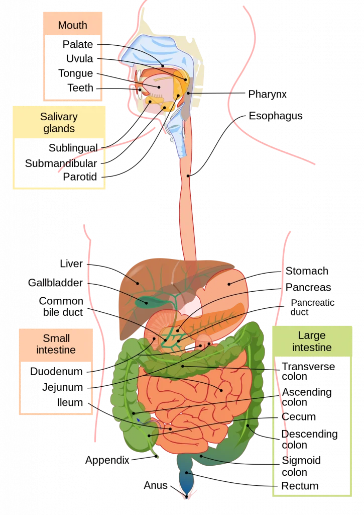
Mouth
Pharyngs
Esophagus
Stomach
Small intestine, which includes the duodenum, jejunum, and ileum.
Large intestine, which includes the cecum, ascending colon, transverse colon, descending colon, sigmoid colon, rectum, and anal canal.
Accessory Organs of the Digestive Tract…
These organs do not come in the main track of the digestive tract but are located on the side which pour their secretions into the alimentary canal and help in the process of digestion hence they are called accessory organs. They help in digestion with the special secretion of their glands which organs are as follows..
3 pairs of salivary glands
Liver and biliary tract
Gall bladder
Pancreas.
The above organs are accessory organs which help in digestion.
Stomach….

It is an organ of the digestive system. It is an organ of the shape that. It forms the widest part of the alimentary canal. The stomach is mainly located in the epigastric region and some of it is located in the left hypochondriac region.
In its front part, some part of the liver is located, in the back part, the abdominal aorta, pancreas and spleen are located. In the upper part, the diaphragm is located and in the lower part, the intestine is located.
Structure..
The structure of the stomach is divided into three parts
- fundus
- body and
- pylorus..
Fundus….
It forms the uppermost part of the stomach. It is dome-shaped. It is located above the level where the esophagus joins the stomach. It is sometimes filled with gas. Here, the esophagus joins the stomach, and between them are the cardiac sphincter muscles.
Body…
The part of the stomach below the fundus is called the body, which extends to the lower narrow part of the stomach, the pylorus.
Pylorus…
The part of the stomach that comes down after the body, in which the initial part is called the pyloric antrum, which is the narrow part after the body, from there, going forward towards the duodenum, there is a canal-like structure called the pyloric canal, which opens into the duodenum and between it are sphincter muscles called the pyloric sphincter.
In addition, two curvatures are seen on the body portion of the stomach, one is the lesser curvature that forms the right border of the stomach and the other is the greater curvature that forms the left border of the stomach.
Layers of the Stomach…
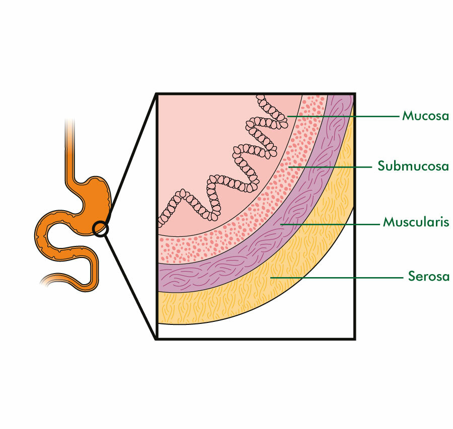
The wall of the stomach, like the basic structure of the digestive tract, is made up of four tissue layers, in which the outermost layer is the peritoneum layer, also called the adventitia layer, which forms the serous lining.
Below it is a layer of muscles. In the stomach, this muscular layer is made up of three layers, in which one is the long tunnel muscle fibers that are arranged superficially, the other is the circular muscle fibers that are arranged in a circular shape, which are located below the superficial layer and these fibers are also important for forming sphincter muscles. Three oblique muscle fibers are mainly found in this layer, which is not found in the basic structure. They are especially found in the wall of the stomach, which is a very important layer for the movement of food in the stomach and mechanical digestion.
Beneath the muscular layer lies the submucosal layer which contains a network of blood vessels and lymph vessels.
The innermost layer of the stomach wall is called the mucosal layer. This layer is made up of columnar epithelium tissue. This layer secretes mucus and also acts as a lubricant. This layer contains folds of mucous membrane which are especially visible when the stomach is empty and is called eructation. When the stomach is full of food, this eructation disappears.
Functions of the Stomach…
The stomach performs the following functions.
The stomach stores food for a short time. Before the food moves into the intestinal tract, it undergoes partial mechanical digestion.
The stomach secretes gastric juice. This gastric juice has an acidic content and the enzymes present in it help in the digestion of food.
The muscular layer in the wall of the stomach is specially arranged, so its contraction causes a churning movement, which helps in the mechanical digestion of food and breaks the food into small pieces and then the food moves on to the duodenum.
Intrinsic factor is secreted from the inner wall of the stomach, which is necessary for the absorption of vitamin B.
Some amount of water, alcohol and some medicines are also absorbed from the wall of the stomach.
It also works to destroy bacteria or viruses present inside the stomach. When these bacteria reach the stomach through mucus and oral cavity, they are destroyed by acidic secretions and are prevented from moving further into the intestinal tract, thus also acting as a protection for the body.
Q-2 Write short Notes (Any Five) 5×5-25
a. Mechanism of Respiration
Respiration is the gas exchange between two surfaces. In which air from the atmosphere enters the lungs. Gas exchange occurs between the lung tissue and blood, which is called external respiration. Gas exchange between each cell tissue of the body and the blood is called internal respiration.
During the act of respiration, oxygen enters the lungs through inhalation and carbon dioxide leaves the body through exhalation.
Usually, the act of respiration is seen 16 to 18 times in a minute.
The following actions are seen in the cycle of respiration.
Inspiration
Expiration
Pause.
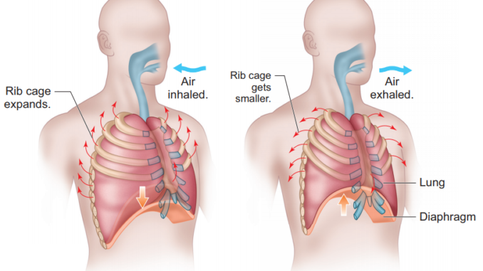
Inspiration
The act of taking air from the atmosphere into the lungs through breathing is called inspiration.
When the brain receives nerve impulses for contraction of the diaphragm and intercostal muscles, the size of the thoracic cavity increases due to contraction of the diaphragm and intercostal muscles. The air pressure inside the thoracic cavity decreases so that air from the outside environment can enter the lungs through the act of inspiration. This act is called inspiration.
When the diaphragm receives contraction impulses, the diaphragm becomes flat and moves downwards and the intercostal muscles contract, causing the ribs and intercostal muscles to move upwards and outwards. So that the size of the thoracic cavity increases and negative pressure is created inside the cavity. Since the air pressure in the outside environment is high and the air pressure in the thoracic cavity is low, the act of inspiration can take place. The act of inspiration is an active process.
Expiration.
The process of expelling air from the lungs into the atmosphere is called expiration. The act of expiration is a passive process that begins after the act of inspiration is completed.
In the act of expiration, the contracted diaphragm and intercostal muscles relax. So that the diaphragm returns to its original position and the ribs come down and inward, the size of the thoracic cavity decreases and the act of expiration takes place. In which air is expelled from the lungs into the atmosphere.
During the act of expiration, the air pressure in the lungs is greater than the pressure of the atmosphere, so that the act of expiration occurs.
Pause.
This is the relaxed stage of the lungs. In which no inspiration or expiration occurs. This period is called the pause period.
b. Functions of large Intestine
Water and Electrolyte Absorption..
Most of the water absorption takes place in the small intestine, but some water and some electrolytes are also absorbed in the large intestine.
The large intestine absorbs water from the stool, which plays an important role in making the stool semi-solid.
Microbial Activity..
Some bacteria are present in the large intestine. These bacteria play an important role in the synthesis of vitamin K and folic acid. These bacteria can be harmful and can also cause infection if they move to other parts of the body.
Due to the activity and fermentation of these bacteria, gas is produced from undigested food, which is passed out of the bowel in the form of flatulence through the anus.
Mass movement…
Peristalsis movement is seen in other parts of the intestine but this type of peristalsis movement is not seen in the large intestine.
After a long interval, a large strong peristalsis movement is seen in the large intestine due to which the content moves towards the descending colon and sigmoid colon in the intestinal tract which is called mass movement.
Defecation…
Due to mass movement in the large intestine, the fecal material moves towards the sigmoid colon and rectum in the interstitium due to which the stretch receptors in the wall of the rectum are pulled and the defecation reflex starts and the process of defecation begins.
In the defecation reflex, the sensory nerve is stimulated due to the stretching of the rectal wall due to the stretching of its receptors. It transmits impulses to the sacrum in the spinal cord. In addition, the pressure of the diaphragm and the abdominal cavity helps in the process of defecation. The sitting position also helps in the process of defecation. Finally, due to the stimulation of the parasympathetic nerve, the internal sphincter relaxes and the stool comes out through it. The external sphincter has voluntary control so that the process of defecation can be stopped for some time through voluntary control, while the internal sphincter has involuntary control. The relaxation of the internal sphincter and the external sphincter causes the stool to pass.
Feces.
Feces are brown in color. This brown color is seen due to the stercobilin present in it. It has semi-solid and solid properties. The composition of feces contains approximately 60 to 70% water. The excess water is absorbed through the wall of the large intestine. The following things are found in the composition of feces.
Dead and live microorganisms.
Dietary fibers.
Epithelium tissue of the intestinal wall.
Mucus and undigested food. etc.
Due to the secretion of mucus through the intestinal wall, stool can pass easily without causing abrasion to the intestinal wall.
c. Thyroid gland
Thyroid Gland

The thyroid gland is an important gland of the endocrine system. It is located in the soft tissue of the neck. This gland is a butterfly-shaped gland.
The weight of this gland is approximately 30 grams. Its length is 5 centimeters and width is 3 cm.
This gland is located from the level of the fifth cervical vertebra to the level of the first thoracic vertebra.
The thyroid gland has one lobe on each side. Which is covered with fibrous tissue around it. The middle part connecting the two lobes is called the isthmus. The lobes of the thyroid gland have a pyramidal shape.
The tissue in the thyroid gland is made up of small structures called follicles. Each follicle is made up of simple cuboidal glandular epithelium tissue. Which is associated with secretion.
The function of the thyroid gland is regulated by the hormone thyroid stimulating hormone released from the pituitary gland.
The thyroid gland secretes the following hormones.
- Triiodothyronine (Triiodothyronine) T3
The main function of this hormone is to maintain normal physical growth and development in the body. This hormone also plays a useful role in maintaining heart rate and some metabolic activities.
- Thyroxin (Thyroxin) T4…
This hormone also performs the same function as the T3 hormone. That is, it maintains metabolic activity in the body and works to maintain normal physical growth and development. This hormone increases the basal metabolic rate.
- Calcitonin
This hormone is secreted by the parafollicular cells of the thyroid gland. It affects the metabolism of calcium in the blood.
d. Structure of ear
Ears are located on either side of the head. They are organs involved in hearing and maintaining body balance.
Structure…
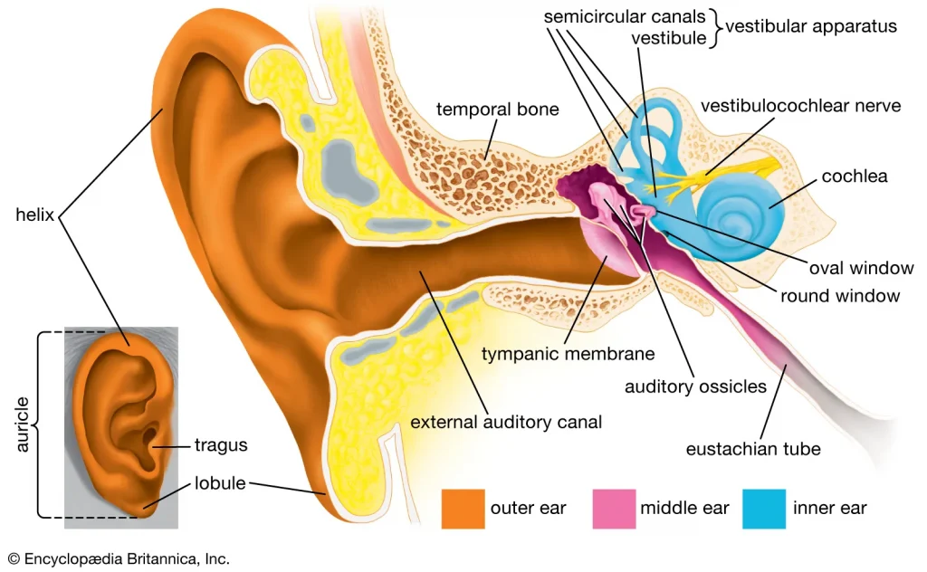
Structurally, the ear is anatomically divided into three parts:
- External or outer ear
- Middle ear
- Inner ear.
External or outer ear
The external ear collects and transfers sound waves to the inside. The external ear consists of the following parts.
Auricle or pinna
External acoustic meatus i.e. auditory canal
Auricle…
It is a flap-like part on both sides of the head which is made of elastic cartilage and is covered with skin on the outside. It collects sound waves coming from the outside environment and transmits them to the inner hole. Its most prominent upper part is called the helix and the soft part hanging down is known as the lobule.
External acoustic meatus…
It is also known as the auditory canal, it is a curved tube in the shape of an S. It is about 2.5 cm long and is an elongated canal on the inside and the glands in its wall secrete a wax-like viscous fluid, also known as cerumen. Some hairs are also present in this canal which, combined with the viscous fluid, prevent dust and foreign bodies from reaching the inner wall, i.e. the tympanic membrane.
Tympanic membrane…
It is a thin membrane that separates the external ear and the middle ear and is oval in shape. This membrane is made up of epithelium tissue and connective tissue, and fibroblast cells are present in it.
Middle ear..
It is a small air-filled cavity located in the temporal bone. The lining of this cavity is made of epithelial tissue and it is separated from the external ear by the tympanic membrane i.e. the ear drum and the inner ear is separated from the middle ear by the oval and round windows.
Its anterior wall has an opening known as the eustachian tube. This tube connects the middle ear to the structures of the nasopharynx and maintains pressure inside the cavity, which prevents the tympanic membrane from rupturing during yawning and swallowing food.
Auditory ossicles…
The bones in the middle ear are called auditory ossicles and include the malleus, incus and stapes bones. Its number is one in each ear, that is, there are a total of 6 auditory ossicles in the body.
Malleus..
It is hammer-shaped, its handle is attached to the wall of the tympanic membrane. The head of the malleus is attached to the incus bone.
Incus..
It is the middle bone which is in the shape of anvil, it is attached to the malleus bone and its head is attached to the stapes bone. There is fibrous tissue in the joint part.
Stapes..
It is a stirrup-shaped bone whose head is attached to the incus bone and its foot plate is attached to the oval window, and the round window is located below it.
Thus, the structure of the part from the tympanic membrane to the oval and round windows is called the middle ear.
- Inner ear…
It is also known as the labyrinth. It has a complex structure and is a part associated with the function of hearing and maintaining balance..
It has two parts, the bony labyrinth and the membranous labyrinth..
The bony labyrinth…
It is a cavity located in the petrous portion of the temporal bone, whose wall is made of a layer of periosteum. On its inner side there is a fluid called perilymph. It has three parts. A vestibule, a cochlea and three semicircular canals.
Vestibule..
It is the middle oval-shaped portion of the bony labyrinth, inside which the utricle and saccular structures are located. Its lateral wall has oval and round windows.
Cochlea…
This part of the bony labyrinth is connected to hearing, it is a lump-like part which is also known as Snell’s cell.
Semicircular canal…
The structure of this canal is connected to balance. The three tubes of the semicircular canal are arranged at right angles to each other.
Membranous labyrinth..
Inside the bony labyrinth there is a membrane tube called the membranous labyrinth which contains a fluid called endolymph. The properties of this fluid are similar to CSF.
- Vestibule which contains the utricle and the saccule.
- Vestibule and Semicircular Canal…
The vestibule and semicircular canals in the inner ear work to maintain the body’s balance.
In which the semicircular canal, the cochlea, and the utricle help to maintain the body’s dynamic equilibrium. The semicircular canal is a tube-like structure that is located behind and above the vestibule and these canals are arranged at an angle to each other. The semicircular canal and the cochlea, whose outer wall is the bony labyrinth and inside it is the perilymph fluid, have another tube inside called the membranous labyrinth, which contains endolymph.
The utricle is a membranous sac which is part of the vestibule and all its ducts open into a dilated portion called the ampulla. The saccule is part of the vestibule and communicates with the utricle and the cochlea.
The walls of the utricle, saccule and ampulla contain small hair-like projections of specialized epithelial cells which contain sensory nerve endings which form the vestibular part and from which the vestibulo-cochlear nerve passes.
- Cochlea
Scala media or cochlear duct
Scala tympani
The cochlear duct is a triangular shaped tube. The bony part of the cochlea is divided into two parts, upper and lower, in which the upper part is called the scala vestibuli and the lower part is called the scala tympani. In the middle part is the cochlear duct and its roof is called the basilar membrane. The organ of Corti, which is called the hearing organ, is located on the basilar membrane.
e. Menstrual cycle
Menstruation cycle is seen in females after puberty phase. In which changes are seen in the function of ovaries and uterus.
Menstruation cycle is seen every 26 to 30 days. This is seen due to changes in the hormone levels of the blood.
The beginning of menstruation cycle is known as menarche.
This cycle is seen continuously in females after puberty age. Which stops temporarily during pregnancy and stops completely after the period of menopause.
The beginning of menstruation is seen due to the degeneration of the corpus luteum layer in the uterus and bleeding comes out through the vaginal cavity.
The following phases are seen in the menstrual cycle.
- Menstrual phase..
This phase occurs every 28 days and lasts for about four days. When the egg is not fertilized in the female, the levels of the hormones estrogen and progesterone that support the wall of the uterus decrease and the level of the hormone oxytocin increases. Due to this, the stimulation of uterine contractions increases and the degeneration of the corpus luteum layer of the wall of the uterus starts and blood drains from the uterus through the vagina. This phase lasts for 1 to 4 days.
This menstrual flow consists of endometrial glands, endometrial cells, blood and parts of the unfertilized ovum. Approximately 100 to 200 ml of blood is released during the 3rd to 5th day of this phase which is called the menstrual phase.
- Proliferative phase..
The menstrual phase ends on the 5th day. After that, the proliferative phase starts from the 6th day and continues for 14 days.
In this phase, follicle stimulating hormone stimulates the ovarian follicles and thus increases the production of estrogen. This estrogen stimulates the proliferation of the endometrium.
The endometrium of the uterus starts developing from the sixth day. Its cells multiply and due to which the mucus secreting glands and blood capillaries increase. Thus, the endometrium of the uterus becomes bulky and vascular.
At the end of this phase, the inner wall of the uterus is ready for the implantation of the fertilized egg. This phase ends with ovulation. At the end of this phase, there is a decrease in estrogen levels.
- Secretory phase.
After the proliferation phase is completed, the secretory phase begins. The secretory phase starts from the 15th day of the menstrual cycle and lasts until the 28th day.
Since the progesterone hormone is important in this phase, this phase is also called the progesterone phase.
When a mature egg is released by the ovary due to ovulation, the amount of estrogen and progesterone hormones decreases, but the wall of the uterus, the corpus luteum, secretes progesterone and maintains the pregnancy.
Due to this mature egg not being fertilized by the sperm, the corpus luteum decreases progesterone and due to the decrease in progesterone hormone, the amount of oxytocin hormone increases and contractions begin in the muscles of the uterus.
Due to the corpus luteum not receiving the fertilized egg and due to the increase in uterine contractions, the next cycle begins at the end of this phase.
f. Functions of skin
The skin forms a continuous covering around the outside of the body, acting as the first line of protection.
It prevents microorganisms from directly entering the body.
It prevents any external injury or harmful elements from entering the body.
It plays a very important role in maintaining normal body temperature.
The skin provides the outer framework of the body. It is the largest organ in the body. It works by covering all the organs on the outside and giving shape to the body.
It synthesizes vitamin D in the body. In which the skin contains a chemical called seven-dihydrocholesterol. Which converts the ultraviolet rays coming from the sun into vitamin D3 and cholecalciferol. Thus it works to synthesize vitamin D.
The skin also works to excrete waste products and harmful substances from the body. It also excretes some waste products from the body through the secretion of perspiration.
Absorption of some substances also occurs through the skin. This is also a route for medication. In which the skin absorbs some ointments and medicines applied on it and sends them to the systemic circulation.
The skin contains sensory nerve endings. Which help in transmitting impulses of touch, temperature and pain etc. to the brain. This allows us to interpret each stimulation.
The skin also serves to store certain nutrient materials such as fat.
The skin plays an important role in wound healing.
g. Functions of Kidney
The kidneys mainly function to form urine.
The kidneys filter the blood and remove waste products through urine.
The kidneys function to maintain a normal balance of electrolytes.
They function to maintain the pH of the blood.
They function to remove waste products accumulated at the end of metabolism in the body from the body.
The kidneys secrete a hormone called erythropoietin, which plays a very important role in the production of RBCs.
The kidneys secrete a hormone called renin, which plays a very important role in maintaining blood pressure.
The kidneys function to maintain water balance in the body.
h. Preventive measures of infection
Standard Precautions:
Standard precautions should be followed to prevent transmission of pathogens through body fluids to break the chain of infection.
- Hand Hygiene:
This is the number one weapon in preventing cross-infection. The spread of microorganisms is mostly through hands, so hand washing should be done before and after every procedure. It is very important to wash hands after touching any contaminated object.
- Aseptic Technique
In which contact precautions and aseptic technique should be used during the procedure. Only sterile items should be used in invasive procedures.
- Environmental Infection Control Measures
The equipment used in the hospital and the surrounding environment should be kept clean and the hospital floor should be cleaned with an antiseptic solution. Bio-medical waste generated during the procedure or work should be properly disinfected and disposed of.
- Droplet precautions
A mask should always be worn while working in the hospital and the infected patient should also wear it, precautions should be taken while coughing, sneezing etc.
- P.P.E
Personal protective equipment can be used to prevent cross infection between the nurse and the patient, whose cap, mask, gown are used. Which should be worn during the care of highly infectious patients.
- Health education:-
To prevent the spread of infection in the hospital, health education is given to the patients and the employees working under them.
Q-3 (A) Fill in the blanks.
1) ………..gives the color of eye (Iris).
2) ……….is stored in the gall bladder. (Bile)
3) Layer of the brain is called………. (Meninges)
4) Islets of Langerhans situated in ……….. (Pancreas)
5) Male sexual hormone situated by testes is……… (Testosterone)
6) 10th Cranial nerve is…….. (Vegas)
7) is the longest & strongest bone in the human body. (femur)
8) …….cartilage is situated in trachea. (Highline)
9) Life span of R.B.C is ……….days. (90 to 120 days)
10) Middle layer of artery is called…….. (Tunica media)
(B) State whether following statements are True or False. 10
1) Pyloric Sphincter is situated between Stomach and Esophagus. ❌
2) Largest organ of the body is Skin.✅
3) Respiratory center is situated in the Medulla oblongata. ✅
4) The Pineal gland is the master gland of the body ❌
5) Normal systolic blood pressure is 80mm of Hg. ❌
6) Pulmonary artery supplies blood to the heart.❌
7) 2d cervical vertebra known as Axis. ✅
8) C.S.F. is circulated in Spinal cord. ✅
9) Uterus is situated in abdominal cavity.❌
10) Causative organism of cholera is Vibrio. ✅
( C) Write Multiple Choice Questions. 10
1.Deglutition is also called as-
a) Bolus –
b) Swallowing –
c) Pain
d) Digestion
2.Integumentary system
a) Cervix –
b) Ureters-
c) Skin –
d) Langs-
3.Part of eye included all of the following except-
a) Stapes –
b) Comes –
c) Lens-
d) SELEM-
4.Hospital acquired infection is called as
a) Endogenous infection
e) Nosocomial infection
b) Exogenous infection
d) Septicemia
5.Normal PH value of the blood is
a) 7.30 to 7.45
b) 7.35 to 7.40
c) 7.15 to 7.45
d) 7.30 to 7.50
6.Normal number of cardiac cycle per minute is –
a) 10-20
b) 90-100
c) 60-80
d) 200-400
7.Movable bone in face is-
a) Maxillary bone –
c) Zygomatic bone
b) Mandible bone –
d) Vomer bone
8.Which human organ system is responsible for exchanging gases with outside environment?
a) Respiratory system
c) Urinary system –
b) Endocrine system –
d) Circulatory system –
9.Total pair of Spinal nerves in human body
a) 30
c) 32
b) 31
d) 28
10.Knee joint is a type of –
a) Hinge joint –
c) Saddle joint –
b) Pivot joint
d) Ball & Socket joint