ENG-BIO SCIENCE (GNM 1st year )PAPER SOLUTION 01/04/2025-paper solution no.10
BIO SCIENCE (GNM 1st year )PAPER SOLUTION 01/04/2025-UPLOAD NO.10
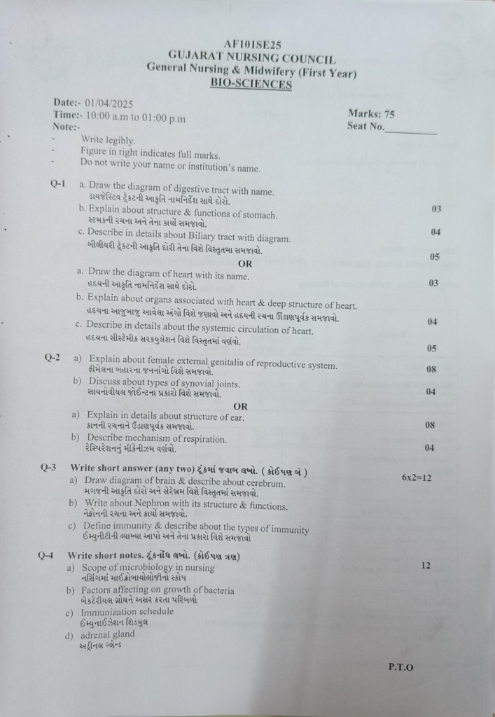
Q-1 a. Draw the diagram of digestive tract with name.
Organs of the digestive system:
The digestive tract is also called the alimentary tract, which is a hollow tube that starts from the mouth and extends to the anus. Its organs are as follows.
- Mouth
- Pharyngs
- Esophagus
- Stomach
Small intestine, which includes the duodenum, jejunum, and ileum.
Large intestine, which includes the cecum, ascending colon, transverse colon, descending colon, sigmoid colon, rectum, and anal canal.
Accessory Organs of the Digestive Tract:
These organs do not come in the main track of the digestive tract but are located on the side which pour their secretions into the alimentary canal and help in the process of digestion hence they are called accessory organs. They help in digestion with the special secretions of their glands which organs are as follows.
- 3 pairs of salivary glands
- Liver and biliary tract
- Gall bladder
- Pancreas.
b. Explain about structure & functions of stomach. 04
stomach

. The stomach is mainly located in the epigastric region and some of it is located in the left hypochondriac region.
In front of it is part of the liver, behind it is the abdominal aorta, pancreas and spleen. In the upper part is the diaphragm and in the lower part is the intestine.
stomach
The structure of the stomach is divided into three parts
- Fund,
- Body and
- Pylorus..
Fund
It forms the uppermost part of the stomach. It is found in a dome shape. It is located above the level where the esophagus joins the stomach. It is sometimes filled with gas. Here, where the esophagus joins the stomach, the cardiac sphincter muscles are located between them.
Body
The part below the fundus of the stomach is called the body, which is located at the bottom of the stomach, i.e. the pylorus.
Pylorus
The part that comes down after the body in the stomach, in which the initial part is called the pyloric antrum, which is the narrow part after the body, from there, going forward towards the duodenum, there is a canal-like structure called the pyloric canal, which opens into the duodenum and between it are sphincter muscles called the pyloric sphincter.
In addition, two curvatures are seen on the body portion of the stomach, one is the lesser curvature, which forms the right border of the stomach, and the other is the greater curvature, which forms the left border of the stomach.
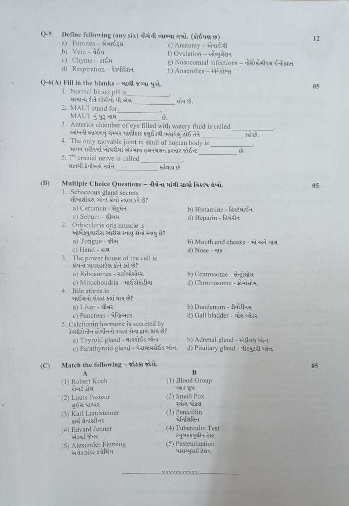
Layers of the Stomach
The wall of the stomach, like the basic structure of the digestive tract, is made up of four tissue layers, in which the outermost layer is the peritoneum layer, also called the adventitia layer, which forms the serous lining.
Below it is a layer of muscles. In the stomach, this muscular layer is made up of three layers, in which one is the long tunnel muscle fibers that are arranged superficially, the other is the circular muscle fibers that are arranged in a circular shape, which are located below the superficial layer and these fibers are also important for forming sphincter muscles. Three oblique muscle fibers are mainly found in this layer, which is not found in the basic structure. They are especially found in the wall of the stomach, which is a very important layer for the movement of food in the stomach and mechanical digestion.
Beneath the muscular layer lies the submucosal layer which contains a network of blood vessels and lymph vessels.
The innermost layer of the stomach wall is called the mucosal layer. This layer is made up of columnar epithelium tissue. This layer secretes mucus and also acts as a lubricant. This layer contains folds of mucous membrane which are especially visible when the stomach is empty and is called eructation. This eructation disappears when the stomach is full of food.
Functions of the Stomach
The stomach performs the following functions.
The stomach stores food for a short time. Before the food moves on to the intestinal tract, it undergoes partial mechanical digestion.
The stomach secretes gastric juice. This gastric juice has an acidic content and the enzymes present in it help in the digestion of food.
The muscular layer in the wall of the stomach is specially arranged, so its contraction causes a churning movement, which helps in the mechanical digestion of food and breaks the food into small pieces and then the food moves on to the duodenum.
Intrinsic factor is secreted from the inner wall of the stomach, which is necessary for the absorption of vitamin B.
Some amount of water, alcohol and some medicines are also absorbed from the wall of the stomach.
It also works to destroy bacteria or viruses present inside the stomach. When these bacteria reach the stomach through mucus and oral cavity, they are destroyed by acidic secretions and are prevented from moving further into the intestinal tract, thus also acting as a protection for the body.
c. Describe in details about Biliary tract with diagram. 05
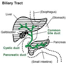
The biliary tract, also known as the biliary system, is an important part of the digestive tract. This system is made up of the liver, gallbladder, and bile ducts, which form, store, and secrete bile. Bile is primarily produced in the liver and is important for the digestion of fats in the digestive tract.
- Liver
The liver is the main organ of the biliary system. The liver forms bile, which then flows as a liquid into bile canaliculi. These canaliculi join to form segmental bile ducts, which then join the hepatic ducts.
- Hepatic Ducts
There are two types of hepatic ducts:
Left hepatic duct: Collects bile from the left liver part.
Right hepatic duct: Collects bile from the right liver part.
These two hepatic ducts collect to form the common hepatic duct.
- Cystic Duct
The cystic duct connects the gallbladder and the common hepatic duct. When a person eats, the gallbladder sends bile through the cystic duct into the common hepatic duct, which then enters the common bile duct.
4.Common Bile Duct
The common bile duct is formed by the union of the common hepatic duct and the cystic duct. This duct carries bile to the duodenum, the first part of the digestive tract. Here, bile helps in the digestion of fats.
5.Pancreatic Duct
The pancreatic duct distributes digestive enzymes from the pancreas into the duodenum. The common bile duct and the pancreatic duct join together to form the hepatopancreatic ampulla, also called the ampulla of Vater.
6.Gallbladder
The gallbladder is a small organ located below the liver, on the right side. This organ stores bile and releases it into the duodenum during the digestion of food. The gallbladder concentrates (compresses) the bile, making it more effective in digestion.
Functions of the Biliary Tract:
- Formation of Bile: The liver produces bile, which is produced by liver cells (hepatocytes).
- Storage of Bile: Bile passes through the cystic duct into the gallbladder, where it is stored.
- Secretion of Bile: During the digestion of food, the gallbladder sends bile through the cystic duct into the common hepatic duct, which then enters the duodenum through the common bile duct.
- Help in Digestion: Bile in the duodenum helps in the digestion of fats.
OR
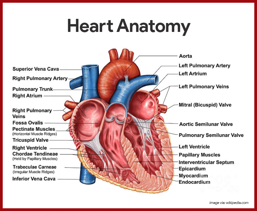
a. Draw the diagram of heart with its name. 03
b. Explain about organs associated with heart & deep structure of heart. 04
The heart is an important organ of the circulatory system. During human life, the heart beats continuously. Due to its beating, blood circulates continuously in the blood vessels.
The heart is an organ made up of a ball and a muscle. Its weight in men is approximately 310 grams and in women its weight is approximately 250 grams. The heart beats approximately one lakh times during a day.
Location of the heart:
The heart is located in the thoracic cavity between the two lungs in the mediastinum space above the diaphragm.
The heart is roughly cone-shaped. Its upper flat part is known as the base and the lower angled part is known as the apex.
The size of the heart is about the size of a human clenched fist. Its length is 12 cm, width is 9 cm and thickness is 6 cm.
The heart is located in the thoracic cavity between the two lungs, slightly tilted to the left.
The heart is found in the middle of the thoracic cavity between the second and fifth intercostal ribs.
Organs associated with the heart:
The heart is located in the thoracic cavity. There are two lungs on both the left and right sides of it.
The diaphragm and the central tendon are located on the lower side.
On the upper side of the heart are the vena cava and aorta and the pulmonary artery and pulmonary vein.
On the back side of the heart are the esophagus, trachea, bronchi and bronchioles, and the descending aorta and thoracic vertebrae.
The sternum bone, ribs, and intercostal muscles are located on the front side of the heart.
(Structure of the heart):
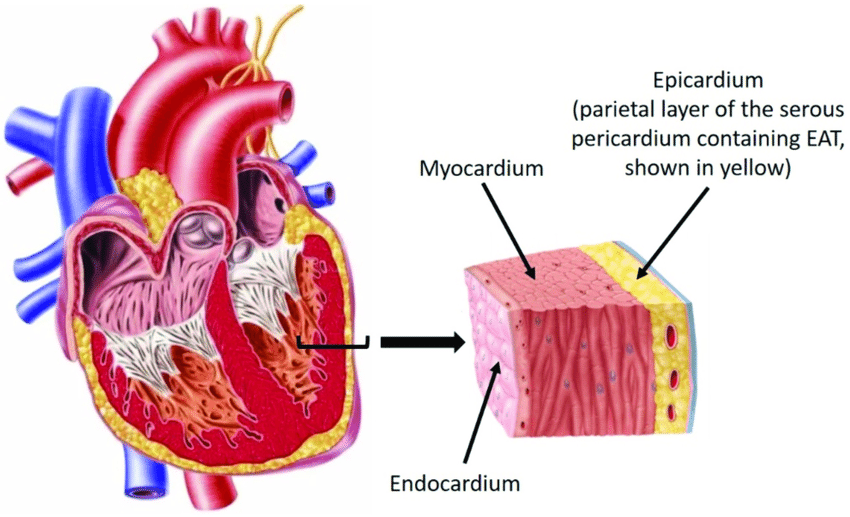
The heart is a semi-muscular organ. Its wall is made up of three types of tissue layers.
The outermost layer of the heart wall is called the epicardium or pericardium.
Epicardium or pericardium.
It is thin and transparent and covers the heart from the outside. It is made up of fibrous connective tissue. In which the outermost layer of fibrous tissue is present and on the inner side of the fibrous tissue is a serous membrane which is found in a double layer. The outer layer of the serous membrane is known as the parietal and the inner layer is known as the visceral pericardium layer.
The space between the parietal and visceral pericardial layers is called the pericardial space. This space contains a fluid called serous fluid or pericardial fluid. This prevents friction between the two layers.
This outer pericardium layer works to protect the heart from the outside and is also found around the vessels coming out of the heart.
Myocardium:
The myocardium is the middle layer of the heart. It lies below the pericardium. It is made up of a special type of cardiac muscle tissue. The pumping action of the heart is seen due to the contraction of these muscles.
This layer of myocardium is thin at the base and thick at the apex. The wall of the left ventricle is also thicker than the wall of the right ventricle.
The contraction of these muscles is an involuntary action, which causes the pumping action of the heart, and is controlled by the autonomic nervous system and the conducting system in the heart.
Endocardium:
It is the innermost layer of the heart. This layer is in contact with the blood. This layer is made up of epithelium tissue and connective tissue. This layer is smooth and shiny, which is important for the smooth flow of blood inside the heart. This layer also covers the valves inside the heart and this layer is also continuous in the inner wall of the blood vessels coming out of the heart.
Chambers of the Heart:
The heart is mainly divided into two parts. Right side and left side.
Between the right and left sides is the septum of the heart. This is again divided into four chambers, above the valve and below the valve by the valve between the right and left sides.
The two chambers above the valve are called atrium or auricle. The two chambers below the valve are called ventricles. Thus, the heart is divided into a total of four chambers.
The right side of the heart contains deoxygenated blood and the left side of the heart contains oxygenated blood. After birth, there is no connection between the right and left sides. Before birth, there is an opening between the two atria, which is called the foramen ovale, but after birth it closes.
Valve of the heart:
The flap of tissue inside the heart is called a valve. There are two types of valves in the heart.
- Atrioventricular valve.
- Semilunar valve.
Atrioventricular valve:
This valve is located between the atrium and the ventricle in the heart. The valve located between the right atrium and the ventricle is called the tricuspid valve. This valve is made up of three tissue flaps.
The valve located between the left atrium and the ventricle is called the bicuspid valve or mitral valve. This valve is made up of two tissue flaps.
The hanging ends of both the bicuspid and tricuspid valves are attached to the inner wall of the ventricle of the heart by the chordae tendineae and papillary muscles. This gives strength to the valve and prevents it from opening in the opposite direction.
Semilunar valve:
This valve is called semilunar valve because it is C-shaped or crescent-shaped. These valves are present in the aorta and pulmonary artery. The valve present in the aorta is called aortic valve and the valve present in the pulmonary artery is called pulmonary valve. Which is present at the opening of its vessels. These valves also open in the same direction.
Both the above atrioventricular valves open when the pressure of the atrium increases i.e. during the contraction of the atrium and blood comes from the atrium to the ventricle and during the contraction of the ventricle the atrioventricular valve closes and the semilunar valve opens and due to this the blood goes to the aorta and pulmonary artery.
Thus due to the opening and closing of these valves, blood circulation can take place in the heart. This valve opens in only one direction so that the blood cannot go in the opposite direction again.
Great vessels associated with heart or Openings of the heart:
The heart is mainly connected to large blood vessels. Through which blood comes from the body to the heart and is pumped by the heart and the blood goes out to the body. The vessels and their openings are as follows.
Superior venacava:
This brings deoxygenated blood from the upper part of the thoracic cavity, head and neck to the right atrium of the heart. It is one in number.
Inferior venacava:
These blood vessels bring deoxygenated blood from the inferior and lower parts of the thoracic cavity of the body and open into the right atrium of the heart. They are one in number.
Pulmonary Artery:
It carries deoxygenated blood from the right ventricle of the heart out of the heart and towards the lungs. Its number is one.
Pulmonary Vein:
Two pulmonary veins from both lungs bring oxygenated blood to the left atrium of the heart. Its number is four.
Aorta:
It takes oxygenated blood from the left ventricle of the heart and circulates it throughout the body. Its number is one.
Thus, a total of eight blood vessels and their openings are directly connected to the heart. Which are connected to the blood circulation in the heart.
Blood supply of the heart:
The oxygenated blood supply to the heart is provided by the aorta, which originates from the left ventricle of the heart. As soon as it emerges from the left ventricle of the heart, two branches, the right and left coronary arteries, arise from it on either side, which supply the heart with oxygenated blood. These coronary arteries are branches of the ascending aorta.
Of the blood that the heart pumps out through the aorta during a single contraction, about five percent of the blood is supplied to the heart through the coronary arteries.
These coronary arteries divide into many branches and supply oxygenated blood to all parts of the heart.
Deoxygenated blood in the heart drains into the coronary sinus through small coronary veins and this deoxygenated blood drains directly into the right atrium of the heart through the coronary sinus.
Functions of the Heart:
- The heart supplies oxygenated blood to all the organs and tissues of the body.
- The heart is an important organ of the cardiovascular system. Without it, the human body cannot survive and it functions as a vital organ.
- The heart circulates the blood towards the lungs so that the blood can be oxygenated and purified.
- Circulations such as pulmonary circulation and systemic circulation are regulated by the heart.
- The heart also regulates the heart rate according to the needs of the body and body temperature.
- Since the heart supplies blood circulation to every part of the body, it also regulates the body’s temperature. This layer is also found continuously in the inner walls of blood vessels.
c. Describe in details about the systemic circulation of heart. 05
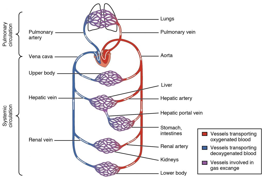
Systemic circulation begins with the left ventricle of the heart. The left ventricle of the heart contains oxygenated blood which is circulated throughout the body through the aorta and oxygenated blood supply to every cell and tissue is provided through the general circulation.
In systemic circulation, the aorta exits the heart and separates into different arteries according to the region and supplies oxygenated blood to different regions.
Aorta…

It is the main vessel of systemic circulation. It is the largest artery in the body. It circulates oxygenated blood from the left ventricle to the entire body.
The aorta that emerges from the left ventricle and is further known by different names according to its location. Such as ascending aorta, arch of aorta and descending aorta.
After reaching the thoracic and abdominal regions, the descending aorta is converted into two right and left common iliac arteries and circulates blood in the lower limbs.
In systemic circulation, the aorta is divided into branches below according to the thoracic and abdominal regions, which supply oxygenated blood to those areas.
•Thoracic Aorta : This part of the aorta is above the diaphragm and is described in three parts:
1.Ascending Aorta
- Coronary arteries
2.Arch of aorta
•Brachiocephalic artery
- Left common carotid artery
- Left Subclavian artery
3.Descending Aorta in the thorax.
•Brachial arteries
•Oesophageal arteries
•Intercostal arteries
- Abdominal aorta :
•Inferior Phrenic
•Renal artery
•Testicular Arteries or Ovarian arteries
•Lumbar arteries
•Median sacral arteries
•Coeliac artery
- Left gastric artery
- Common hepatic artery
- Splenic artery
•Superior mesentric artery
•Inferior mesentric artery
All the above arteries together complete the systemic circulation and supply oxygenated blood to every organ of the body.
Q-2 a) Explain about female external genitalia of reproductive system. 08
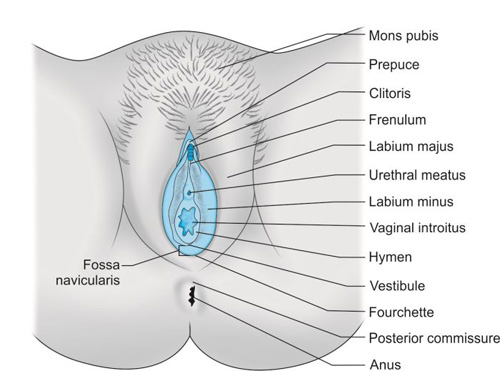
External genitalia of female / VULVA / PUENDENDAM
Mons pubis
Labia majora
Labia minora
Clitoris
Perineum
Vestibule
Mammary gland
MONS PUBIS
The mons pubis is a cushion-like structure made of fat and skin. It lies in front of the symphysis pubis.
During puberty, it becomes covered with hair and forms a horizontal fold.
LABIA MAJORA
There are two thick folds on the front side. This forms the brim of the vulva. (The vulva is the external reproductive organ, also called the vulva).
The labia majora are made up of skin, fat, areolar tissue, and smooth muscle. On the upper surface, there are hairs. On the inner side, there are sebaceous glands.
The round ligament ends at the labia majora.
The labia majora meet anteriorly near the mons pubis and posteriorly near the skin of the perineum. Just as males have testes, females have labia majora.
LABIA MINORA
The labia minora are two small skin folds (which do not have fat or hairs but do have many sebaceous glands) that lie inside the labia majora.
Its upper part meets the clitoris and forms the prepuce (the cover of the clitoris in females, just as the foreskin of the penis in males is present), and the lower part forms the floor (frenulum) of the clitoris.
CLITORIS
It is cylindrical in shape and triangular in shape. It is made up of erectile tissue (stimulating tissue).
Just as the penis in males is present, the clitoris in females is present.
It contains sensory nerve endings. It functions as a stimulus during sexual intercourse.
PERINEUM
This is an area that extends from the base of the labia majora to the anal canal. It is roughly triangular in shape.
It is made up of connective tissue, fat, and muscle. It provides attachment to the muscles of the pelvic floor.
VESTIBULE
Vestibular glands (Bartholin gland) are 2 and are located on either side of the vaginal opening. They are about the size of peas. They have ducts that open into the vagina and secrete mucous. This keeps the vulva moist and the vaginal cavity wet.
b) Discuss about types of synovial joints.
Types of Synovial Joint:
Synovial Joint is one of the softest and most mobile joints found in the body. Such joints have a synovial cavity between two bones, which is filled with synovial fluid. This synovial fluid reduces the friction between the bones and helps the patient for easy and effective movement. Synovial Joint is of different types according to its special structure and work.
1.Hinge Joint:
Example: Elbow, Knee, Interphalangeal Joint
This joint works like a hinge — it allows movement like flexion and extension in only one direction, forward and backward. This is a uni-axial joint. It is very important for the movement of the hands and feet in the daily life of the patient.
2.Ball and Socket Joint:
Example: Shoulder, Hip
In this joint, the head of one bone is like a ball and the other bone has a cup-like opening. This is a Triaxial Joint, in which movements in all directions like Flexion, Extension, Abduction, Adduction and Rotation are possible. It is the joint that does the most work for the major movement of the patient’s body.
3.Pivot Joint:
Example: Atlantoaxial Joint
In this joint, one bone fits into the ring-like bone of another and rotates around it. It is a uni-axial joint. It is the main joint for the patient to turn his head left and right.
4.Saddle Joint:
Example: First Carpometacarpal Joint
In this joint, both bones look like a saddle. This is a Biaxial Joint in which movements like Flexion, Extension and Opposition are possible. Works specifically for the movement of the thumb.
5.Plane Joint:
Example: Intercarpal Joint, Intertarsal Joint
In this joint, the surface of the bones is flat and they glide over each other. Limited but important movement occurs in this. It is necessary for small movements in the hand and foot.
6.Condyloid Joint:
Example: Radiocarpal Joint (Wrist)
This joint is biaxial in which the Oval Head bone fits into the groove of the other. In this, movements like Flexion, Extension, Abduction and Adduction occur. Wrist Joint works especially for fine movements in the patient’s hand.
General Features of Synovial Joint:
Joint Capsule covers the joint
Synovial Fluid reduces friction between bones
Articular Cartilage covers the head of the bone
Ligaments and Muscles provide stability to the joint
Synovial Joint plays a major role in the movement of the body. Each type of joint has its own specific structure and function.
a) Explain in details about structure of ear.
Ears are located on either side of the head. They are organs involved in hearing and maintaining body balance.
Structure…
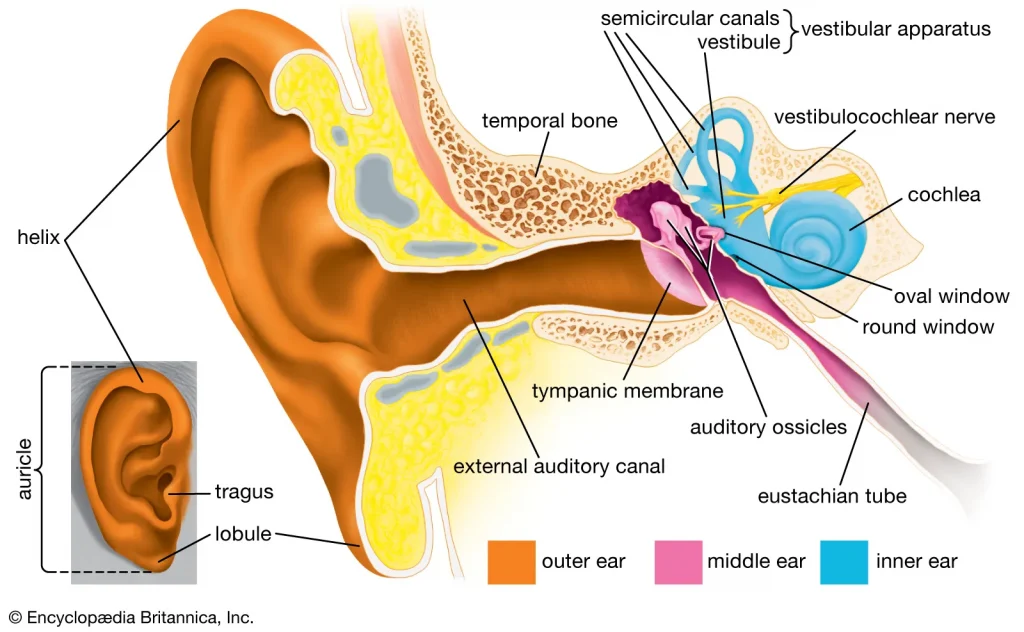
Structurally, the ear is anatomically divided into three parts
External or outer ear
Middle ear
Inner ear. 1.External or outer ear
The external ear collects sound waves and transfers them to the inside. The external ear consists of the following parts.
Auricle or pinna
External acoustic meatus i.e. auditory canal
Auricle…
It is a flap-like part on both sides of the head which is made of elastic cartilage and is covered with skin on the outside. It collects sound waves coming from the outside environment and transmits them to the inner hole. Its most prominent upper part is called the helix and the soft part hanging below is known as the lobule.
External acoustic meatus…
It is also known as the auditory canal, it is an S-shaped curved tube. Its length is about 2.5 cm. It is an elongated canal on the inside and the glands in its wall secrete a wax-like viscous fluid, also known as cerumen. Some hairs are also present in this canal which, combined with the viscous fluid, do not allow dust and foreign bodies to reach the inner wall, i.e. the tympanic membrane.
Tympanic membrane…
It is a thin membrane that separates the external ear and the middle ear and is oval in shape. This membrane is made up of epithelium tissue and connective tissue, and fibroblast cells are present in it.
Middle ear..
It is a small air-filled cavity located in the temporal bone. The lining of this cavity is made of epithelial tissue and it is separated from the external ear by the tympanic membrane i.e. the ear drum and the inner ear is separated from the middle ear by the oval and round windows.
Its anterior wall has an opening known as the eustachian tube. This tube connects the middle ear to the structures of the nasopharynx and maintains pressure inside the cavity, which prevents the tympanic membrane from rupturing during yawning and swallowing food.
Auditory ossicles…
The bones in the middle ear are called auditory ossicles and include the malleus, incus and stapes bones. Its number is one in each ear, that is, there are a total of 6 auditory ossicles in the body.
Malleus..
It is hammer-shaped, its handle is attached to the wall of the tympanic membrane. The head of the malleus is attached to the incus bone.
Incus..
It is the middle bone which is in the shape of anvil, it is attached to the malleus bone and its head is attached to the stapes bone. There is fibrous tissue in the joint part.
Stapes..
It is a stirrup-shaped bone whose head is attached to the incus bone and its foot plate is attached to the oval window, and the round window is located below it.
Thus, the structure of the part from the tympanic membrane to the oval and round windows is called the middle ear.
- Inner ear…
It is also known as the labyrinth. It has a complex structure and is a part associated with the function of hearing and maintaining balance..
It has two parts, the bony labyrinth and the membranous labyrinth..
The bony labyrinth…
It is a cavity located in the petrous portion of the temporal bone, whose wall is made of a layer of periosteum. On its inner side there is a fluid called perilymph. It has three parts. A vestibule, a cochlea and three semicircular canals.
Vestibule..
It is the middle oval-shaped portion of the bony labyrinth, inside which the utricle and saccular structures are located. Its lateral wall has oval and round windows.
Cochlea…
This part of the bony labyrinth is connected to hearing, it is a lump-like part which is also known as Snell’s cell.
Semicircular canal…
The structure of this canal is connected to balance. The three tubes of the semicircular canal are arranged at right angles to each other.
Membranous labyrinth..
Inside the bony labyrinth there is a membrane tube called the membranous labyrinth which contains a fluid called endolymph. The properties of this fluid are similar to CSF.
Vestibule which contains the utricle and the saccule.
Vestibule and Semicircular Canal…
The vestibule and semicircular canals in the inner ear work to maintain the body’s balance.
In which the semicircular canal, the cochlea, and the utricle help to maintain the body’s dynamic equilibrium. The semicircular canal is a tube-like structure that is located behind and above the vestibule and these canals are arranged at an angle to each other. The semicircular canal and the cochlea, whose outer wall is the bony labyrinth and inside it is the perilymph fluid, have another tube inside called the membranous labyrinth, which contains endolymph.
The utricle is a membranous sac that is part of the vestibule and all its ducts open into a dilated portion called the ampulla. The saccule is part of the vestibule and communicates with the utricle and the cochlea.
The walls of the utricle, saccule and ampulla contain small hair-like projections of specialized epithelial cells, which contain sensory nerve endings, forming the vestibular part, through which the vestibulocochlear nerve passes.
- Cochlea
The cochlea, also known as the snail, is a coiled structure resembling a snail. Inside the cochlea is a membrane called the cochlear duct, which is mainly involved in the process of hearing. The following parts are seen in the cross section of the cochlea.
Scala vestibuli
Scala media or cochlear duct
Scala tympani
The cochlear duct is a triangular-shaped tube. The bony part of the cochlea is divided into two parts, upper and lower, in which the upper part is called scala vestibuli and the lower part is called scala tympani. In the middle part, the cochlear duct is located and its roof membrane is called basilar membrane. The organ of Corti, which is called the hearing organ, is located on the basilar membrane.
b) Describe mechanism of respiration. 04
Mechanism of respiration:
Respiration is the gas exchange between two surfaces. In which the air of the atmosphere enters the lungs. Gas exchange occurs between the tissues of the lungs and the blood, which is called external respiration. The gas exchange that occurs between each cell tissue of the body and the blood is called internal respiration.
During the act of respiration, oxygen enters the lungs through inhalation and carbon dioxide leaves the body through exhalation.
Usually, the act of respiration is seen 16 to 18 times in a minute.
The following actions are seen in the cycle of respiration.
Inspiration
Expiration
Pause
Inspiration…
The act of entering the lungs through inhalation of atmospheric air is called inspiration.
When the brain receives nerve impulses for contraction of the diaphragm and intercostal muscles, the size of the thoracic cavity increases due to the contraction of the diaphragm and intercostal muscles. The air pressure inside the thoracic cavity decreases so that air from the outside environment can enter the lungs through the act of inspiration. This act is called inspiration.
When the diaphragm receives impulses of contraction, the diaphragm becomes flat and moves downwards and the intercostal muscles contract, causing the ribs and intercostal muscles to move upwards and outwards. This increases the size of the thoracic cavity and creates negative pressure inside the cavity. Since the air pressure in the outside environment is high and the air pressure in the thoracic cavity is low, the act of inspiration can take place. The act of inspiration is an active process.
Expiration…
The process of expelling air from the lungs into the atmosphere is called expiration. The act of expiration is a passive process that begins after the act of inspiration is completed.
In the act of expiration, the contracted diaphragm and intercostal muscles relax. So that the diaphragm returns to its original position and the ribs come down and inward, the size of the thoracic cavity decreases and the act of expiration occurs. In which the air from the lungs is expelled into the atmosphere.
In the act of expiration, the air pressure in the lungs is higher than the atmospheric pressure so that the act of expiration occurs.
Pause… This is the relaxed stage of the lungs. In which no inspiration or expiration occurs. This period is called the pause period.
Q-3 Write short answer (any two) 6 × 2 = 12
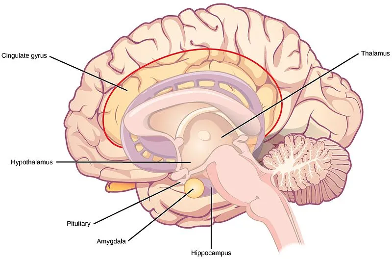
a) Draw diagram of brain & describe about cerebrum.
The cerebrum is made up of the right and left cerebral hemispheres.
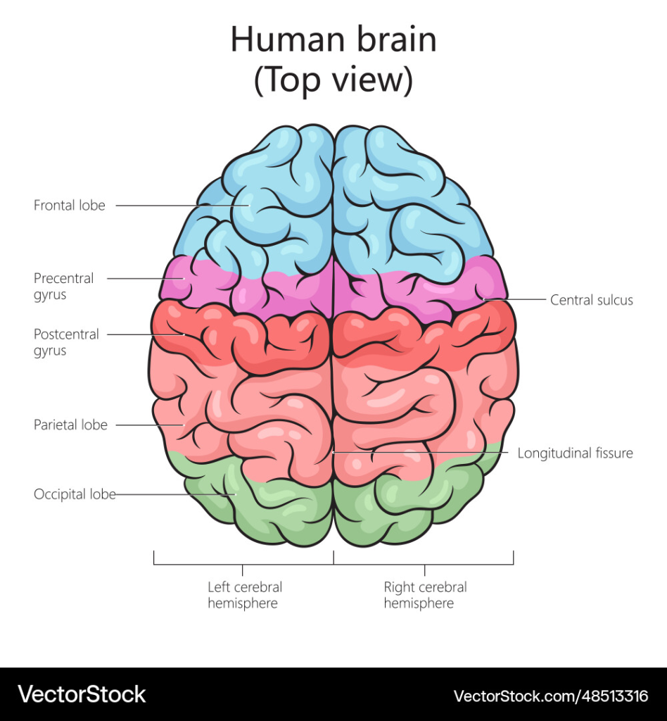
The two cerebral hemispheres are connected by the corpus callosum. Each hemisphere has a cavity called the lateral ventricle.
The superficial structure of the cerebrum is made up of gray matter called the cerebral cortex. The deep part of the cerebrum is made up of white matter.
The cerebral cortex is made up of raised areas called gyri or convolutions and is separated by grooves called fissures or sulci.
The deepest of these fissures is the longitudinal fissure which separates the right and left hemispheres.
The hemispheres of the cerebrum are divided into different lobes which are as follows.
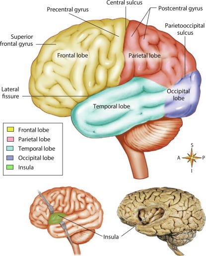
•Frontal lobe
•Parietal lobe
•Temporal lobe
•Occipital lobe..
•Important fissure in the cerebrum;
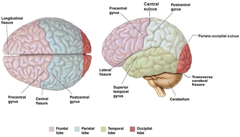
•Longitudinal sulcus or fissure which is the deepest and divides the cerebrum into two hemispheres
•Central sulcus which is located between the frontal and parietal lobes.
•Lateral sulcus which is a deep groove and separates the temporal lobe from the frontal and parietal lobes.
•Parieto-occipital sulcus which separates the occipital lobe from the two parietal lobes….
•The inner part of the cerebrum contains nerve fiber tracks such as
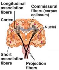
•Association Fibers
•Commissural Fibers
•Projection Fibers
These fibers are connected to each other and connect one area to another and help in the transmission of impulses.
FUNCTIONS OF THE CEREBRUM.
- The cerebrum controls intelligence, memory, reasoning, thinking, speaking, reading, writing, etc.
- The cerebrum controls sensory perception such as pain, temperature, touch, sight, hearing, taste, smell, etc.
- The control of skeletal muscle contraction is controlled by this area.
Functional areas of the Cerebrum.
There are many areas in the hemisphere of the cerebrum which are as follows.
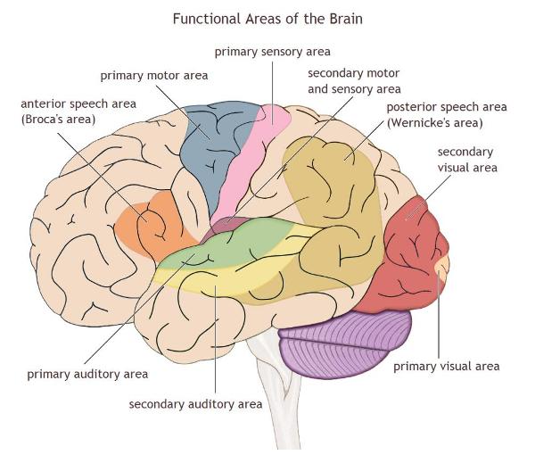
•Motor areas
Which are mainly located in the front part of the cerebral hemisphere. In which the following areas are found.
- Primary motor area which is located in the frontal lobe and controls the voluntary contraction of muscles.
- Motor speech area which is also called Broca’s area which is located in the frontal lobe.
•Sensory areas of cerebrum
- Primary somatosensory area or general area which is located in the parietal lobe behind the central sulcus which is the sensory area for touch pain and temperature.
- Primary visual area which is located in the occipital lobe which interprets vision.
- Primary auditory area which is located in the temporal lobe and is associated with the function of hearing.
•Primary gustatory area located in the parietal cortex behind the central sulcus and associated with the testes.
•Primary olfactory area located in the temporal lobe. Area associated with smell.
OTHER AREAS OF CEREBRUM.
There are some special areas in the cerebrum which are involved in the transmission of impulses and other important functions which are as follows.
- Basal ganglia..It is an area in each cerebral hemisphere which works to coordinate muscle tone, posture and voluntary muscle movements..
- Thalamus..It is located in the middle of the brain at the top of the brain stem. It acts as a relay station for sensory impulses coming towards the sensory areas of the cerebral cortex which allows proper interpretation of sensations and some movement control.
- Hypothalamus…The hypothalamus is made up of a group of nerve cells located below the thalamus, whose functions are as follows.
•The hypothalamus controls the following functions.
•Body temperature
•Hunger and thirst
•Emotional reactions
•Autonomic nervous system
•Sexual behavior
•Biological clock or circadian rhythm…
•Secretion of certain hormones
All the above are done by the hypothalamus.
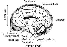
BRAIN STEM.
The brain stem includes the following parts
1.Midbrain
2.Ponse Vertebrate
3.Medulla Oblongata
•Midbrain
It forms the upper part of the brain stem. On its side, the cerebral aqueduct connects the third and fourth ventricles.
The upper part of the midbrain contains important centers for vision and hearing.
The lower part of the midbrain contains the motor pathways that run from the pons vertebrate and medulla oblongata to the spinal cord.
The midbrain contains the control centers for balance and eye movement.
•Ponse Vertebrate
It forms the middle part of the brain stem.
It also contains ascending and descending nerve pathways like the midbrain and many of its nerve tracks are connected to the cerebellum and cerebellar cortex
It helps in the transmission of impulses.
•Medulla oblongata
It forms the lowest part of the brain stem and connects the pons veroli to the spinal cord
It is approximately 2.5 cm long and contains the following centres.
1.Respiratory center
2.Cardiovascular center
3.Vasomotor center
4.Reflex center for vomiting, coughing and swallowing..
Some special functions are found in the medulla oblongata which are as follows
- The descending motor pathway crosses from the medulla and passes to the spinal cord which mainly provides impulses to skeletal muscles.
- Like the motor pathway, the sensory pathway also crosses from the medulla and passes to the brain.
- The medulla contains the cardiovascular center which controls the rate and force of the heart. Sympathetic stimulation increases the heart rate and force while parasympathetic stimulation decreases the heart rate and force.
- The medulla contains the respiratory center which controls the rate and depth of respiration, in which inspiration and expiration are observed by receiving nerve impulses to the intercostal muscles and diaphragm.
- The medulla contains the vasomotor center which controls the diameter of the blood vessels, due to which vasoconstriction and vasodilation are observed.
- The reflex center in the medulla controls vomiting, coughing and hiccups, which is also a protective response.
b) Write about Nephron with its structure & functions,
Structure Of Nephron
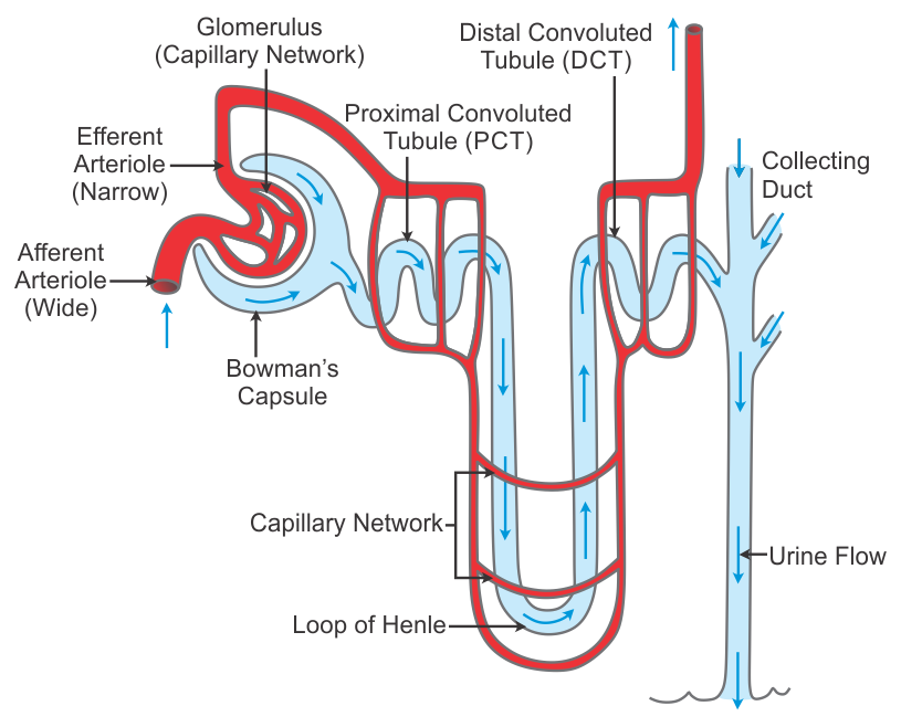
Looking at the microscopic structure of the kidney, many compositions are seen. In which, when the kidney is viewed under the microscope, the microscopic functional structure i.e. nephron is seen, which is the main working unit of the kidney. There are millions of nephrons in the kidney.
The microscopic structure of the nephron includes the following structures.
- Bowman’s capsule..
There is a cup-shaped mouth at the front of the nephron, which is called Bowman’s capsule. In the wall of this capsule, there are special types of epithelial cells called podocytes. Both the parietal and visceral layers of this Bowman’s capsule are seen.
The cup-shaped part of the Bowman’s capsule is surrounded by a network of arterial capillaries called the glomerulus. The podocyte cells present here help in the filtration process. The three-centimeter long part anterior to the Bowman’s capsule is called the tubular part. This tubular part can be divided into the following parts:
- Proximal convoluted tubule..
The initial tubular portion anterior to the Bowman’s capsule is known as the proximal convoluted tubule. It is the initial part of the tubules of the nephron. Its wall is lined with epithelial cells. This part is located in the cortex of the kidney.
- Loop of Henle..
The U-shaped part after the proximal convoluted tubule is called the loop of Henle. It is also called the medullary loop. The loop of Henle consists of an ascending and descending loop, with a sharp bend in the middle, forming a U-shaped part. The loop of Henle is located in the medulla of the kidney.
- Distal convoluted tubules.
The tube-shaped part of the nephron after the loop of Henle is called the distal convoluted tubule. This tube-shaped part forms the descending limb. Which further joins with the collecting duct..
- Collecting duct..
The collecting duct is a straight tube. Which connects the distal convoluted tubule of many nephrons. This tube is known as the large duct. These collecting tubes connect with each other and open into the minor calyx of the renal pyramid.
When the renal artery enters the kidney from the hilum, it then divides into many smaller arterioles. When these arterioles enter the Bowman’s capsule, these arterioles are called afferent arterioles. The arterioles that divide into a network of capillaries in the glomerular capsule are called glomeruli. The arterioles of this capillary network that exit the Bowman’s capsule are called efferent arterioles.
The diameter of the efferent arterioles is smaller than the diameter of the afferent arterioles, due to which pressure is created on the glomerular membrane and the process of filtration is accelerated and the process of urine formation begins.
The efferent arterioles again divide into small capillaries and absorb nutrients and water and mix them with the blood circulation. These small vessels drain the blood out of the kidney through the renal veins.
Main Functions of Nephron:
- Filtration:
Blood plasma is filtered from the glomerulus and enters the Bowman Capsule.
- Reabsorption:
Nutrition, water, glucose, amino acids, etc. are taken back into the blood from various parts of the tubule.
- Secretion:
Hydrogen ions, potassium, nitrogenous toxins, etc. are released from specific tubule parts into the urine.
- Osmoregulation:
Nephron controls the amount of water and salts in the body so that blood pressure and electrolyte balance are maintained.
- Acid-Base Balance:
Nephron controls the pH level of the blood so that the patient’s homeostasis is maintained.
Nephron is a very important functional unit of the kidney which plays a major role in the health of a person by removing toxic substances from the body, forming urine and maintaining homeostasis. Therefore, proper functioning of the nephron is extremely important for health.
c) Define immunity & describe about the types of immunity
The resistance shown by the host against the condition caused by the micro organism and its products (toxins) is called Immunity
or
Immunity means when any antigen or micro organism enters our body and our body resists it and provides protection, it is called immunity.
Immunity:
Immunity is a type of resistance that is activated by the host’s body.
When any foreign body (antigen) enters the host’s body, the antibody that fights against that antigen is called immunity. When any antigen (foreign body) enters the body, the ability of the body to fight against it is called immunity.
There are two main types of immunity:
1) Innate immunity
2) Acquired immunity
(1) Innate immunity
Innate immunity is the immunity that a person has by birth. This is a type of natural immunity.
(2) Acquired immunity
The immunity that the body gets during life after birth is called acquired immunity.
Classification of immunity:
there are two types of immunity .
1)INNATE IMMUNITY ( IN BORN),
2)ACQUIRED IMMUNITY
AFTER BORN.
TYPES OF INNATE IMMUNITY:
1)SPECIES IMMUNITY
2)RACIAL IMMUNITY
3)INDIVIDUAL IMMUNITY
1)species immunity
2)Racial immunity
3)individual immunity
There are two types of acquired immunity.
1)ACTIVE IMMUNITY
2)PASSIVE IMMUNITY
1)Active immunity
(Active immunity: The immunity that is produced by the body itself is called active immunity)
There are also two types of active immunity
A)Active natural immunity:
(Active natural immunity: When a person is exposed to an infection, immunity is seen in that person.)
B)Active artificial immunity:
(Active artificial immunity: When a person is immunized and immunity develops in that person, it is called active artificial immunity.)
2)PASSIVE IMMUNITY
(Passive Immunity: The immunity that is developed by someone else is called passive immunity. This immunity does not develop in the own body.)
There are also two types of passive immunity.
A) Passive natural immunity: When the fetus is in the mother’s womb and the immunity that the fetus gets from the mother is called passive natural immunity.)
B) Passive artificial immunity: (Passive artificial immunity: When antibodies are made from someone else and transferred to the body of someone else, it is called passive artificial immunity.)
the full information about AQUIRED IMMUNITY :
Full information about Acquired Immunity:
The immunity that the body gets after birth is called acquired immunity.
The immunity that the body gets after birth during whole life is called acquired unity.
There are two types of acquired immunity.
1)ACTIVE IMMUNITY
(Active immunity),
2)PASSIVE IMMUNITY
1)ACTIVE IMMUNITY
When any antigen enters the body, the body produces antibodies against the antigen on its own and it is called active immunity. (active immunity develop after entry of antigen)
There are (2) types of active immunity.
1)Active natural immunity
This immunity is natural and active, hence it is called active natural immunity.
This immunity is produced when an antigen enters the body and antibodies are produced in the body against that antigen, then immunity is seen in the body and this immunity is long-lived.
A)humoral immunity
Humoral immunity develops from the body’s plasma.
B)cell-mediated immunity
Cell-mediated immunity mainly develops from T-lymphocytes.
T- lymphocytes provide defense against any infection in the body, against any microorganism that is chronic bacterial infection.
2)Active artificial immunity
Artificial means (man made) is artificially made and is not related to nature.
When an immunity is provided by a vaccine and the body produces antibodies against it, it is called artificial active immunity.
Ex:=BCG( Baccilus callmate guarine),
hepatitis,
DPT( Deptheria, Pertussis, Tetanus),
DT( Deptharia, Tetanus),
pentavelent etc.
2)PASSIVE IMMUNITY
Passive immunity is the immunity that is transferred to a ready recipient and is called passive immunity.
There are two types of passive immunity
1)Passive natural immunity
2)Passive Artificial immunity
1)Passive natural immunity
This immunity is given to the fetus through the mother and is called passive natural immunity.
This immunity is also called congenital immunity.
Ex: 1) IgG (immunoglobulin G) is given to the fetus from the placenta.
2) IgA (immunoglobulin A) is given to the fetus from breast milk.
These two antibodies IgG and IgA are given to the fetus from the mother, so it is called passive natural immunity.
2) Passive Artificial immunity (passive artificial immunity).
This immunity is artificial (man made).
In this, the antibody is directly administered into the body of the recipients.
Given by:
1)antitoxin,
2)antibacterial,
3)antiviral,
Ex:=Tetanus toxoid ( TT),
Gas gangrene antitoxin,
Anti venom sera,
Anti lymphatic serum,
These antibodies provide immediate protection and are for a temporary period.
Use of passive immunization
1)Passive immunization provides immediate immunity in the body.
2) Suppresses active immunity.
3) If there is a serious infection, it provides immediate immunity to treat it.
Q-4 Write short notes. 12
a) Scope of microbiology in nursing
Microbiology is a branch of science that deals with the study of microorganisms such as bacteria, viruses, fungi, protozoa, and algae. Knowledge of microbiology is very important in the nursing field as it forms the basis for the prevention, control, and treatment of infections during patient care.
Scope and Importance of Microbiology in the Nursing Field:
- Infection Control and Prevention:
Nurses are exposed to potential threats from various pathogens while caring for patients. Knowledge of Microbiology teaches them the proper use of Hygiene, Septic Technique and Antiseptic methods.
2.Disease Diagnosis:
Nurses become more capable of understanding the reports of microbiological tests like Blood Culture, Urine Culture, Skin Swab taken in the laboratory with knowledge of Microbiology.
3.Antimicrobial Therapy:
It is important for nurses to know about the proper use, side effects and resistance of medications like Antibiotics, Antifungals, Antivirals etc. – and this becomes possible with the study of Microbiology.
- Sterilization and Disinfection:
Knowledge of microbiological techniques is essential to keep medical instruments and the hospital environment free from microbial contamination.
- Epidemiology and Public Health:
Nurses can gain a deeper understanding of epidemics and pandemics through microbiology to understand the transmission and control of infectious diseases.
6.Understanding Immunology:
Microbiology plays an important role in gaining knowledge about the functioning of the patient’s immune system, the use of vaccines, and hypersensitivity reactions.
7.Hospital Infections and Nosocomial Infection:
Knowledge of microbiology is essential for identifying, preventing, and managing healthcare-associated infections (HAIs) in hospitals.
8.Patient Education and Community Health:
Nurses provide information to patients about personal hygiene, food safety, and disease prevention, in which the contribution of microbiology is important.
In the field of nursing, microbiology is not just a science, but a practical tool that prepares nurses for patient safety, transmission control, and quality care. Therefore, a deep knowledge of microbiology is essential for every nurse.
b) Factors affecting on growth of bacteria
1) Moisture
Every bacteria needs water for growth, just like nourishing food. In fact, bacteria cannot get food in the absence of water, because all food elements need to be in a liquid state to pass through the bacterial wall. Every type of bacteria grows well in an aqueous medium, a completely moisture-free environment prevents its growth. Or destroys it. In addition, cells cannot survive in low or high humidity.
2) Light
Most of the bacteria are killed by direct contact with ultraviolet rays present in sunlight.
3) Temperature:-
Temperature is a very important factor affecting the growth of bacteria. Optimal temperature along with food and water is necessary for bacterial growth.
Different bacteria have different optimal (favorable) temperatures.
For bacteria growing in the human body, 37° C is the optimal temperature.
However, many bacteria are mesophilic ( meso = middle, phille = loving ). The optimal temperature for them is 25 to 39* C.
Most bacteria grow in this way.
Apart from psychrophilic ( Psychro = cold ) bacteria grow better between 4 C to 10° C, some
Thermophilic {Therma – Heat ) are also found. Their growth is better between 55° C to 75 C.
Temperatures above 75 C are fatal for bacteria. In fact, high temperatures are created to destroy bacteria in various ways.
Such as moist heat (steam), boiling water, pasteurization & autoclaving.
Many species can survive at very low temperatures. Such as yeast, mould, viruses & Rickettsia, spirochetes (76* C can survive for years).
(4) Oxygen
O2 also plays an important role in the life of bacteria. Many types of bacteria can live or grow only in the presence of O2. They are called Aerobes (EX.Sarcina).
On the contrary, Anaerobes can live or grow in the absence of 02. E.g. Closteridium tetani-
Apart from this, there are also bacteria. Which can live in the presence or absence of 02. They are known as facultative anaerobes. E.g. Salmonella typhi.
Microaerophils grow more in less oxygen than present in the air.
(5)Hydrogen Ion Concentration: (Acidity and Alkalinity) PH Medium
The acid or alkali concentration of the medium in which bacteria grow affects their growth.
This is measured by the hydrogen ion concentration index.
PH – 0 (Zero) is the most acidic,
PH – 14 is the least acidic.
PH – 7.) is neutral,
PH <7 is acidic
and PH >7 is alkaline
. Most bacteria grow best between pH 5.0 and 8.5. There are some exceptions to this.
6) Osmotic pressure :-
The life of bacteria also depends on more or less osmotic pressure. If bacteria are immersed in a liquid whose osmotic pressure is very high or very low, the bacterial cell collapses or becomes dormant due to the fluid escaping from it.
Carbon Dioxide is also necessary for the growth of bacteria.
c) Immunization schedule
Immunization is a very important service in preventive pediatric services. By giving vaccines, specific protection is developed against specific diseases.
Immunization provides specific protection against diseases that are seen as dangerous infections in children such as poliomyelitis, diphtheria, pertussis, measles, rubella, hepatitis B, pneumonia, viral diarrhea, etc. Immunization provides specific protection against diseases that are particularly responsible for mortality and morbidity in children.
Immunization produces immunity against specific diseases in children, and children can be protected from specific diseases or infections.
The following immunization schedule is followed under the Universal Immunization Program.

d) adrenal gland
Adrenal glands
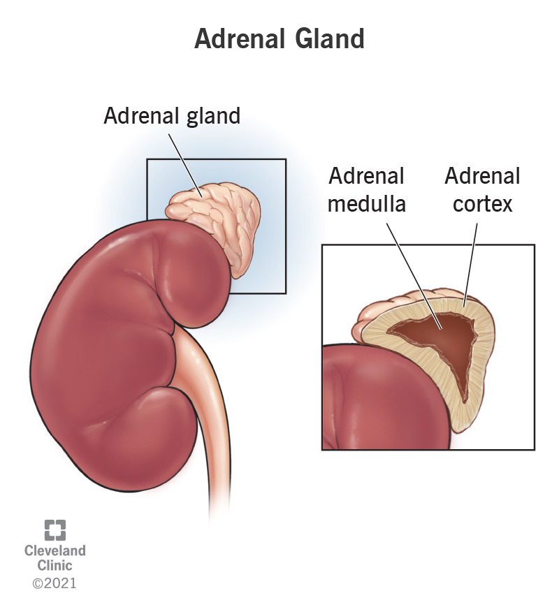
This gland is also called the suprarenal gland because it is located above the kidneys on both sides. It is found in the number of 2. Its size is 3 to 5 centimeters long, 2 to 3 centimeters wide and 1 centimeter thick. Its weight is approximately 3 to 5 grams.
The outer part of the adrenal gland is known as the adrenal cortex and the inner part is known as the adrenal medulla.
Adrenal cortex.
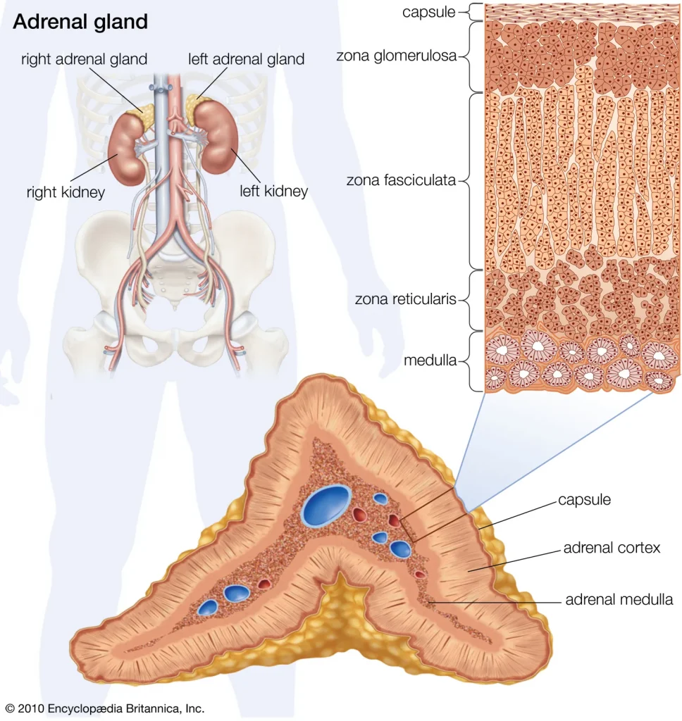
This part is located towards the outer part of the adrenal gland. The cells in this part secrete steroid hormones.
The following steroid hormones are secreted by the cortex part of the adrenal gland.
A. Glucocorticoids.
This hormone is secreted by the Zona Fasciculata cells in the adrenal gland.
ACTH secreted from the anterior lobe of the pituitary gland stimulates the adrenal cortex part and causes the secretion of glucocorticoid hormones.
This hormone helps in the metabolism of protein, glucose and fat in the body.
It provides resistance to stress in the body. Due to which the heart rate and blood pressure increase during stress. Also, an increase in blood sugar is seen.
The effect of this hormone is seen in every cell of the body. Its highest level in the blood is seen in the morning and its level gradually decreases till night.
This hormone also gives anti-inflammatory effect in the body. It also depresses the immune system of our body. Due to which our immune response decreases.
It includes cortisol and cortisone hormones.
B. Mineralocorticoid (Mineralocorticoids).
This hormone is secreted by the Zona Glomerulosa cells in the adrenal gland.
Secreted from the adrenal cortex, this hormone mainly includes the aldosterone hormone. This hormone works to maintain the level of mineral salts in the body. Due to which the fluid and electrolyte balance is maintained in the body.
Aldosterone hormone reabsorbs sodium and excretes potassium through urine. Due to which water retention occurs in the body and blood pressure is maintained due to maintenance of body fluid.
This mechanism is maintained by the renin angiotensin aldosterone system (RAAS).
C. Androgens (Androgens).
This hormone is secreted by the Zona Reticularis cells in the adrenal gland.
It is also known as a sex hormone. Testosterone in males and estrogen hormone in females are considered as androgen steroid hormones.
Which perform important functions in developing sex characteristics, hair growth and muscular structure in males and females.
Adrenal Medulla (Adrenal Medulla).
The middle part of the adrenal gland is known as the adrenal medulla. This part secretes catecholamine hormones. In which adrenaline and noradrenaline are mainly secreted, i.e. epinephrine and norepinephrine.
These hormones maintain the fight and flight response of the sympathetic nervous system of the autonomic nervous system during stress.
These hormones perform the following functions in the body.
Increases blood pressure and increases heart rate
Constricts the diameter of blood vessels
Increases blood sugar and also increases the rate of cellular metabolism
Dilates the pupils
Dilates the airways of the lungs, etc. Functions are seen through these hormones. Response to stress
Q-5 Define following (any six) 12
a) Fomites
Fomites are non-living objects that can transmit infectious microorganisms such as bacteria, viruses, or fungi from one person to another upon contact. Fomites include hospital bed sheets, stethoscopes, thermometers, nursing tables, door handles, etc. They are known to be important means of transmission that increase the likelihood of person-to-person transmission of diseases.
b) Vein
Vessels that carry deoxygenated blood from different parts of the body to the heart are called veins. Small venules join to form larger veins that carry deoxygenated blood to the right atrium of the heart. Among all the veins in the body, the superior and inferior vena cava are the main veins. Which carry deoxygenated blood from all parts of the body to the heart.
The pulmonary vein, which carries oxygenated blood from the lungs, carries oxygenated blood to the left atrium of the heart. This vein is the only one in the body that carries oxygenated blood.
c) Chyme
Chyme is a semi-conductor substance that is formed in the stomach in the form of a paste during the digestion process. When food mixes with the enzymes and gastric juice of the stomach, it is transferred to chyme. This chyme then moves to the small intestine where further digestion and absorption takes place.
d) Respiration
Respiration is the gas exchange between any two surfaces. In respiration, oxygen enters the body from the atmosphere through inspiration and carbon dioxide, which is a waste product of the body, is released through expiration. Thus, the process of respiration is seen.
There are two main types of respiration in the body.
The exchange of oxygen and carbon dioxide between each cell tissue of the body and the surrounding blood capillaries is called internal respiration. This respiration is seen everywhere in the body.
The gas exchange of oxygen and carbon dioxide between the lung tissue, i.e. the alveoli, and the surrounding blood capillaries is called external respiration. This respiration occurs in the lungs.
e) Anatomy
Anatomy is the study of the structure of the organs in the body and its scientific study is called anatomy.
Anatomy includes the structure of any organ in the body, the location of the organs around it, and its complete study.
Anatomy is the scientific study of the structure of human body organs.
f) Ovulation
Ovulation is the process of release of secondary Oocytes from the mature Graafian follicle.
The process of development and release of mature ovum is called ovulation.
Usually women have a menstrual cycle of 28 days and ovulation occurs in the mid-period of the menstrual cycle. That is, ovulation occurs on the 14th day.
This process of ovulation starts during menarche, that is, the first menstrual cycle of a woman, and this process stops with the menopause period. That is, the process of ovulation lasts from menarche to menopause period.
Usually, only one secondary Oocytes is released from one ovary.
g) Nosocomial infections
A nosocomial infection, also called a hospital-acquired infection or healthcare-associated infection (HAI), is an infection that a patient acquires after being admitted to a hospital and was not present at the time of admission. These infections usually occur in healthcare facilities and are spread through contact between patients, healthcare staff, visitors, medical equipment, or the surrounding environment.
Some common types of nosocomial infections are:
- Urinary tract infection (UTI): Caused by catheter use.
- Surgical site infection: Caused by surgical site infection after surgery.
- Respiratory infection: Caused by patient on ventilator.
- Bloodstream infection: Caused by IV catheter use.
h) Anaerobes
Anaerobes are microorganisms that can survive and obtain nutrition without oxygen. Many types of bacteria are known as anaerobic pathogens and thrive in the absence of oxygen. Anaerobes are mostly found in human body cavities such as the intestine, oral cavity, and wounds and can sometimes cause serious infections.
Q-6(A) Fill in the blanks05
1.Normal blood pH is …… Answer: 7.35 to 7.45
2.MALT stand for …… MALT Answer: Mucosa Associated Lymphoid Tissue
3.Anterior chamber of eye filled with watery fluid is called …… Answer: Aqueous humor
4.The only movable joint in skull of human body is …… Answer: Temporomandibular joint
5.7th cranial nerve is called …… Answer: Facial nerve
(B) Multiple Choice Questions 05
1.Sebaceous gland secrets.
a) Cerumen
b) Histamine –
c) Sebum –
d) Heparin –
c) Sebum – સીબમ
Sebaceous gland produces sebum, an oily substance that lubricates skin and hair.
2.Orbicularis oris muscle is
a) Tongue –
b) mouth and check –
c) Hand –
d) Nose –
b) mouth and check –
Orbicularis oris is a circular muscle around the mouth that controls lip movements like speaking.
3.The power house of the cell is
a) Ribosomes –
b) Centrosome –
c) Mitochondria –
d) Chromosome –
c) Mitochondria –
Mitochondria generate energy (ATP) through cellular respiration, hence known as the powerhouse of the cell.
4.Bile stores in….
a) Liver –
b) Duodenum –
c) Pancreas –
d) Gall bladder –
d) Gall bladder –
Bile is produced by the liver and stored in the gallbladder until needed for fat digestion.
5.Calcitonin hormone is secreted by
a) Thyroid gland –
b) Adrenal gland –
c ) Parathyroid gland
d) Pituitary gland –
a) Thyroid gland –
Calcitonin is secreted by the parafollicular cells (C-cells) of the thyroid gland and helps lower blood calcium levels.
(C) Match the following 05
| A | B | Correct Match |
|---|---|---|
| (1) Robert Koch – | (4) Tuberculin Test – | (1) → (4) |
| (2) Louis Pasteur – | (5) Pasteurization – | (2) → (5) |
| (3) Karl Landsteiner – | (1) Blood Group – | (3) → (1) |
| (4) Edward Jenner – | (2) Small Pox – | (4) → (2) |
| (5) Alexander Fleming – અલેકઝાંડર ફલેમિંગ | (3) Penicillin – | (5) → (3) |