—ENGLISH-ANATOMY (PART :1) SKELETON SYSTEM UNIT 13
SKELETON SYSTEM:
INTRODUCTION (Introduction):
Skeletal system means the bones in the body and their structure related study. In this system, we will see the study of all the small and large bones in the body and their structure. Bones provide the framework for the body. They work to provide structural support to our body. Due to which the structure of our body is maintained. Since bones form the framework in the body, they also work to protect the organs in the cavity.
The scientific study of bones and the study of their structure and disorders is known as Osteology. The skeletal system provides connection, protection, and a fixed shape to our body and body parts.
The skeletal system is found inside and outside the body. Which has two types.
1.Exo-skeleton (Exo – Skeleton)
2.Endo-skeleton (Endo – Skeleton)
1.Exo-skeleton (Exo – Skeleton):
This is the outside of the body. Found, which gives the body a certain shape and provides protection to the body. Which includes Skin and skin derivatives.
2.Endo-skeleton (endo – skeleton)::
It is found inside the human body, which gives the body a fixed shape.
It is made up of skeleton bone and cartilage.
SKELETON TISSUE (skeleton tissue):
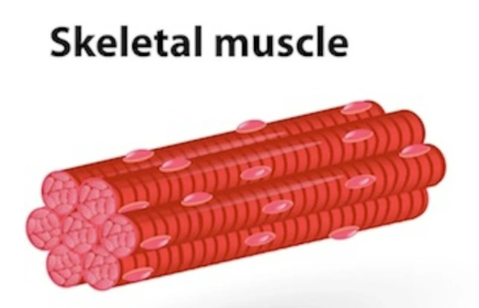
Skeleton tissue is a type of connective tissue.
There are two types of skeleton tissue:
1: Cartilage (Cartilage)
2: Bone
1: Cartilage:
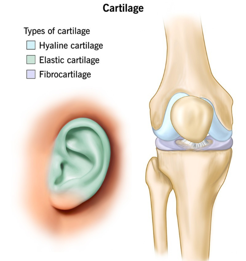
- Cartilage is a rigid form of connective tissue.
- There are 3 types of it.
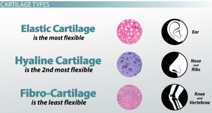
1. Hyaline Cartilage (Hyaline Cartilage):
2. Fibro Cartilage (Fibro Cartilage):
3. Elastic Cartilage:
1. Hyaline Cartilage (Hyaline Cartilage):
- Hyaline cartilage is flexible and smooth.
- Hyaline cartilage is made up of transparent blue tissue and cells and is embedded in a matrix of protein called chondrin.
- chondrocyte cells are found in cartilage and they produce chondrin.
- Hyaline cartilage is found on the surface area of the bone and it forms cartilage joints.
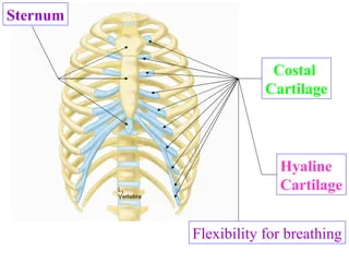
- Hyaline cartilage is the costal cartilage that connects the ribs and the sternum.
- It is found in the lower part of the body.
- -anterior part of the nose
- -larynx
- -trachea
2. Fibro Cartilage:
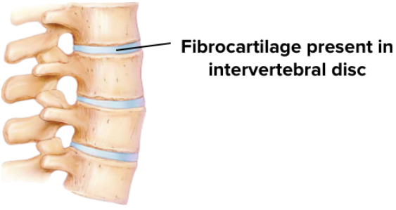
- Fibro cartilage is also known as white cartilage because white collagen fibers are found in it.
- This cartilage is somewhat flexible (also flexible) and firm (firm).
- This cartilage is found in the intervertebral disc.
- It is also found in the symphysis pubic.
3. Elastic cartilage (Elastic Cartilage):
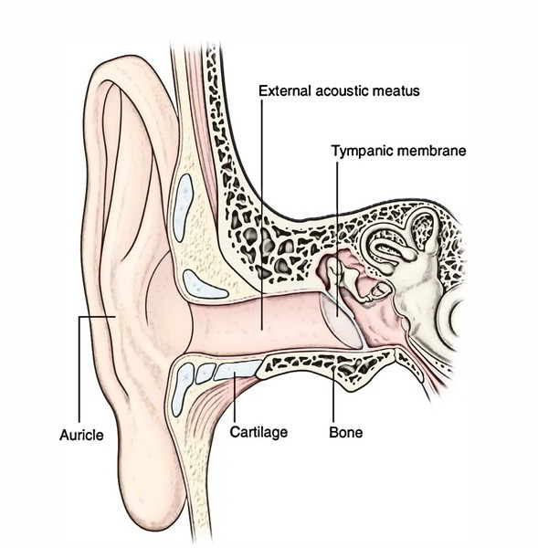
- Elastic cartilage is known as yellow cartilage because of the presence of yellow elastic fibers.
- This is a flexible (bendable) type of cartilage that has elasticity.
- In the human body, it is found in the following parts:
- ear pinnae
- In epiglottis (part of larynx)
2. BONE (bone):
- Bone is a hard connective tissue, in which the endo-skeleton is connected to the joint in the human body.
- Bone is made up of the following substances:
- -Inorganic substance (50%)
- -organic matter (30%)
- -water(20%)
According to the structure of the bone, there are 2 types of bones:
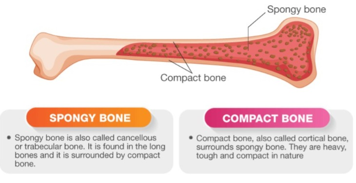
1. Compact bone
2. Cancellous bone or spongy bone
1. Compact bone (compact bone):
- Compact type has strong bone.
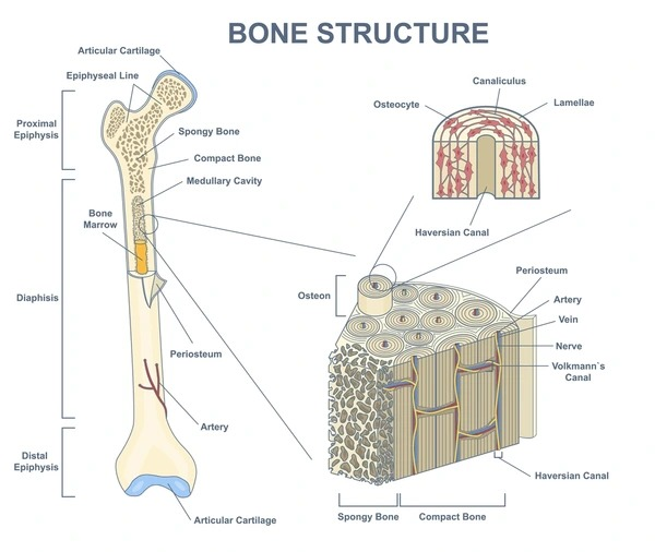
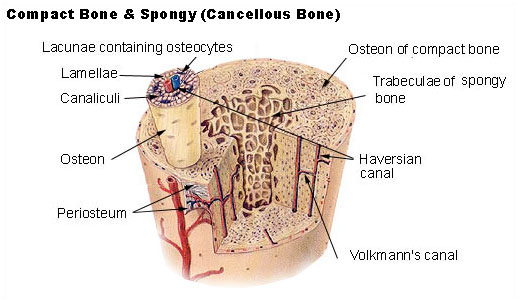
- These bones are made up of tube-like units called osteons or Haversian systems.
- An osteon is made up of a central canal, also called a Haversian canal, which is surrounded by a circular ring-like structure.
- This central or Haversian canal contains blood vessels, nerves, and lymphatic channels.
- These bones are found in flat bones and long bones.
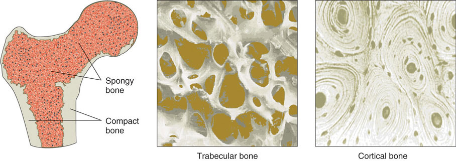
2. Cancellous bone or spongy bone
- This bone is flexible (can bend) and is found at the edges (tops) of long bones and small bones.
- When a cross section of spongy bone is taken, it shows many small ducts called trabeculae, which are made up of lamellae and osteocytes that are interconnected by canaliculi, an interstitial fluid.
- The space between the trabeculae is filled with red bone marrow.
- Red bone marrow produces red blood cells and white blood cells and makes bone cells. provides nourishment.
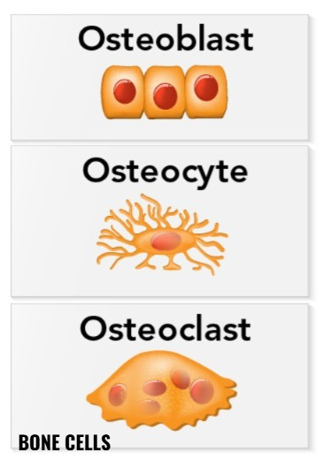
BONE CELLS:
Bone cells are responsible for the formation of new bone.
A protein called Matrix is found in the bone around the Haversian system.
Bone cells are found in the Matrix, which helps in the formation of bone.
There are 3 types of bone cells.
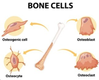
- Osteoblast
- Osteoclast
- Osteocyte
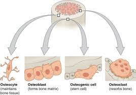
1.Osteoblast:
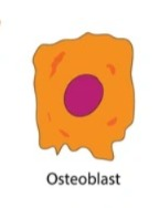
New bone cells are found in the osteoblast cell. These cells are found in the deeper layer of the periosteum, which is found at the fracture site and in the bone marrow of long bones.
Osteoblast cells release collagen and have only one nucleus.
These cells are helpful in the deposit of inorganic salts. e.g. Calcium, Phosphate NOTE:
Periosteum: The fibrous tissue membrane overlying the bone
Collagen: An insoluble protein found in white fibers found in bone.
2.Osteoclast:
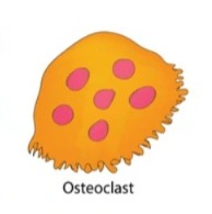
This is a large cell that contains more than one nucleus (more than 50).
This cell absorbs bone tissue, which causes it to break down and releases calcium and phosphate.
This cell is located on the upper surface of the bone.
It continuously remolds bone tissue.
3.Osteocyte (Osteocyte):
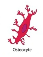
These cells are located on the inner side of the bone.
Osteocyte cell is a type of osteoblast cell in which mature osteoblast cell is known as osteocyte cell.
BONE TYPES :
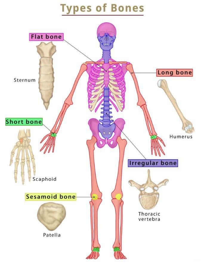
- Long bone (Longbone)
- Short bone (short bone)
- Irregular bone
- Flat bone
- Sesamoid bone
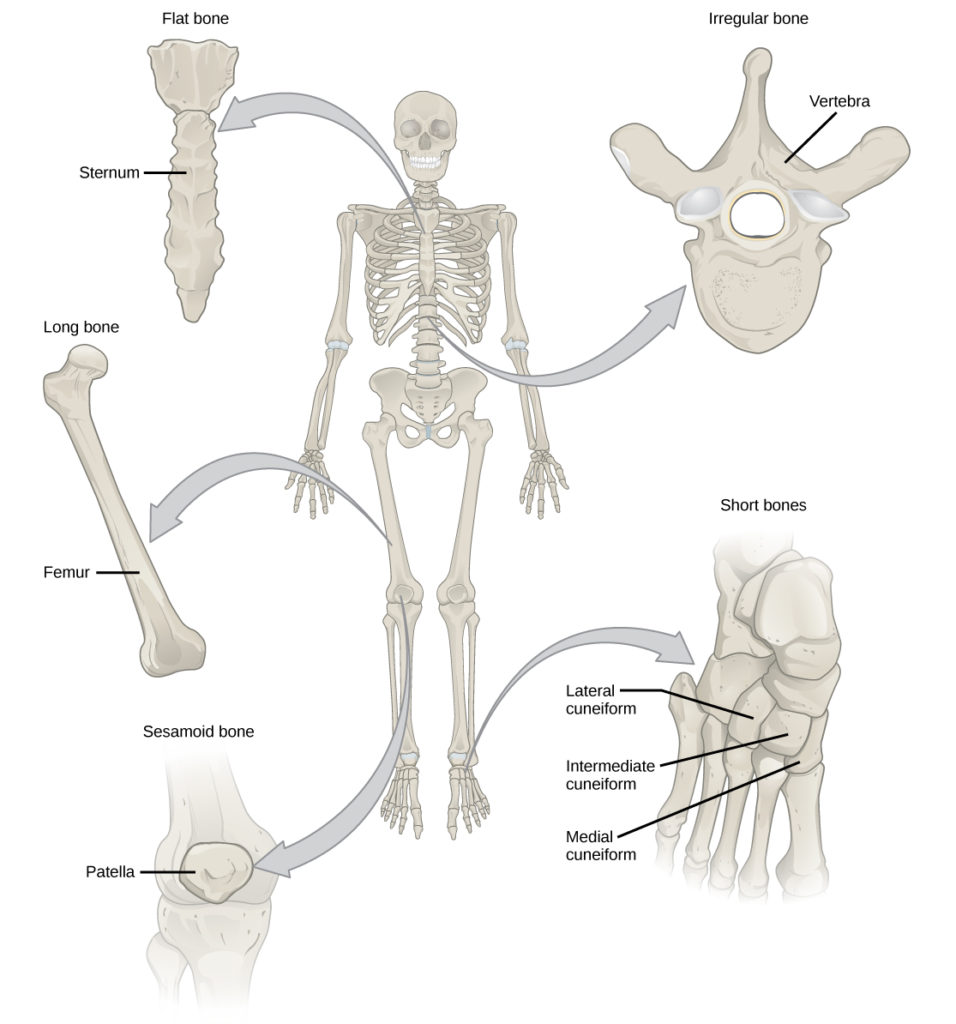
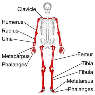
1. Long bone:
- This type of bone is long in size with the epiphysis at the edges and the shaft in the middle. is.
- In this type of bone, its length is greater than its width.
- This type of bone is found in the hands and feet, such as humorous, radius, ulna in hand ,femur, tibia, fibula in leg are long bones.
2. Short bone:
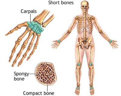
- This type of bone is small in size.
- The shaft part is absent in this bone.
- This type of bone is found in the wrist of the hand as the carpal bone.
3.Irregular bone:
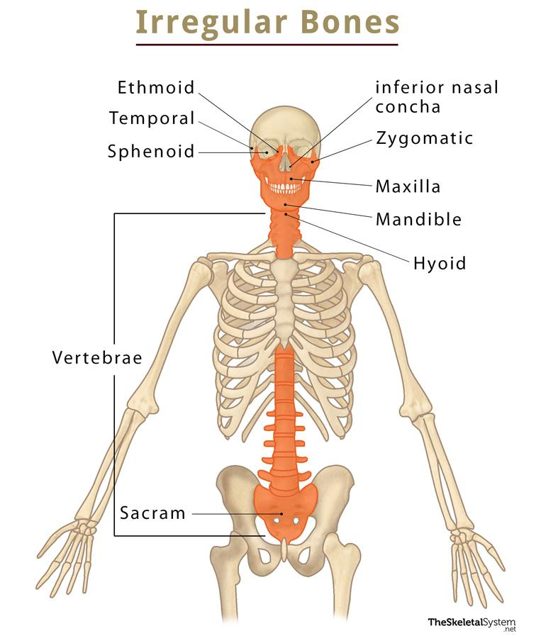
- This type of bone does not have a fixed size, so it is called irregular bone.
- This type of bone is found on the face and in vertebrates is.
- The shaft is absent in the bone of this type.
4. Flat bone
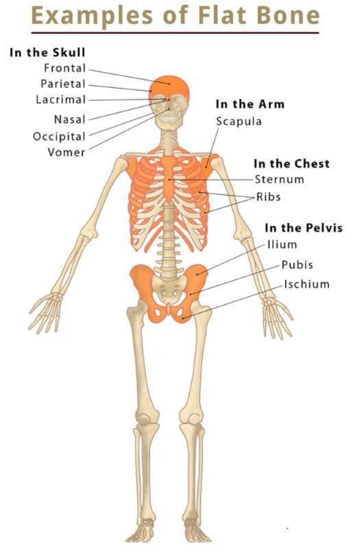
- This type of bone is flat in shape.
- Such bones are found where organs are located in cavities inside. These bones are present for protection of such organs, such bones are present there.
- Such bones are skull, chest bone-sternum and ribs.
5.Sesamoid bone (Sesamoid bone):
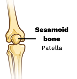
This type of bone is not found in the shape of sesame.
This bone is found around some joints.
Example.. knee cap
DEVELOPMENT OF BONE :
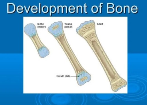
The development of bone in the human body begins at birth and continues until the age of 21 years.
The process of bone development is known as ossification is.
Bone development happens in 2 ways:
1.membranous ossification
2.cartilagenous ossification
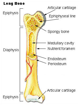
Structure of Long Bone:
Different body shapes Bones are found in which long bones, short bones, irregular bones, flat bones and sesamoid bones are found.
Long bones are cylindrical shaped bones in the body such as humerus bone, femur bone etc.
The length of these types of bones is more than the thickness. In which the following characteristics are found.
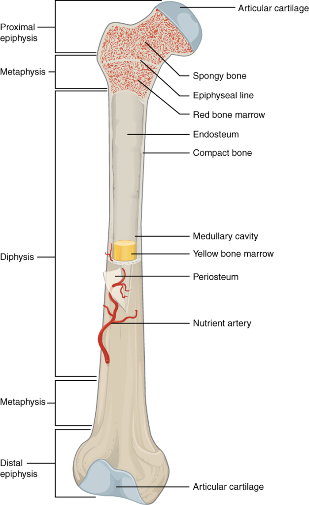
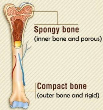
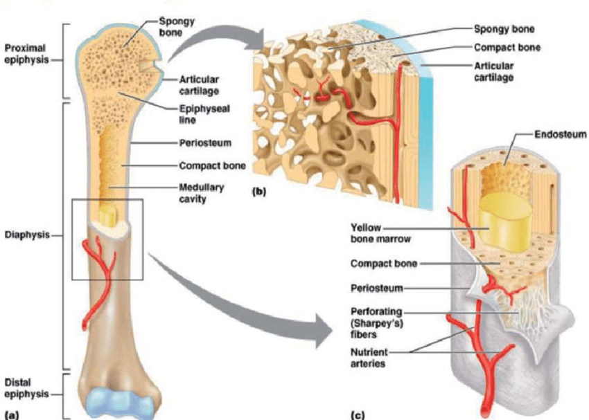
1.Epiphysis (Epiphysis):
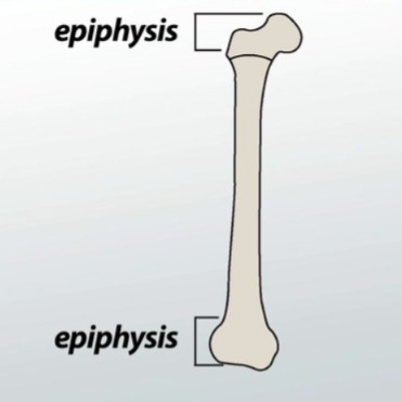
- These are the ends of the long bone. The distal and proximal parts of the long bone are known as the epiphysis. It consists of compact bone tissue on the outside and spongy bone on the inside. Red bone marrow is found at this end of the bone.
2.Diaphysis:
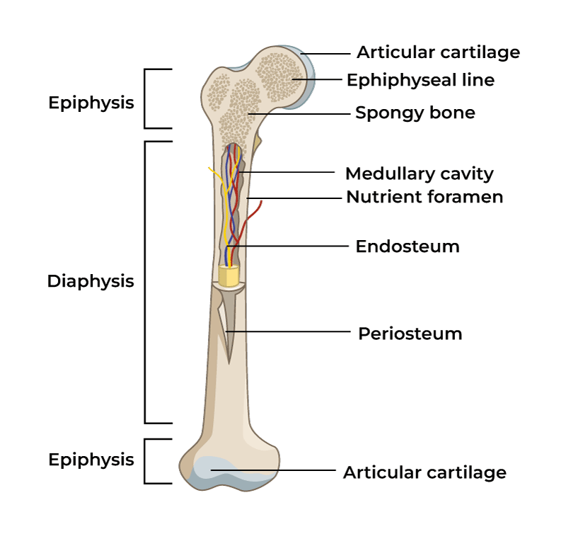
This is the middle part of the long bone. It is also known as the soft part of the bone. This is the main part of the bone in which compact tissue is mainly found and yellow bone marrow is found in this middle part of the bone. There is a canal-like structure in the middle of this part, it is known as the medullary canal or medullary cavity. In which we will see more details during the microscopic structure of the bone.
3. Metaphysis:
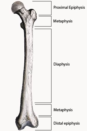
The part of a long bone where the epiphyseal plate joins the diaphysis is called the metaphysis.
4.Articular Cartilage:
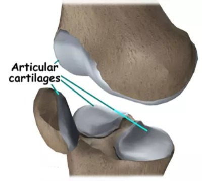
The covering on the lining of the epiphysis is called articular cartilage. It is said. With the help of this cartilage, shock and jerk are absorbed during the joint of the bone and the movement becomes painless.
5.Periosteum (periosteum):
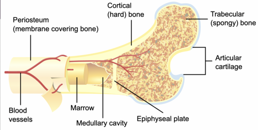
The outer layer on all parts of the bone except the end of the long bone which is covered by articular cartilage is called periosteum. This periosteum is made up of fibrous connective tissue. It has the structure of blood vessels, lymph vessels and nerves. Since this layer is strong, it also works to provide protection to the bone from the outside. Since its outer surface is rough, it can provide good attachment to muscles.
In addition, the medullary cavity is present in the shaft of the bone. In which yellow bone marrow is present, the outer lining of this medullary cavity is called endosteum. Which contains basic bone cells and osteoclast cells.
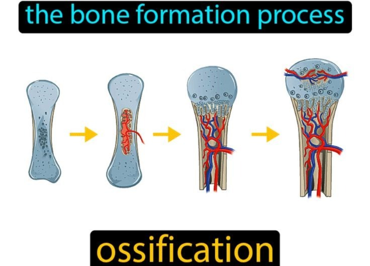
Ossification:
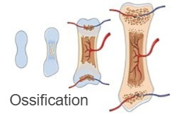
The process of bone formation and bone maturation is called ossification. Bone growth begins when the child is in the mother’s uterus and continues throughout childhood and adolescence. Bones in children are soft and weak, but as they grow older, they become hard, strong and mature.
FUNCTIONS OF BONE:
- The functions of Bone are as follows:
- Bone provides stability and support to our body.
- Bone provides protection to our body. Example. The thoracic rib cage protects the lungs and heart, while the skull protects the brain.
- Bone helps in many movements of muscles and joints.
- Bone acts as a reservoir in our body, which stores minerals like calcium and phosphorus.
- Bone cells form from bone.
- Red bone marrow is found in the epiphysis of long bones, from which red blood cells are formed. The process of making these blood cells is known as hematopoiesis.
TENDON:
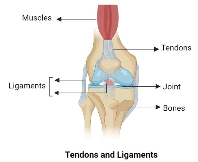
- A tendon is a structure made of fibrous connective tissue that connects a muscle to a bone.
- A tendon secures a muscle to a bone, which helps in the movement of the muscle.
- A tendon holds parts and surrounding organs in a fixed position.
LIGAMENTS:
- LIGAMENTS are made up of connective tissue that attach one bone to another.
- Ligaments hold organs together and provide support.
- Ligaments hold joints together.
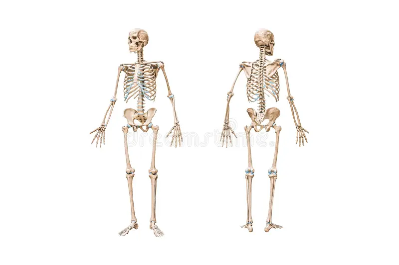
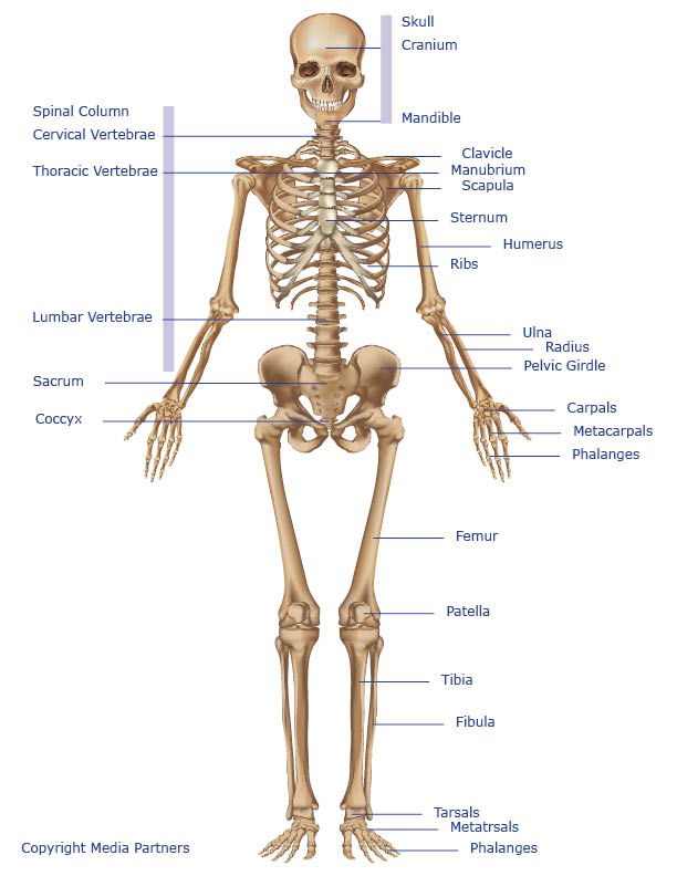
Human Skeleton:
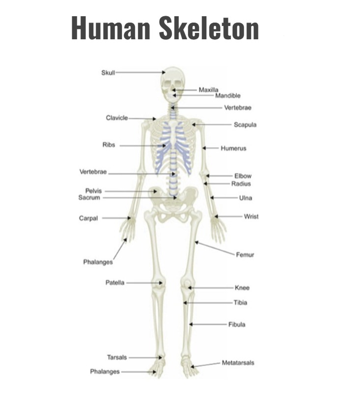
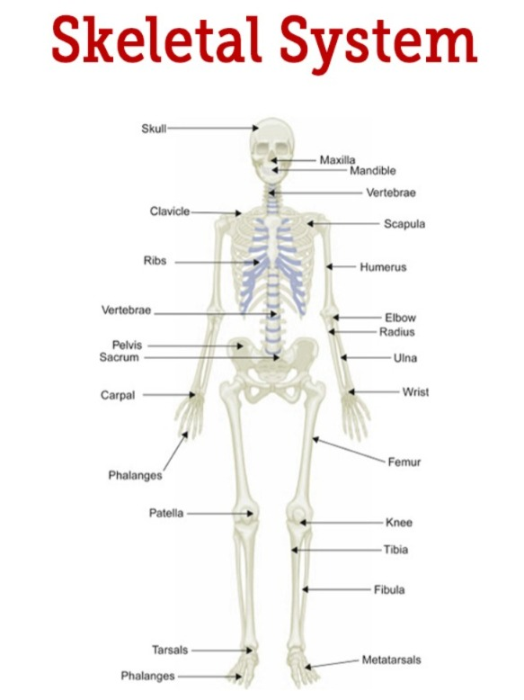
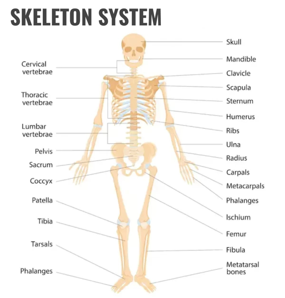
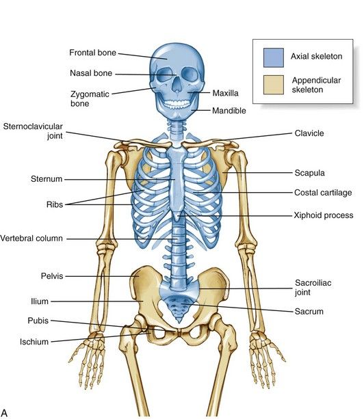
The human skeleton is made up of many bones. There are a total of 206 bones in the adult human body. The skeleton in the human body is divided into 2 parts:
1. Axial skeleton
2. Appendicular skeleton (appendicular skeleton)
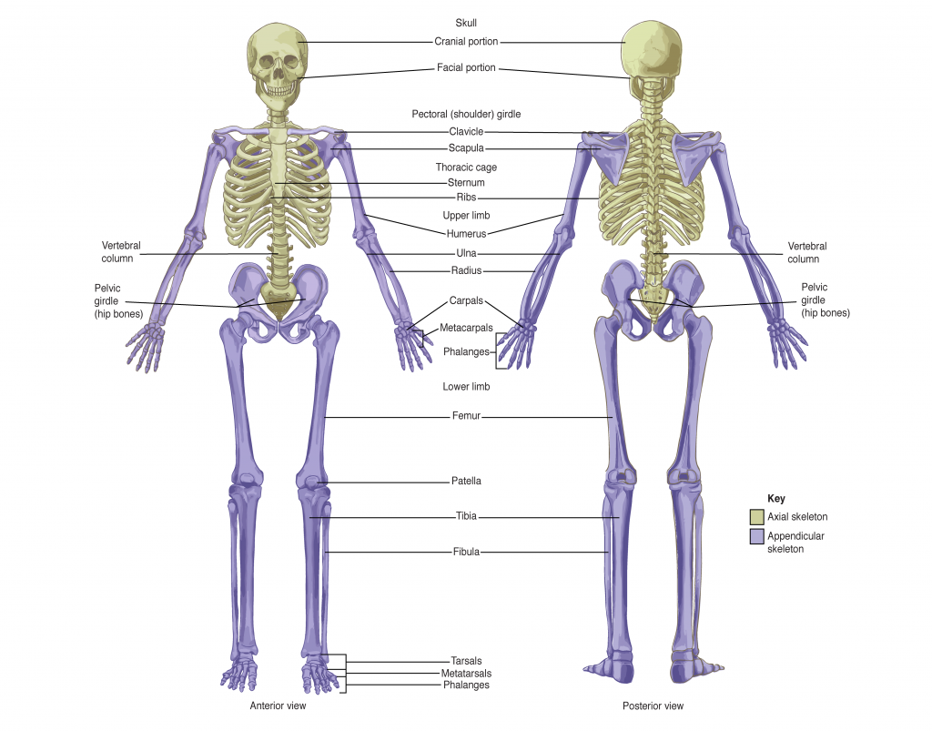
1. Axial skeleton
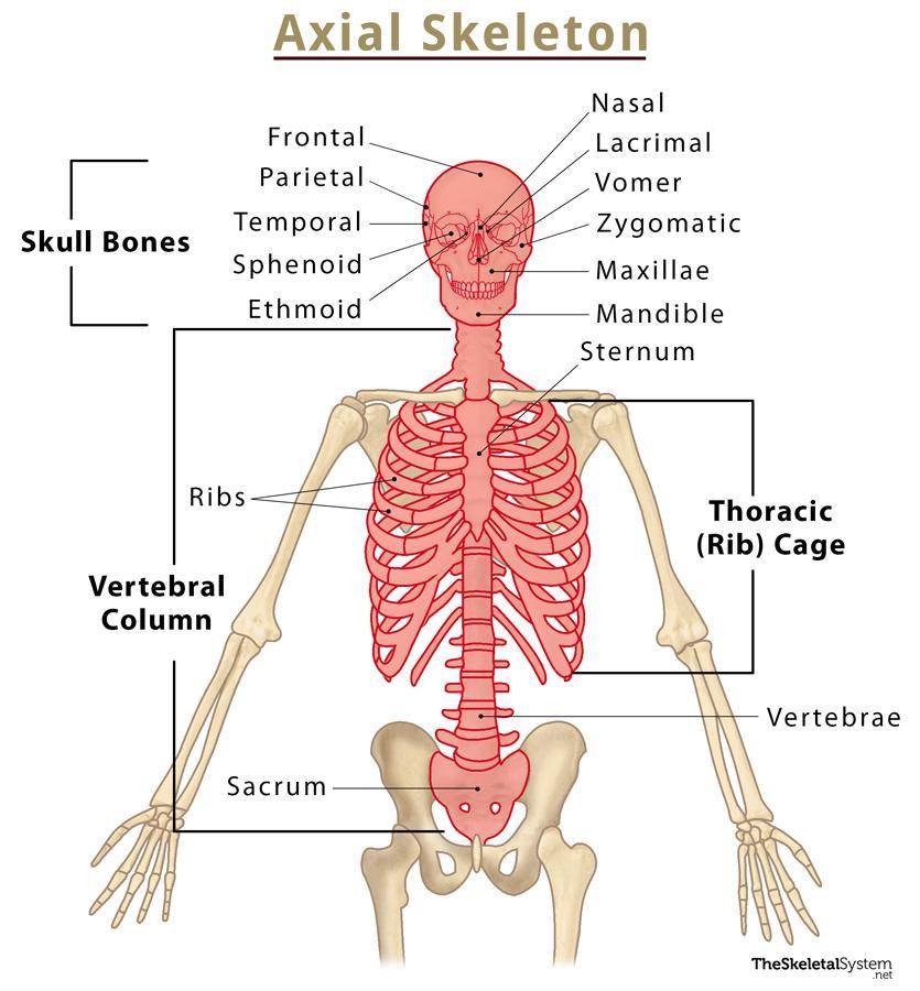
The axial skeleton is found in the center of the human body.
The following bones are found in the axial skeleton: skull, vertebral column, ribs, sternum, etc.
Axial skeleton has a total of 80 bones.
- There are a total of 22 bones in the skull.
- Cranium cavity 8
- Face 14
- Total number of vertebrae 26
- Total number of ribs 24
- Total number of ear bones 6.
In addition The sternum bone and the hyoid bone are included in the axial skeleton. They are both numbered 1.
1.SKULL:
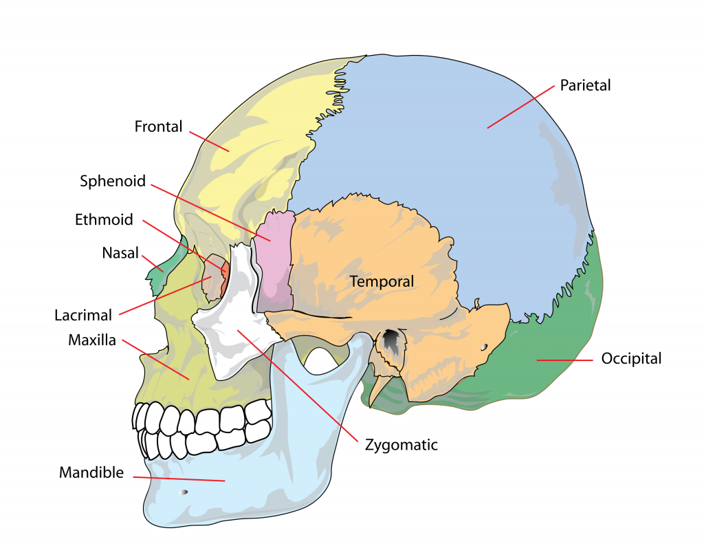
The skull is found in the upper part of the body, above the vertebral column.
It has the following 2 parts.
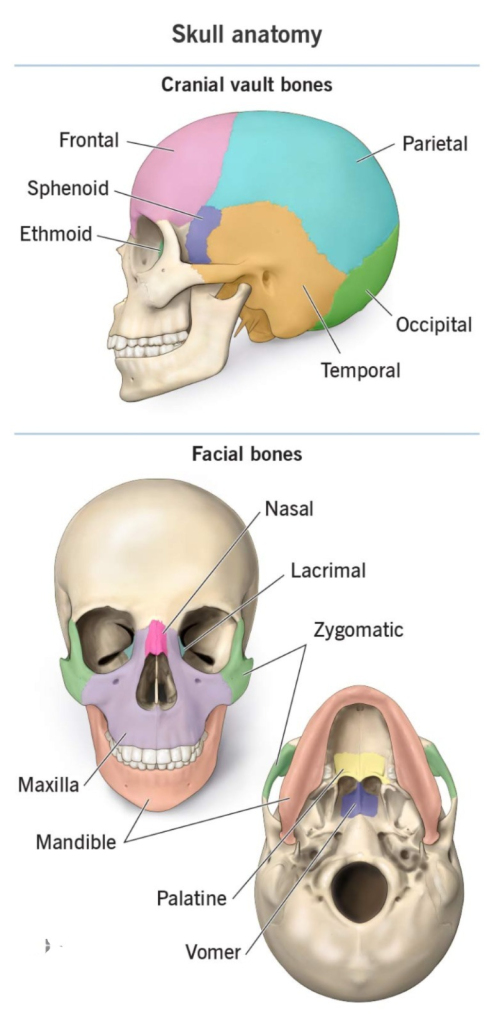
1. cranium (cranium)
2. face (face)
1. cranium (cranium):
- The upper part of the skull bone is known as cranium.
- The cranial cavity is made up of 8 irregular cranial bones.
- The human brain is located in the cranial cavity.
- The lower part of the cranium is known as the base and the upper part is known as the vault. Its shape is like a dome.
- Cranial bones join with each other to form immovable joints known as sutures.
Sutures of the cranium bone (Sutures in the cranium cavity):
The cranium cavity is made up of irregular bones. These flat bones join to form immovable joints. These joints are known as sutures. The sutures in the cranial cavity are as follows.
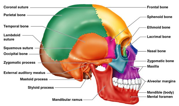
Coronal suture (Coronal suture) Suture):
The suture between the frontal bone and the 2 parietal bones is called the coronal suture.
Sagittal suture:
The suture between the 2 parietal bones is called the sagittal suture.
Lambdoidal suture Suture):
2 The suture between the parietal bone and the occipital bone is called the lambdoidal suture.
Squamous suture:
The suture between the parietal bone and the temporal bone is called the squamous suture.
The above sutures form depression-like structures called fontanelles. These fontanelles are present in the infant skull in number of 2.
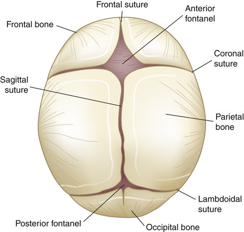
The Anterior Fontanelle is located at the junction of the coronal and sagittal sutures at the front of the skull. It is also known as the bregma. It is large in size. By the time the baby is 1.5 years old, it closes due to skeletal maturity (growth). It is diamond shaped.
At the back of the skull, where the sagittal and lambdoidal sutures join, there is a Posterior Fontanelle near the junction. It can also be known as lemda. It is small in size. By the time the baby is 6 to 8 weeks old, it closes due to skeletal maturity (growth). It is triangular shaped.
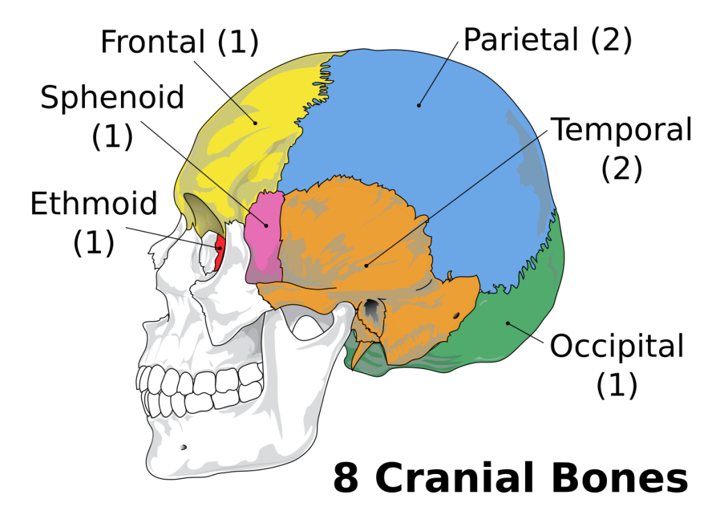
The cranium in the skull is made up of many different flat and irregular bones. The cranium contains a total of 8 bones, which are as follows:
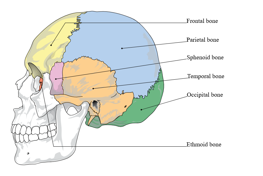
1. Frontal bone 1
2. Parietal bone 2
3. Temporal bone 2
4. Occipital bone 1
5. Sphenoid bone 1
6. Ethmoid bone 1
Frontal bone:
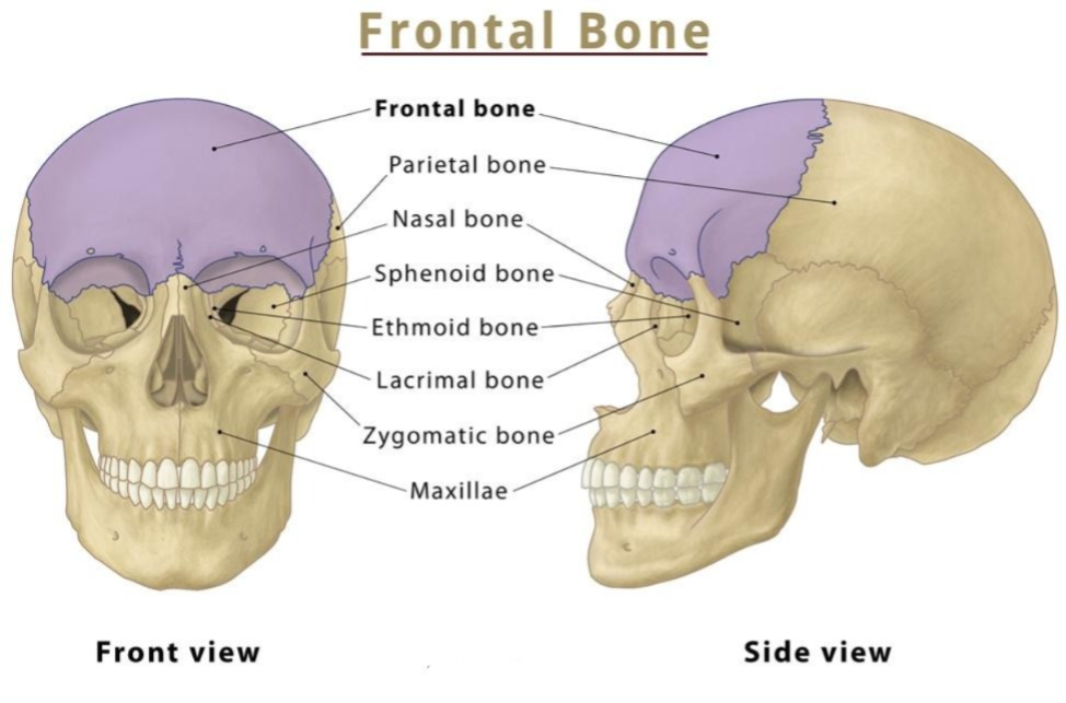
This bone is located at the front of the cranial cavity at number 1. It is also called the forehead bone. It is located above the orbital cavity and the nasal cavity. The frontal bone has a raised margin on both sides above the orbital cavity, which is called the supra orbital margin. This margin also has a foramen. It is called the supra orbital foramen. The area between the two orbital margins is known as the glabella.
Parietal bone:
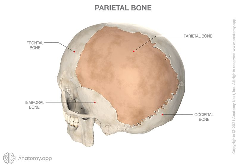
These bones are found in the cranial cavity in numbers of 2 and they form the roof of the cranial cavity. It is the bone connected to all the sutures of the cranial cavity.
Temporal bone:
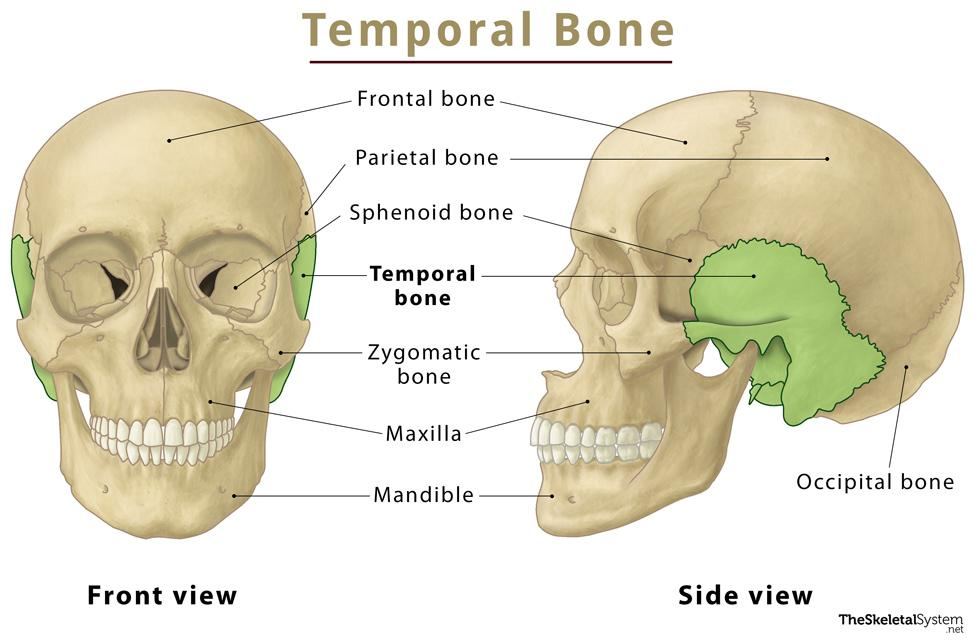
They are 2 in number. They are located on either side of the cranial cavity, near the ear. The temporal bone is connected to the zygomatic bone by the zygomatic process. It also connects to the mandible bone to form the temporomandibular joint, the only movable joint in the skull. The mastoid portion of the temporal bone is connected to the middle ear by air cells.
Occipital bone:
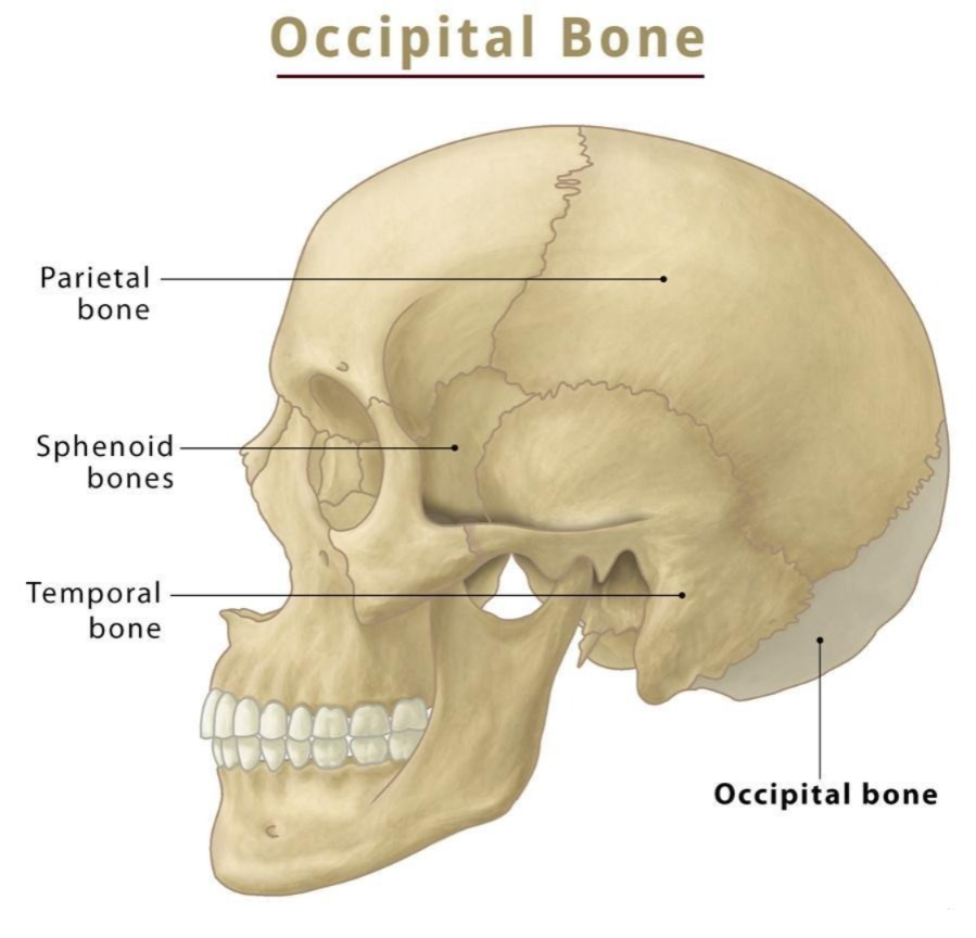
It is the number 1 bone in the cranial cavity. It forms the back and base of the skull. At the bottom of this bone is a large foramen, known as the foramen magnum. Through which the spinal cord exiting the brain passes.
On either side of this foramen are two raised parts, called condyles. These form a hinge joint with the atlas, the first vertebra of the vertebral column. The most prominent part of the occipital bone is called the occipital protuberance.
Ethmoid bone:
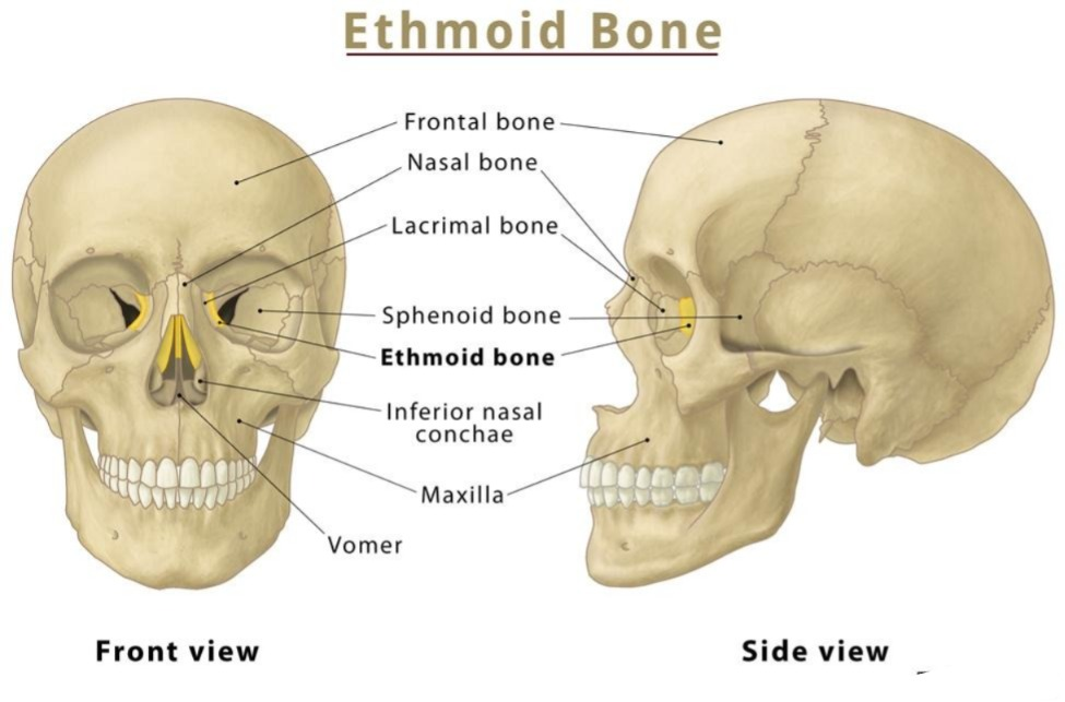
It is the bone located at the front of the base of the cranial cavity. It is numbered 1. It is a cubical-shaped bone. The olfactory nerve passes from the nose to the brain through its path.
Sphenoid bone:
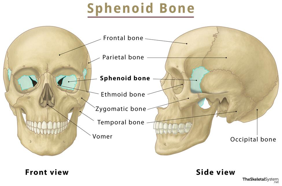
It is numbered 1. It is located in front of the occipital bone at the base of the cranial cavity. It is a butterfly-shaped bone. There is a depression in its middle. It is known as the hypophysial fossa or Sella tarsica. This part contains the pituitary gland. This bone is connected to all the bones of the cranium cavity.
Facial bones:
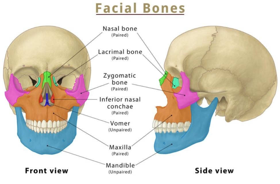
The bones in the face are called facial bones. They are 14 in total. These bones are found as follows.
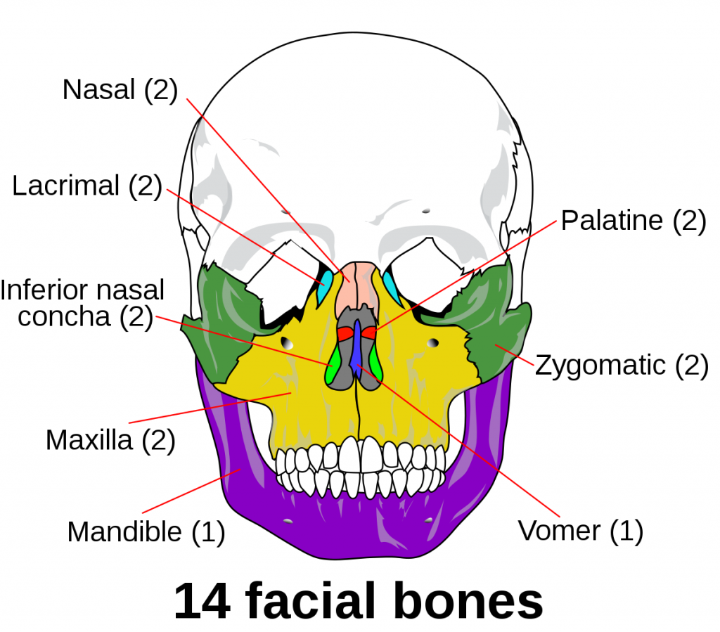
Maxilla bone Bone):
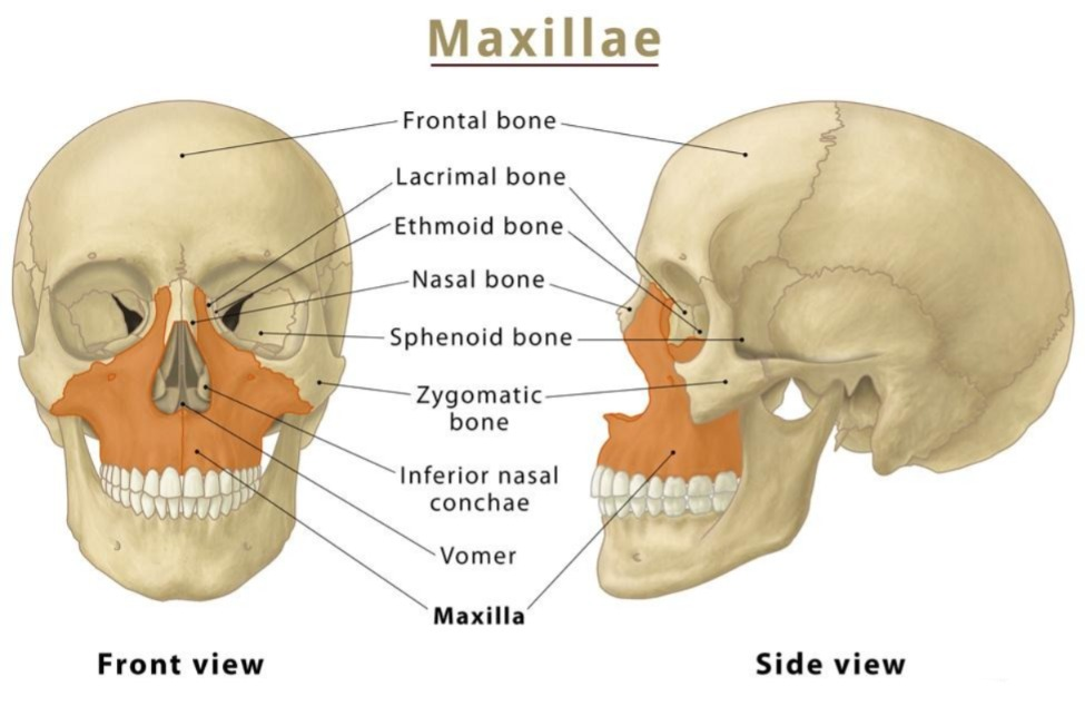
It is also known as the upper jaw bone and is 2 in number. The upper teeth are located on its margin. This bone forms the roof of the mouth and the floor of the nasal cavity and orbital cavity. Some parts of this bone also form the hard palate.
Zygomatic bone:
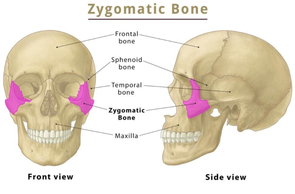
It is known as the cheekbone. It is found in the number of 2. It forms the prominent part of the cheek. This bone articulates with the maxilla bone at the front and with the temporal bone at the back.
Mandible bone:
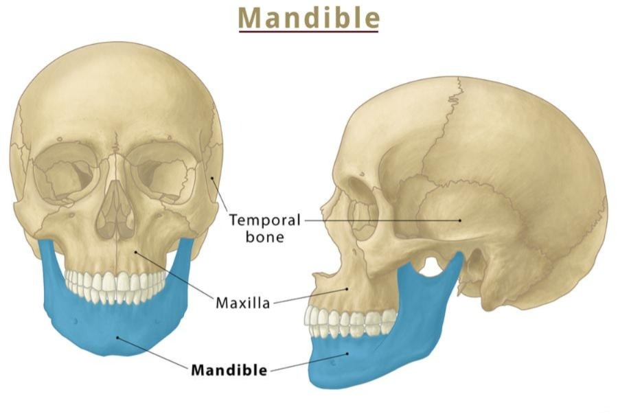
It is the bone at the bottom of the face. It is the only movable and strong bone of the skull. It plays a very important role in the act of chewing. It is numbered 1.
The upper edge of this bone is known as the alveolar ridge. The teeth are arranged on this ridge.
The front part of this bone is called the body. The curved part is called the angle. The flat part on the top is called the ramus. The front part is divided into two processes. In which the coronoid process and condylar process are found.
Muscles attach to the coronoid process while the temporal bone attaches to the condylar process to form the tempo mandibular joint.
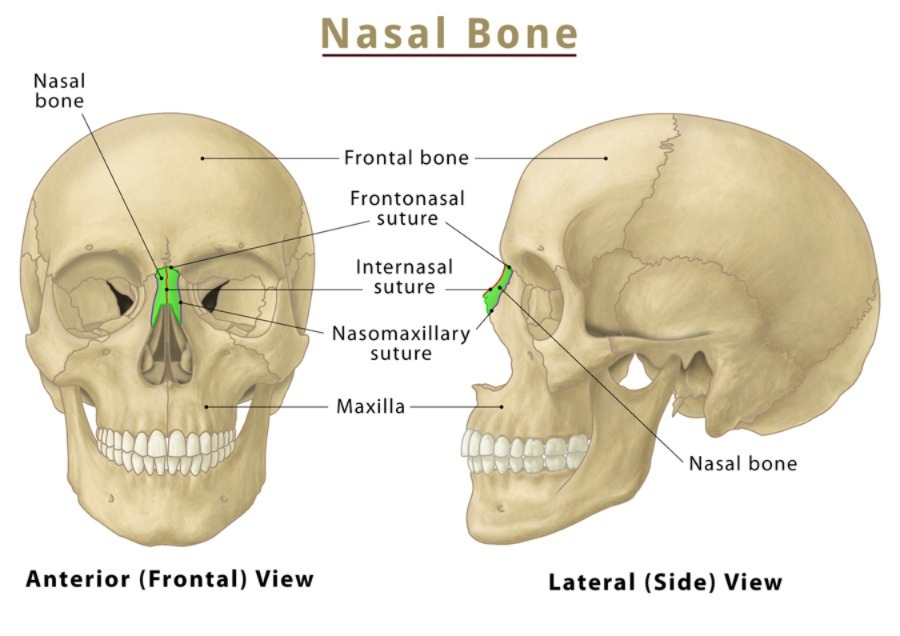
Nasal bone:
They are flat bones, numbered 2. They form the bridge of the nose. They form the superior and lateral walls of the nasal cavity.
Lacrimal bones (Lacrimal bone):
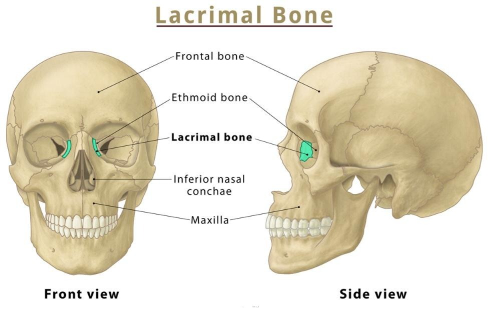
They are 2 bones in number. They form the medial wall of the orbital cavity and carry the eye secretions to the nose through the nasolacrimal duct. This bone is shaped like a finger nail.
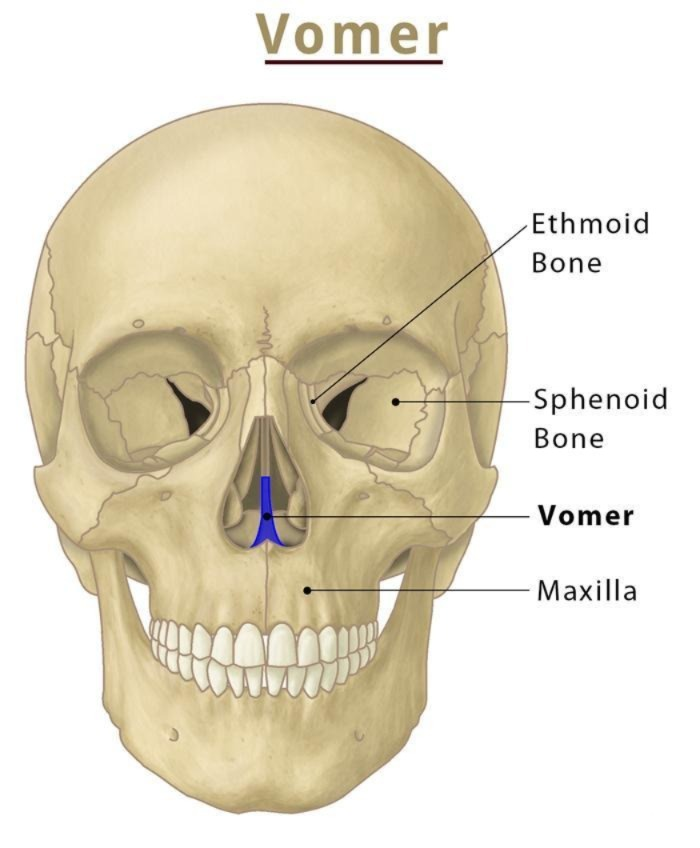
Vomer bone (Vomer bone):
It is the bone numbered 1. It is the bone located in the middle of the nasal cavity. It is connected to the septum of cartilage in the front. It separates the nasal cavity into 2 parts. Above it is connected to the ethmoid bone.
Palatine bones:
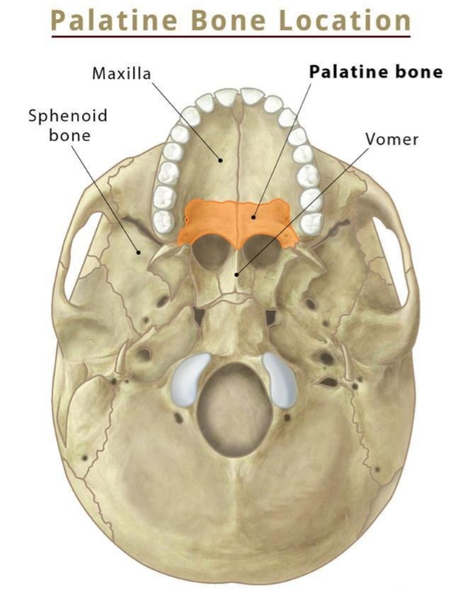
It is a bone in the number of 2. It forms a hard pellet. In it, 2 L-shaped bones are connected to each other and form part of the pellet.
Inferior conchae:
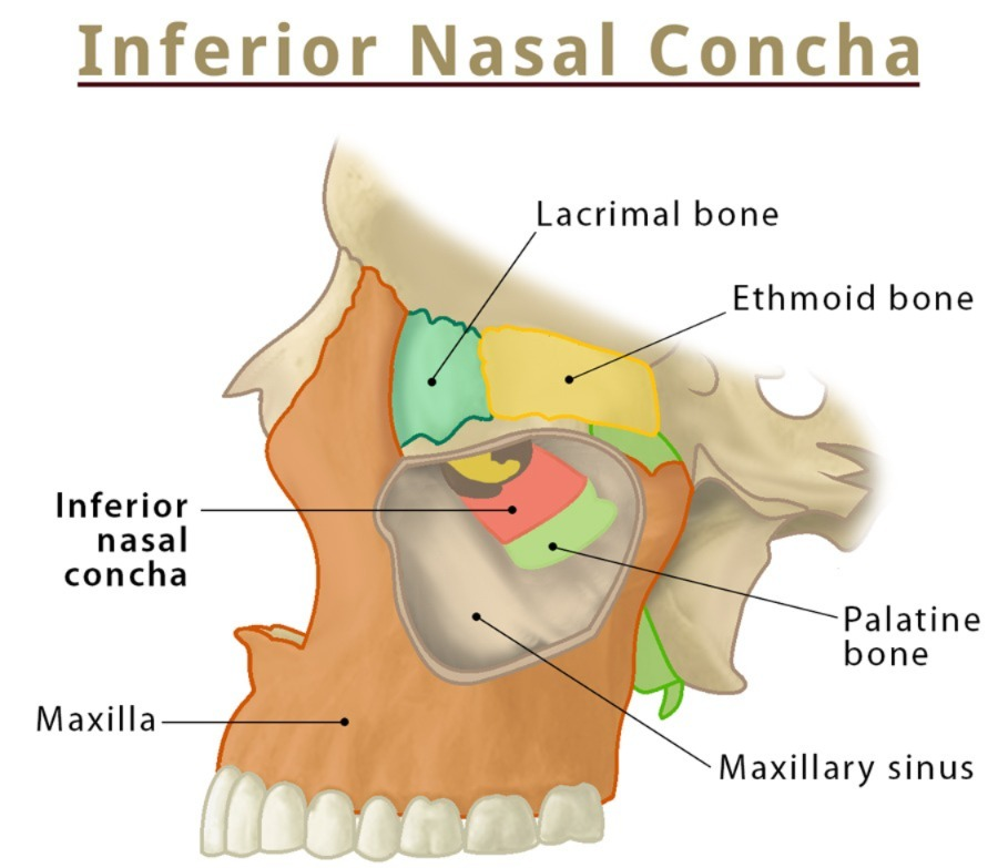
They are 2 in number. They are strip-shaped bones located in the lateral wall of the nasal cavity. It works by filtering and warming the incoming air.
Vertebrae and Vertebral Column (vertebrae connect to each other):
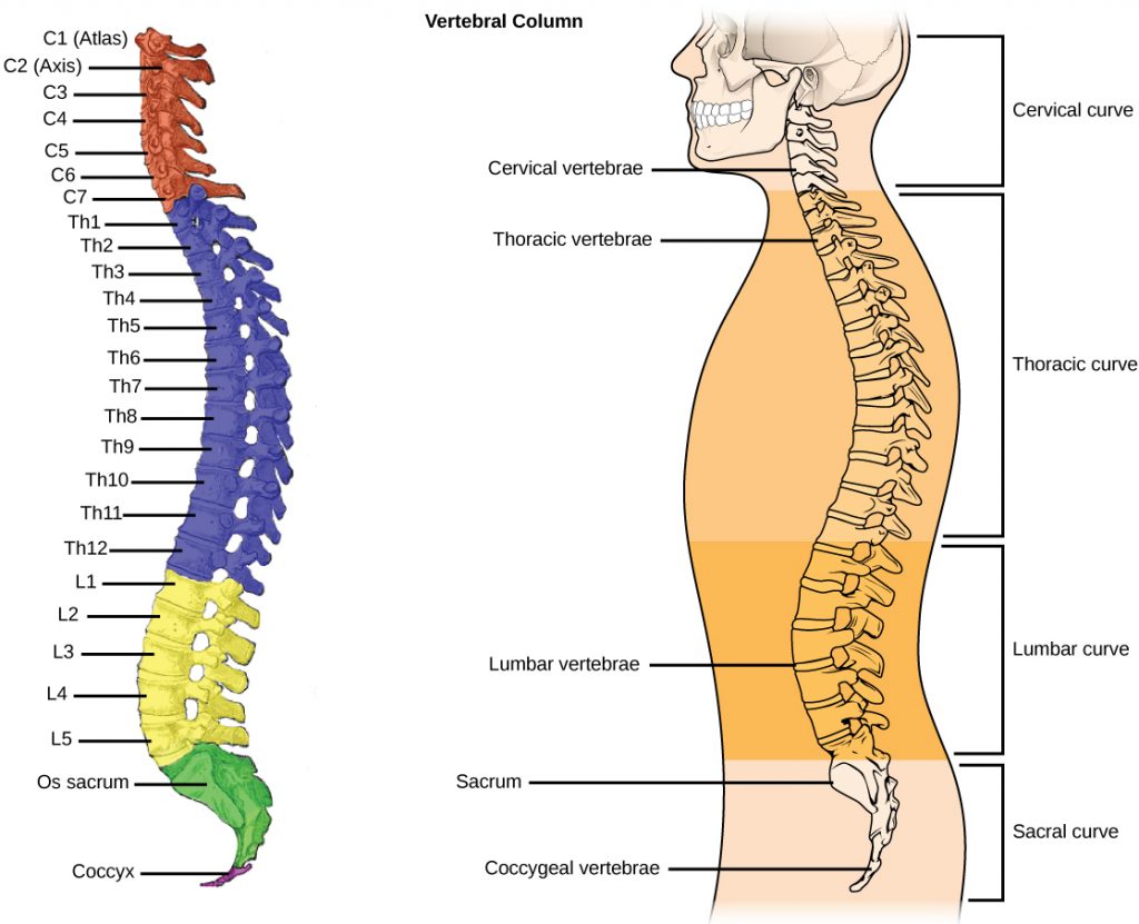
- Below the foramen magnum of the occipital bone, irregularly shaped vertebrae connect to form a column, which is called the vertebral column.
- Vertebrae are strong and irregularly shaped bones. Each of these vertebrae connects to form a vertebral column, creating a canal-like structure between them. Through this canal, the structure of the spinal cord continuously passes from the brain to the lower side. The vertebrae protect the spinal cord.
- The approximate length of the vertebral column is about 70 centimeters in men and 60 cm in women.
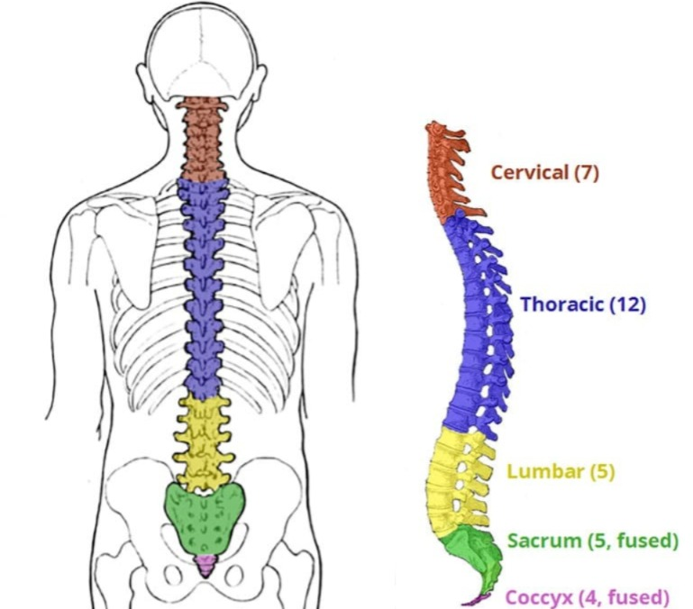
- Different types of vertebrae in the vertebral column join together to form different vertebral regions. Its number is as follows.
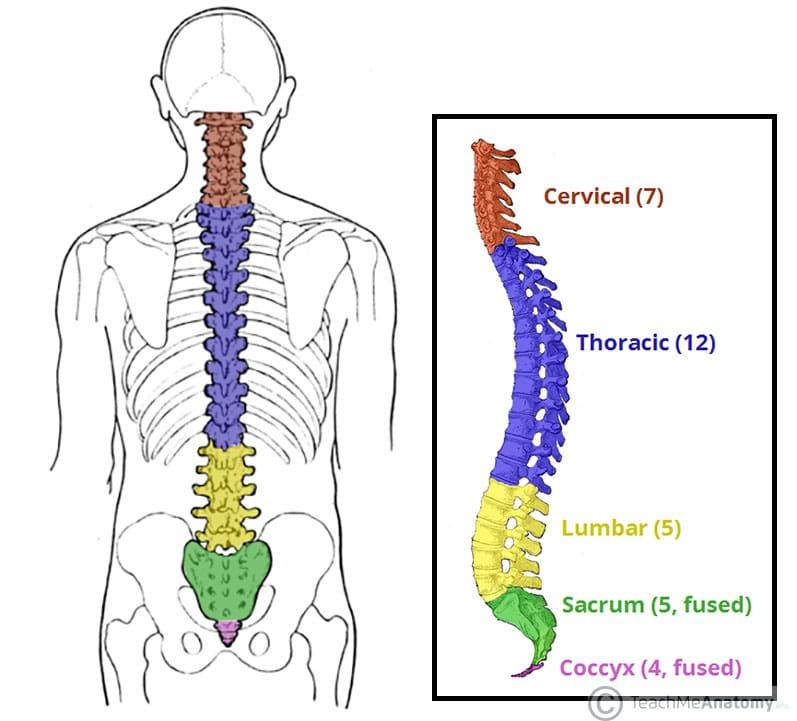
- Cervical Vertebrae. 7
Thoracic Vertebrae. 12
Lumbar Vertebrae. 5 - Sacral Vertebrae which is five vertebrae fused to form 1 Sacral Vertebrae.
- Coccygeal Vertebrae which is four vertebrae fused to form 1 Coccygeal Vertebrae.
- Thus according to all the above regions in the vertebral column A total of 26 typical vertebrae are found. In the middle of the body of each vertebra is a disc made of fibrous cartilage, called the intervertebral disc.
Common Characteristics of Vertebrae:
The following common characteristics are found in all the vertebrae of the vertebral column.
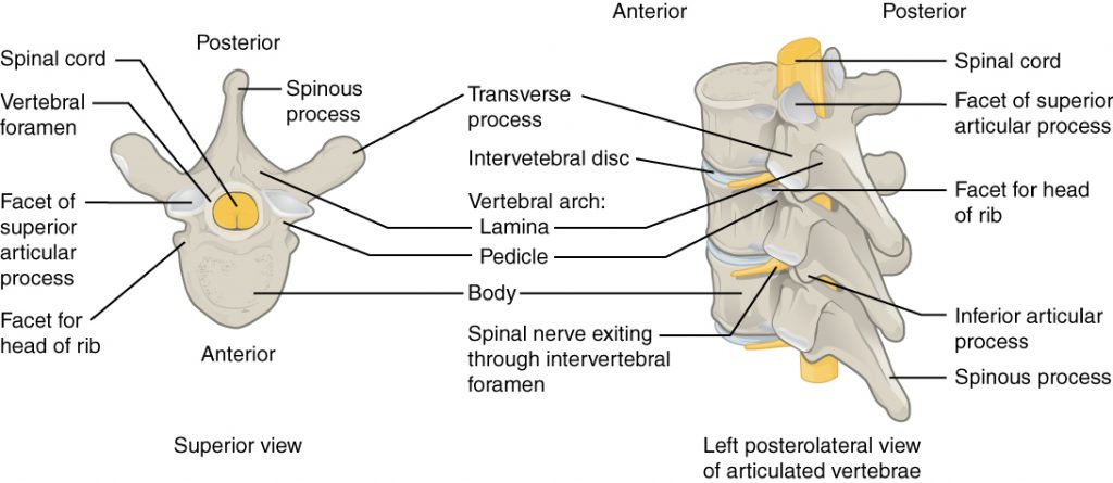
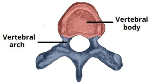
Body:
The vertebra has a flattened body part at the front. Its size varies according to the vertebra of each region. For example, the body part is the smallest in the cervical vertebra and the body size increases towards the thoracic and lumbar vertebrae. The body is the largest and thickest in the lumbar vertebra.
Process of Vertebrae:

- The two processes that extend posteriorly from the body of the vertebra are called pedicles. These pedicles extend posteriorly and form a V-shaped portion, which is known as the lamina. From the junction of these laminae, a straight process arises posteriorly, called the spinous process.
- Near the junction of the pedicle and the lamina of the vertebra, a horizontal process arises on either side, called the transverse process.
- A foramen is formed between all these processes, called the vertebral arch or neural arch. The spinal cord passes through this arch. This arch is the largest in the cervical vertebrae. The opening of this arch becomes smaller as it moves towards the thoracic and lumbar vertebrae.
- In addition, the superior and inferior surfaces of each vertebra have articulating processes to connect the vertebrae.
Specific Characteristics of Vertebrae (Specific Characteristics of the Vertebral Column):
Each vertebra of the vertebral column has specific characteristics according to its region. These characteristics according to the region are seen as follows.
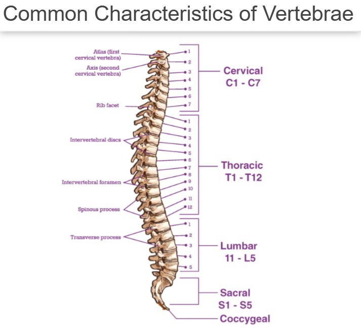
Cervical Vertebrae (Cervical Vertebra):
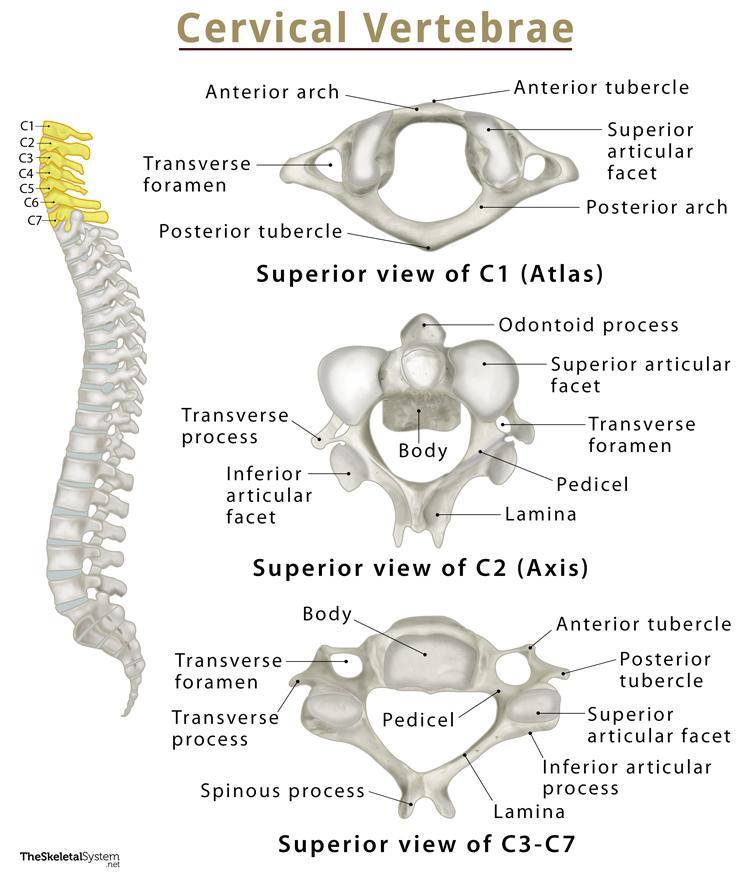
- Cervical vertebrae are found in number of 7. These vertebrae are ring-shaped. The body is the smallest and the neural arch is the largest. The processes of the cervical vertebrae are short and smooth.
- The first vertebra of the cervical vertebrae is known as the atlas. This atlas vertebra articulates with the condyle around the foramen magnum of the occipital bone to form a hinge joint. This vertebra supports the skull, hence it is called the atlas vertebra.
- The second vertebra of the cervical vertebrae is known as the axis. The superior surface of this vertebra has a pointed process, called the odontoid process. This process forms a pivot joint with the atlas vertebra. This joint between the atlas and axis vertebrae allows all types of rotational movements of the head.
- In addition, on both sides of the cervical vertebrae, there is a foramen on the lateral side, called the transverse foramen. Nerves and vessels pass through these foramen.
Thoracic Vertebrae:
- The thoracic vertebrae are 12 in number. The body of the thoracic vertebra is larger than that of the cervical vertebra and smaller than that of the lumbar vertebra.
- The neural arch of this vertebra is smaller than that of the cervical vertebra and larger than that of the lumbar vertebra.
- The processes of the thoracic vertebrae are quite long and pointed.
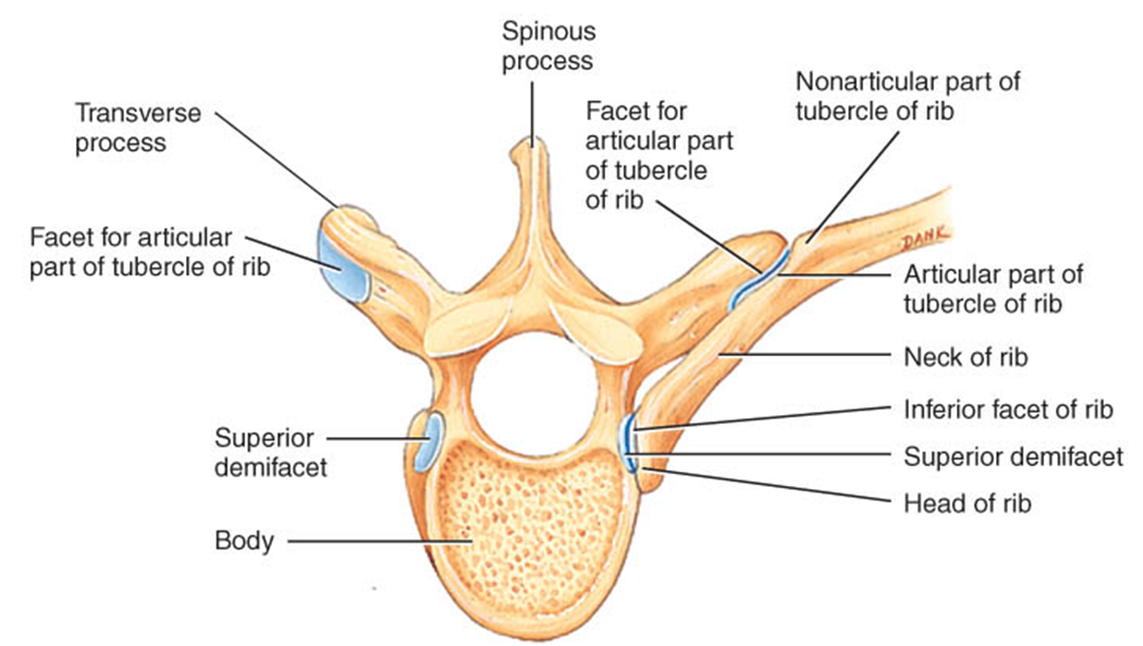
- The lateral inferior side of the body of this vertebra has one facet on each side. The heads of the ribs on both sides attach to this facet. Therefore, the thoracic vertebrae play an important role in forming the thoracic cage.
- These vertebrae are stronger than the cervical vertebrae.
Lumbar vertebrae:
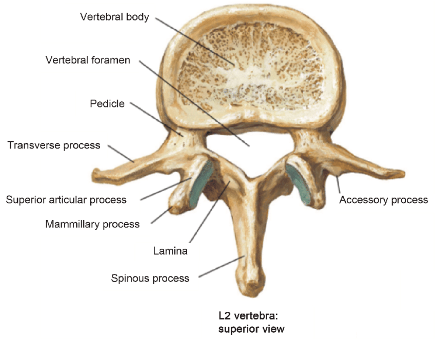
- The lumbar vertebrae are 5 in number. This vertebra is the strongest and largest of all the vertebrae.
- The body of the lumbar vertebra is the largest of all the vertebrae and its neural arch is the smallest.
- The processes of the lumbar vertebrae are short and quite thick.
- The fifth lumbar vertebra articulates with the sacrum vertebra below it to form the lumbosacral joint.
Sacrum Vertebrae:
The sacrum is a bone located below the fifth lumbar vertebra and formed by the 5 vertebrae joining together. This bone is triangular in shape. The following characteristics are found in this bone.
- Base (base)
- Apex (apex)
- Anterior Surface (anterior surface)
- Posterior Surface (posterior surface)
- Lateral Mass (Lateral Mass)
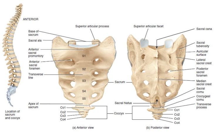
- Sacrum The upper broad part of the bone is the base, which articulates with the fifth lumbar vertebra to form the lumbosacral joint. This base has a wing-like structure on either side known as the sacral ala.
- The apex of the sacrum bone is the triangular-shaped part at the bottom. Which joins the coccygeal vertebra and forms the sacrococcygeal joint.
- The front surface of the sacrum bone is called the anterior surface. It is smooth and concave. This surface has a raised part on the medial side, which is important in midwifery as an obstetric landmark. It is called the sacral promontory. This surface is formed by the connection of five vertebrae, forming a horizontal line in the middle, which is known as the transverse line, and the foramen found in this surface is called the anterior sacral foramen.
- The back surface of the sacrum bone is called the posterior surface. It is an irregular and rough surface. The raised and rough part of it is called the sacral tuberosity. The transverse line is not clearly visible on the posterior surface, but the posterior transverse foramen can be seen.
- The transverse processes of each fused vertebra in the sacrum bone come together to form the lateral side of the sacrum bone, known as the lateral mass. These two sides articulate with the ilium of the innominate bone to form the sacroiliac joint.
- In females, the sacrum bone is short, broad, and curved.
Coccyx Vertebrae:
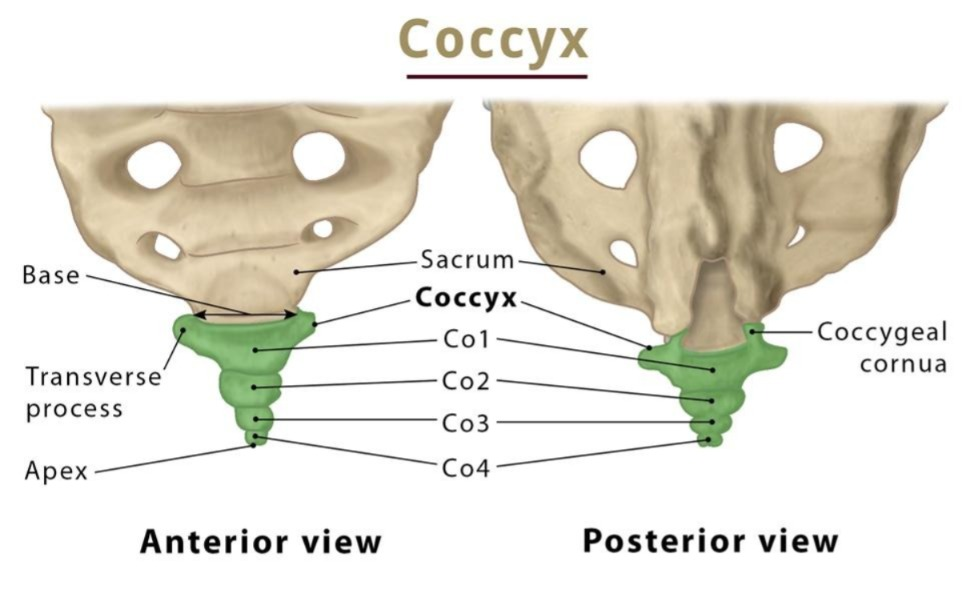
- The coccygeal vertebra is a triangular bone at the end of the vertebral column. It is formed by the 4 coccygeal vertebrae fused together.
- This bone articulates with the apex of the sacrum bone at the top to form the sacrococcygeal joint.
- There are 26 separate vertebrae in the vertebral column, and each of these vertebrae articulates with each other to form the vertebral column. In this vertebral column, there is no movement between the separate 2 vertebrae, but there is some movement of the entire vertebral column. Which is as follows.
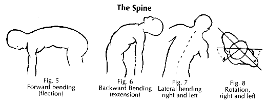
- Flexion – The forward bending movement of the vertebral column.
- Extension – The movement of bending the vertebral column to the back.
- Lateral Flexion – The movement of bending the vertebral column to the sides.
- Rotation – The movement of rotation in a semicircle is seen in the vertebrae of the cervical and lumbar regions.
Functions of Vertebral column:
- The skull is located at the top of the vertebral column, supporting the skull. Provides.
- In the middle of the body of two vertebrae is an intervertebral disc which absorbs shock during movement and protects the brain from jerks.
- A small canal-like strong structure is formed in the middle of the vertebral column through which the spinal cord passes. This serves to protect the spinal cord.
- The transverse processes or pedicles of the vertebrae form the foramen on both sides. Nerves, blood vessels and lymph vessels pass through this foramen.
- Provides attachment for other bones to connect. Provides framework for the body. So many moments are possible.
- It provides a place for the ribs to connect. Thus, the thoracic cage is formed and also provides attachment for the formation of the solder joint and the pelvic joint.
- It connects the axial skeleton with the appendicular skeleton.
- Due to this, the posture of the body is maintained in different movements, sitting and walking.
Hyoid Bone:
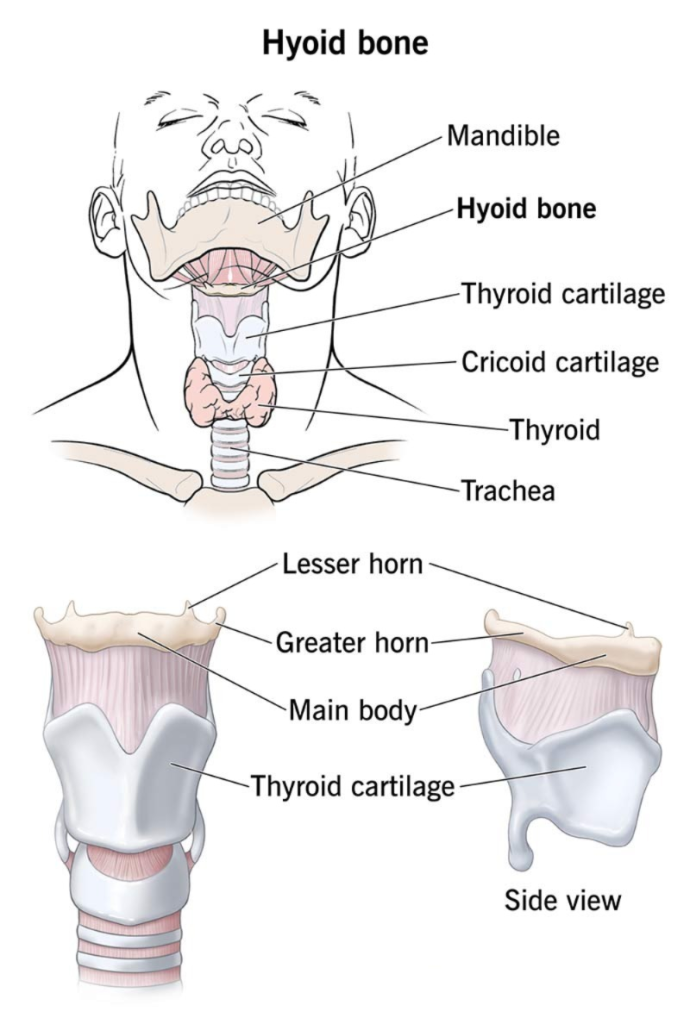
- This is a bone of the axial skeleton. It is located in the soft tissue of the neck above the laryngeal cartilages. Which is a U-shaped bone and is attached to the base of the tongue and provides support to the tongue.
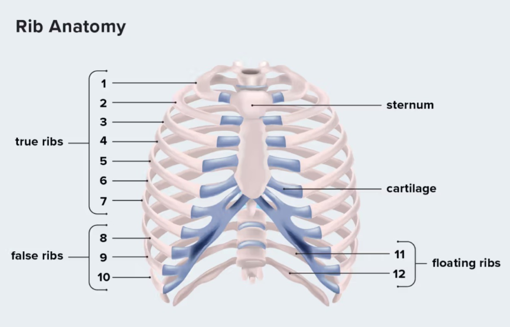
Thoracic Cavity:
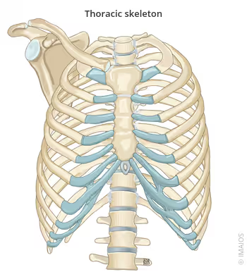
- The chest is known as the thoracic cavity. This part is a cavity of the concept. In which the superior side is narrow and the inferior side is wide. This thoracic cavity is a cavity made of bone. In which organs like lungs and heart are protected. The following bones are connected to form this cavity.
- Sternum Bone 1
Thoracic Vertebrae 12
Ribs. 12 pairs - In addition, the sternum bone and clavicle bone of the thoracic cavity are connected to the solder joint and upper extremity, i.e. the appendicular skeleton, with the help of the appendicular skeleton.
Sternum Bone (Sternum Bone):
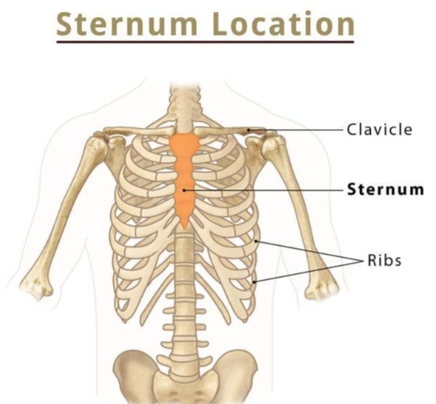
- The sternum bone is a bone of the axial skeleton. It is the middle bone located in the front of the thoracic cavity.
- It is the bone immediately below the skin. The sternum bone is also known as the breast bone. The sternum is a flat type of bone.
- The ribs are attached to the sternum bone with the help of costal cartilage. Therefore, it is a very important bone for forming the thoracic cage.
The sternum bone is divided into three parts as follows.
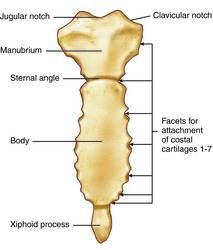
1. Manubrium:
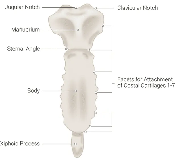
- The uppermost part of the sternum bone is called the manubrium. It forms the upper broad part of the sternum. It is a roughly triangular part.
- The notch in the middle of the uppermost part of the manubrium is called the suprasternal notch. The suprasternal notch is also known as the second jugular notch.
- Below the suprasternal notch of the manubrium on either side is a small notch called the clavicular notch. The clavicular bone joins this groove and forms the sternoclavicular joint.
- The first pair of ribs are attached to the manubrium by means of costal cartilage.
- The second pair of ribs are attached to the manubrium by means of costal cartilage near the angle between the manubrium and the body.
2. Body:
- The body forms the middle, longest part of the sternum bone. It is formed by the manubrium on its upper side and the xiphoid process on its lower side. The part between these two is known as the sternal body.
- There are many small grooves on either side of the body. The third, fourth, fifth, sixth and seventh pairs of ribs on either side of this groove are attached with the help of costal cartilage.
3. Xiphoid Process:
The lowest part of the sternum bone is called the xiphoid process. This part is connected to the abdominal wall muscles and the diaphragm.
Ribs:
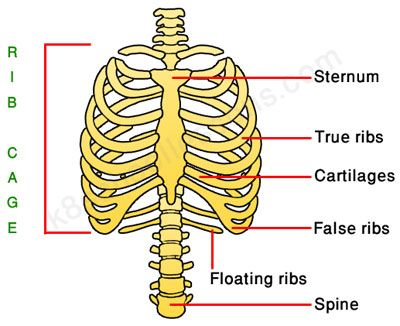
- Ribs are important for forming the thoracic cage. They are found in 12 pairs i.e. 24 in total. At the front, each pair of ribs is connected to the sternum bone with the help of costal cartilage and at the back, each rib is connected to the thoracic vertebra. Thus, each rib is connected to form the thoracic cavity.
- There are a total of 12 pairs of ribs. In which the first 7 pairs of ribs are called true ribs, because each of these ribs is connected directly to the sternum bone with the help of costal cartilage on the front side and is connected to the thoracic vertebra on the back side.
- These three pairs of ribs 8, 9 and 10 are called false ribs, because they are connected to the thoracic vertebra on the back side but are not connected directly to the sternum on the front side but the 10th rib is connected to the 9th rib, the 9th rib to the 8th rib and the 8th rib to the 7th rib, hence they are called false ribs.
- The 11th and 12th pairs of ribs are known as floating ribs. This pair of ribs connects to the thoracic vertebrae on the back side, but is not connected to the front side anywhere, so it is known as a floating pair of ribs.
Characteristics of Rib:
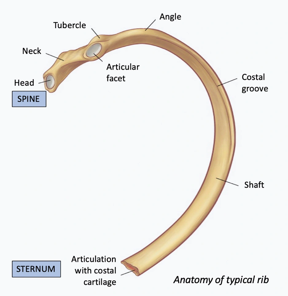
- A rib is a flat bone. This bone has two extremities and a flat part.
- The posterior part of the rib towards the vertebral end has a flat part, which is called the head of the rib. This part of the head is connected to the body of the thoracic vertebra.
- The narrow part after the head is called the neck. Then there is a raised part which is called the tubercle. This part of the tubercle is connected to the facet of the transverse process of the thoracic vertebra.
- The curved part of the rib after the tubercle is known as the angle of rib.
- The middle part of the rib is called the shaft part. The superior border of this part is smooth. Also, its inferior border is curved. It has a groove, which is called the costal groove. The costal nerve and blood vessels pass through this groove.
- The front part of the rib shaft is called the anterior surface. The intercostal muscles attach to it. While the posterior surface of the rib is in contact with the pleura of the lung.
- The end towards the anterior side of the rib i.e. this end is also called the sternal end. Where the anterior side of the rib is connected to the sternum bone with the help of costal cartilage.
- The space between two ribs is called the intercostal space. The intercostal muscles are attached to this area. The contraction and relaxation of these intercostal muscles causes the act of respiration and the diameter of the thoracic cavity fluctuates.
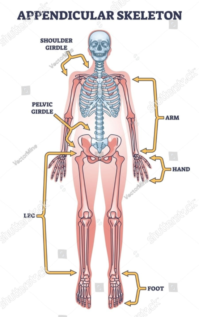
Appendicular Skeleton:
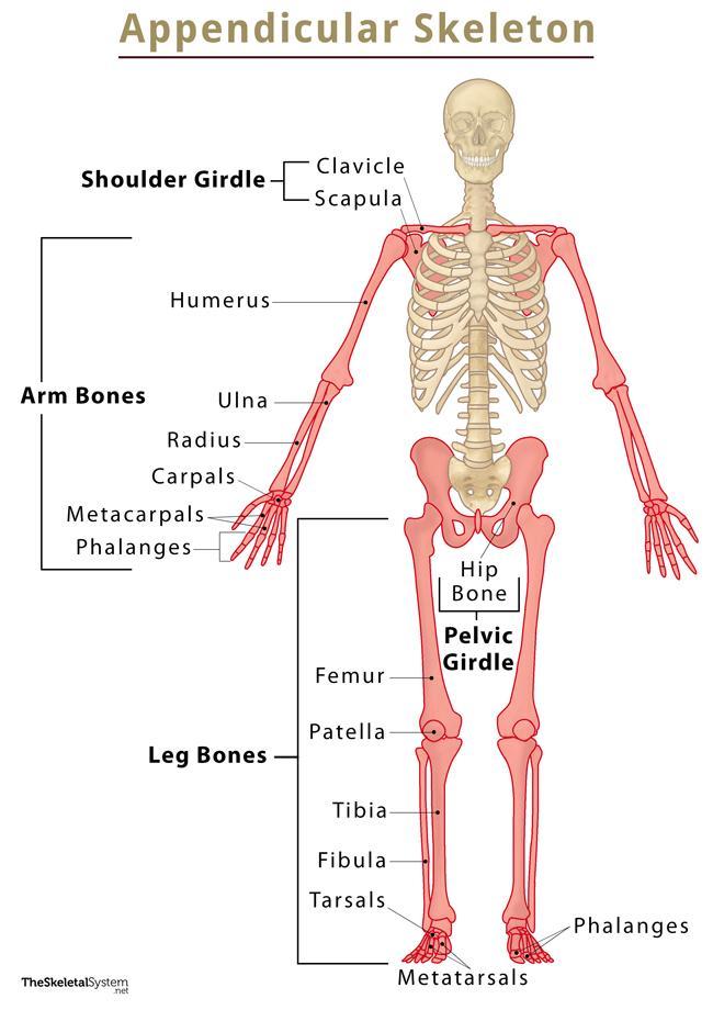
- The appendicular skeleton includes the ankle joint, upper limb, pelvic joint, and lower limb.
- The appendicular skeleton contains a total of 126 bones.
- The upper extremity contains 60 bones.
Lower extremity 60 bones are provided.
Shoulder Joint (Solder Joint):
Solder joint is also called solder girdle. The bones that make up the solder joint are as follows.
- Scapula Bone. 1
- Clavicle Bone. 1
- Humorous Bone. 1
The solder joint is formed when the head of the humerus bone articulates with the glenoid cavity of the scapula bone. The clavicle bone does not form a direct solder joint.
Clavicle Bone:
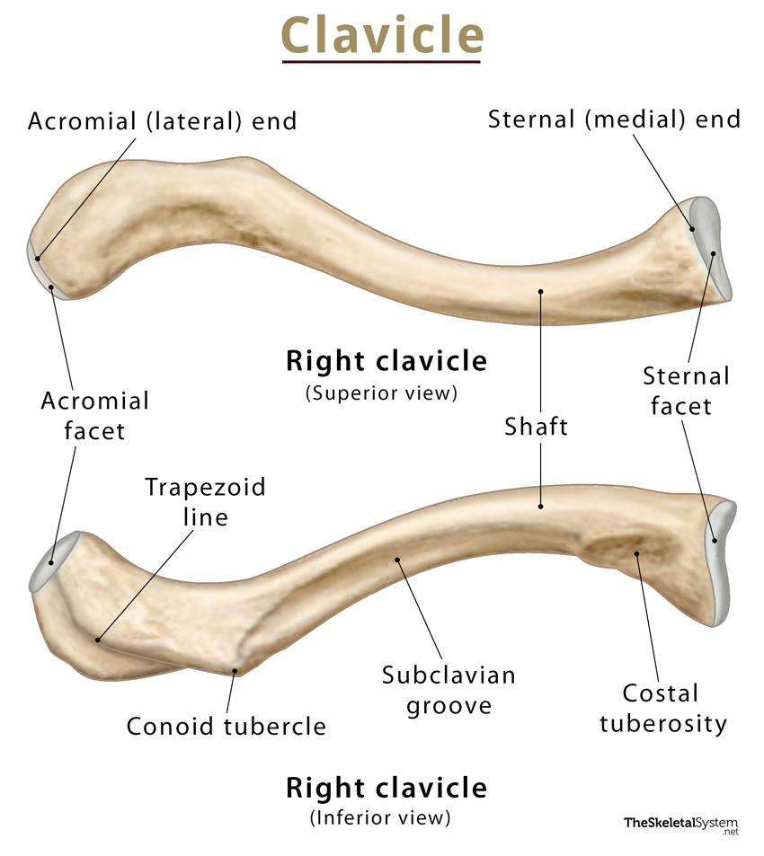
This bone is also called the collarbone. It is an S-shaped long bone. It has two ends or two extremities. In which the medial end of the clavicle bone articulates with the clavicular notch of the manubrium of the sternum bone to form the sternoclavicular joint.
The lateral end of the clavicle bone articulates with the acromion process of the scapular bone to form the acromioclavicular joint.
Scapula Bone
Humorous Bone
Ulna Bone:
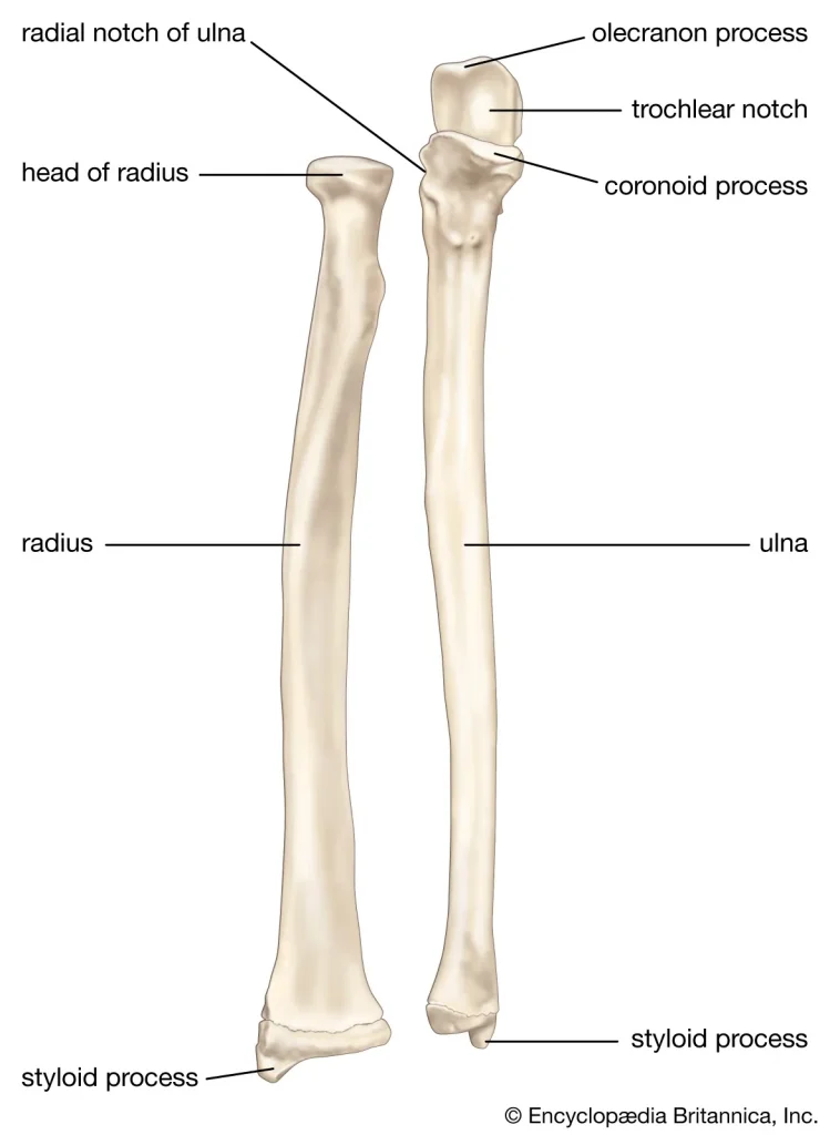
- It is a long bone in the forearm of the upper limb. This bone is also connected to the radius bone that accompanies it. In the forearm, the ulna bone is located on the medial side and the radius bone is located on the lateral side.
- The ulna bone has two extremities and a shaft.
- The upper extremity of the ulna bone has a fang-like process called the olecranon process and a smaller process on its lower side is called the coronoid process. Between these two processes is a groove called the trochlear notch. The trochlea of the humerus bone is attached to this groove, forming the elbow joint. This joint is also a type of joint.
- The diameter of the ulna bone decreases from the upper extremity towards the shaft. The shaft is where the muscles of the forearm are located.
- The lower extremity of the ulna bone has a pointed process at the end. This is known as the styloid process of the ulna. This part articulates with the proximal carpal bone to form the wrist joint.
- The ulna bone articulates with the radius in the upper extremity to form the proximal radioulnar joint. Also, the ulna and radius bones join in the lower extremity to form the distal radioulnar joint.
Radius Bone:
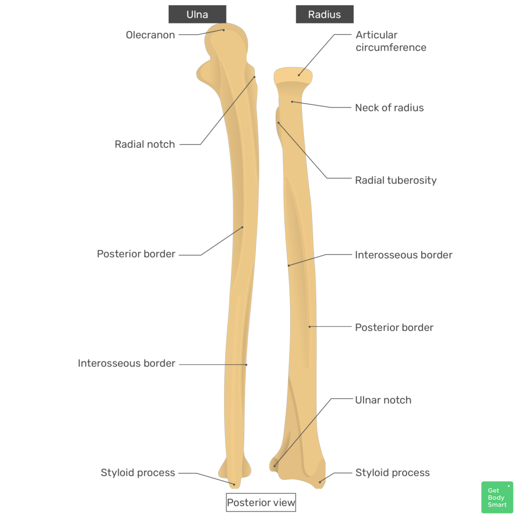
- It is a long bone in the upper limb. It is a bone located on the lateral side of the forearm.
The radius bone has two extremities and a shaft. - The radius bone has a rounded part at its upper extremity, which is called the head. The next narrow part is called the neck and the next protruding part is called the radial tuberosity.
- The radius bone increases in diameter from the upper extremity downwards.
- The soft part is cylindrical in shape.
- The lower extremity has a pointed process, called the styloid process of the radius. This part joins the proximal carpal bones to form the wrist joint.
Carpals Bones:
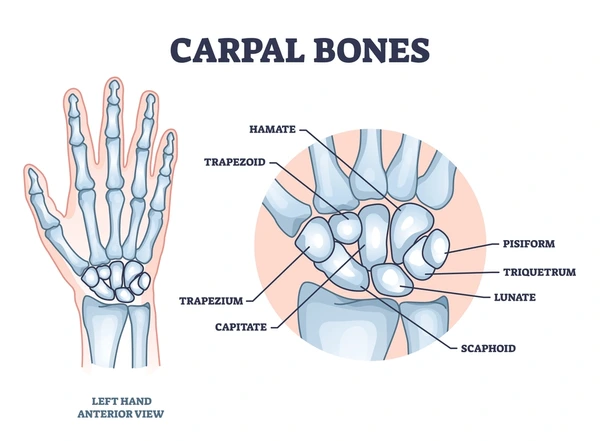
It is also called the wrist bone. It is arranged in two rows of four in a figure of eight in such a way that,
Proximal Row:
The four carpal bones are arranged in the upper row. They include the scaphoid, lunate, triquetrum and pciform bones. The carpal bones of this proximal row articulate with the ulna and radius at the top to form the wrist joint.
Distal Row:
The carpal bones in the distal row include the trapezium, trapezoid, capitate, and hamet.
The carpal bones of the distal row articulate with the proximal carpal at the top and attach to the metacarpal bones at the bottom. The carpo forms the metacarpo joint.
Metacarpal Bones:
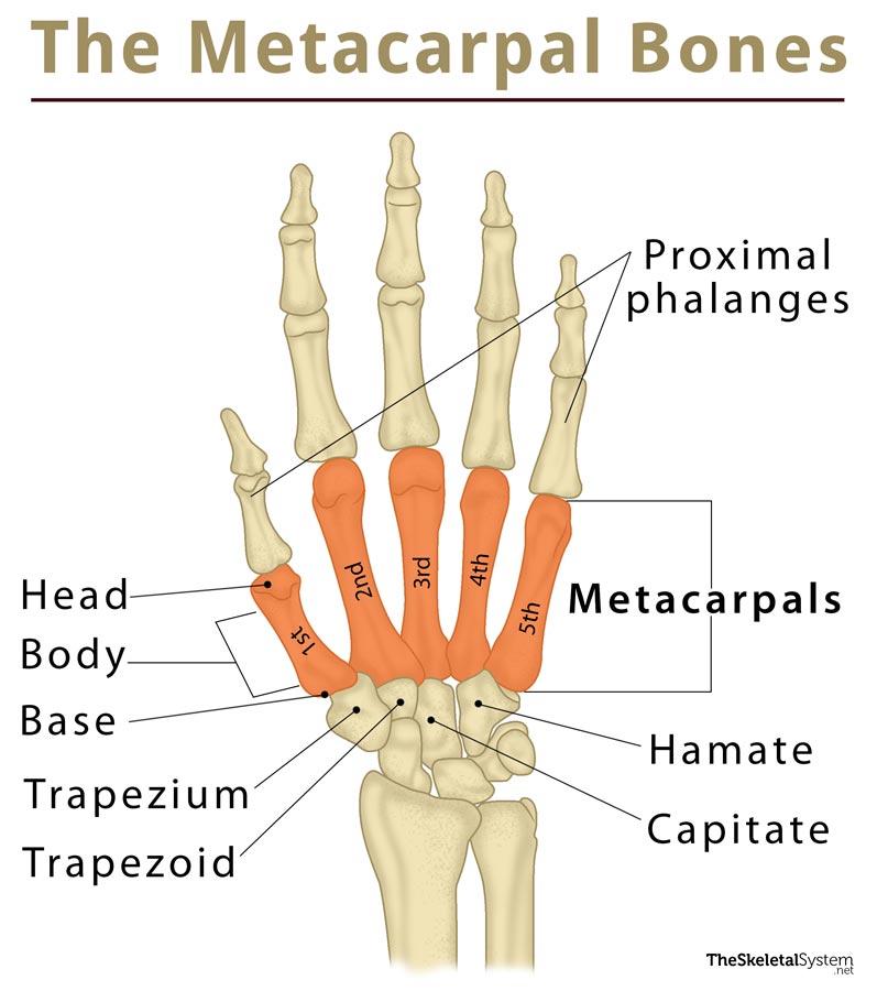
These bones form the palm of the hand. The number 5 of metacarpals are seen. In which the metacarpal of the thumb side is considered the first metacarpal.
The metacarpal connects to the carpal of the distal row on the upper side. Also connects to the proximal phalanges at the bottom.
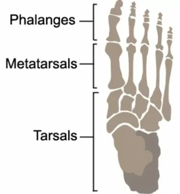
Phalanges:
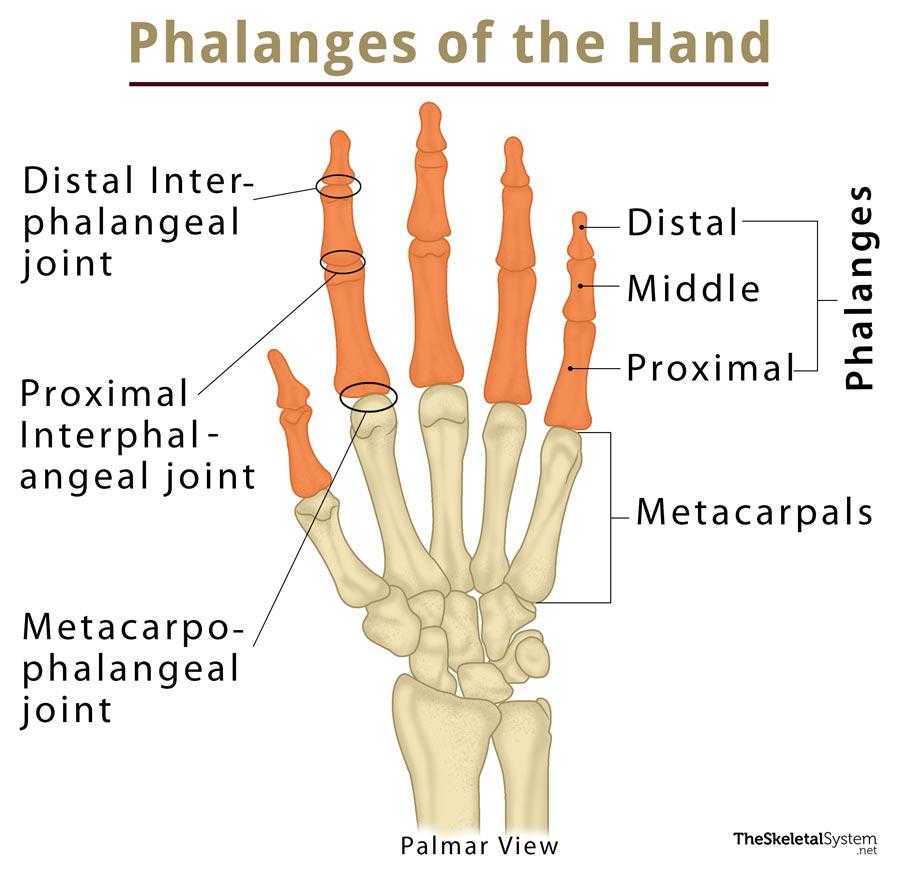
The small bones in the fingers are called phalanges. They are found in number of 14. The thumb has two phalanges. The remaining fingers have three phalanges.
The upper phalanges are called the proximal phalanges, the middle phalanges are called the middle phalanges, and the distal phalanges are called the distal phalanges.
Pelvic Joint (Pelvic Girdle):
The pelvic joint is also called the pelvic girdle. The bones that make up the pelvic joint are as follows.
- Innominate Bone. 2
- Sacrum Bone. 1
- Femur Bone (Femur Bone). 1
The head of the femur bone articulates with the acetabulum cavity of the innominate bone to form the hip joint.
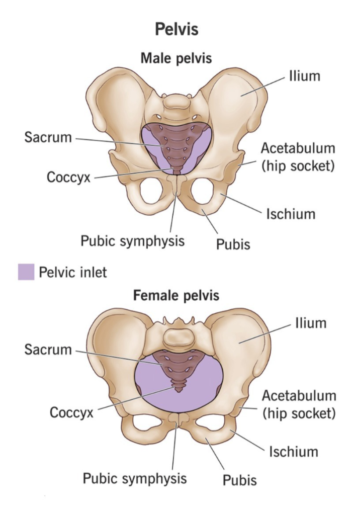
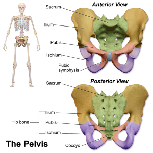
Pelvis:
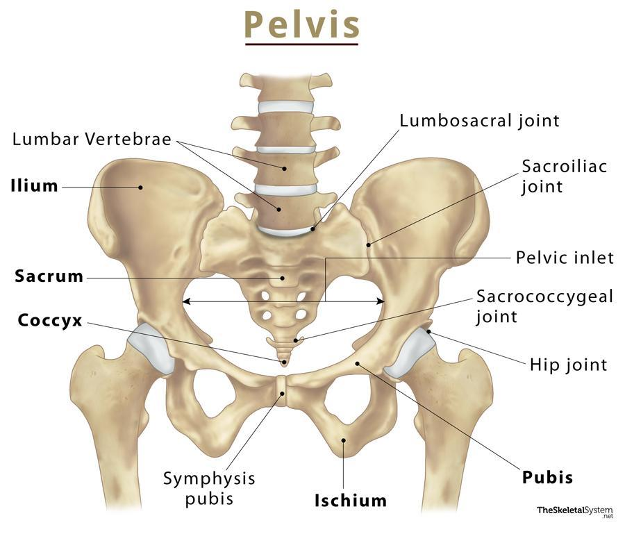
The pelvis is a basin-shaped cavity. It is formed by two innominate bones, a sacrum, and a coccyx.
This forms a rounded part in the middle of the pelvis, which is called the pelvic brim. The part below this pelvic brim is called the true pelvis because of its importance from an obstetric point of view. The upper part of the pelvic brim is called the false pelvis.
Innominate Bone:
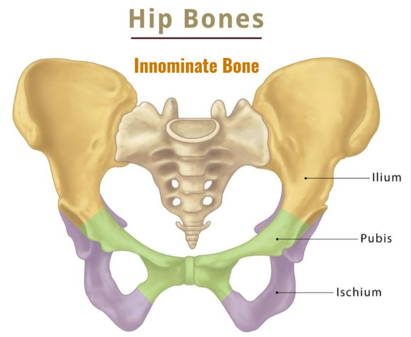
The innominate bone is a bone of the pelvic girdle. It is also known as the Hip Bone.
There are two in the body. They are located on both the right and left sides of the pelvic cavity. Both innominate bones connect to the sacrum bone at the back and form the pelvic cavity.
The innominate bone is a large bone. It is a flat and irregular type of bone. Each innominate bone consists of three bones.
- Illium (Illium)
- Ischium
- Pubis (Pubis)
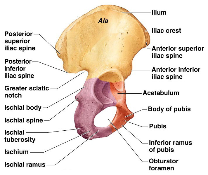
1. Illium:
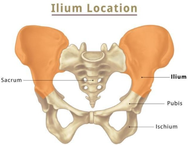
- The ilium is the flat bone on the upper side of the innominate bone. It has a ridge at its top called the iliac crest. The iliac crest is divided into the following parts:
- Anterior superior iliac spine (front top)
- Anterior inferior iliac spine (front bottom)
- Posterior superior iliac spine (back top)
- Posterior inferior iliac spine (back bottom)
- The ilium bone joins the sacrum bone at the back to form the sacroiliac joint.
- There is a large groove at the bottom of this joint. This groove is called the greater sciatic notch. Through it, the sciatic nerve and blood vessels pass to the lower extremity.
- The gluteal muscles attach to the posterior surface of the ilium bone. and forms the gluteal region.
- The front surface of the ilium bone is known as the iliac fossa. This part has a depressed part where the muscles are attached.
2. Ischium:
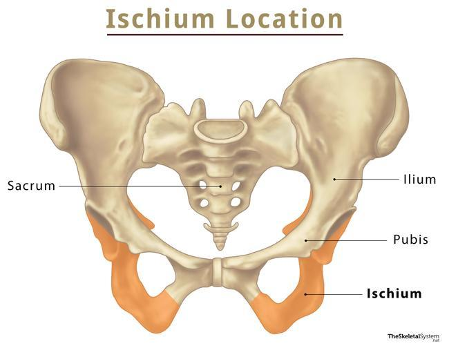
- The ischium bone is located below and behind the ilium bone.
- A protruding pointed part is located on the back side between the ilium bone and the ischium bone. This part is called the ischial spine.
- Below it is a small groove called the lesser sciatic notch.
- At the bottom of this less sciatic notch is a strong thick process called the ischial tuberosity. When we sit in a sitting position, the weight of the body comes on this part. This is a strong structure that bears weight.
Pubis:
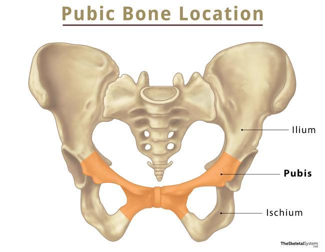
The pubic bone forms the anteriormost part of the innominate bone. The pubic bones of both innominate bones join at the anterior end to form the symphae pubis joint.
There is a large foramen at the bottom of this pubic bone. It is called the obturator foramen. From which nerves and blood vessels pass downwards towards the extremity.
The three bones in the hip bone, the ilium, ischium and pubis, all come together to form a cavity-like structure, this cavity is called the acetabulum cavity. The head of the femur bone joins this cavity and forms the hip joint.
Femur Bone (Femur Bone):
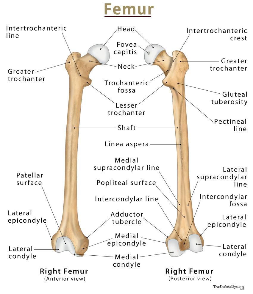
- The femur bone is a bone in the lower extremity. It is the longest and strongest of all the bones in the body.
- This bone has two extremities and a shaft. which is as follows.
Upper Extremities:

The upper one-third of the femur bone is called the upper extremity. Which has the following structure.
Head :
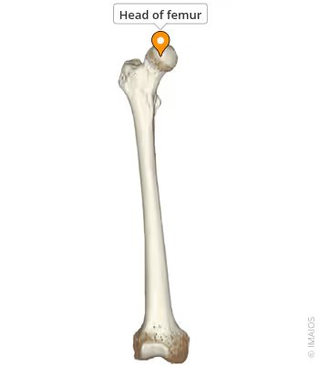
- The frontmost part of the femur bone is a round bone. The part called the head of the femur.
- This rounded part joins the acetabulum cavity of the innominate bone to form the hip joint.
- The hip joint is a synovial joint so all the characteristics of a synovial joint can be seen here.
Neck :
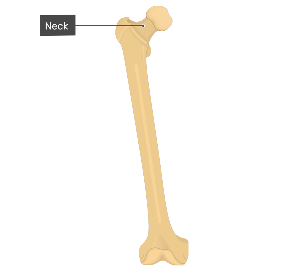
The narrow part after the head is called the neck. Which is four to five centimeters long and round in shape.
Greater Trochanter and Lasser Trochanter:
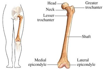
- At the end of the neck, there are two rough and raised large parts of the bone. These are called trochanters.
- The larger part on the outside is called the greater trochanter. While the smaller part on the inside is called the lesser trochanter. The line connecting these parts of the trochanter is called the intertrochanteric line. This part is where the muscles are attached.
- The middle part of the bone after the upper extremity is completed, i.e. the middle part of the bone, is known as soft. whose structure is as follows.
Linea aspera (Linea aspera):
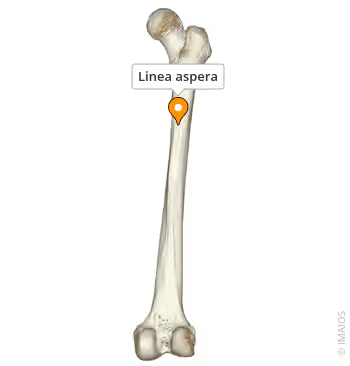
The posterior side of the soft part has a raised bone edge. Which is called the linea aspera. This part is where the muscles are attached. The shaft of the femur bone is cylindrical and round in shape.
Lower Extremity:
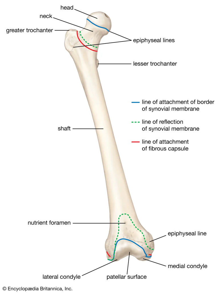
- The lower one-third of the femur bone is called the lower extremity. In which two rounded bony prominences are seen on the lower side. These are called condyles. The condyle on the medial side is the medial condyle and the lateral side is the lateral condyle. The part separating the two condyles is called the intercondylar notch.
- The front surface of this condyle is a smooth surface called the patellar surface, where the patella bone attaches.
- A triangular surface is formed at the back of the lower extremity of the femur bone. This is called the popliteal surface. The popliteal vessels and nerves are found in this area.
Patella Bone:
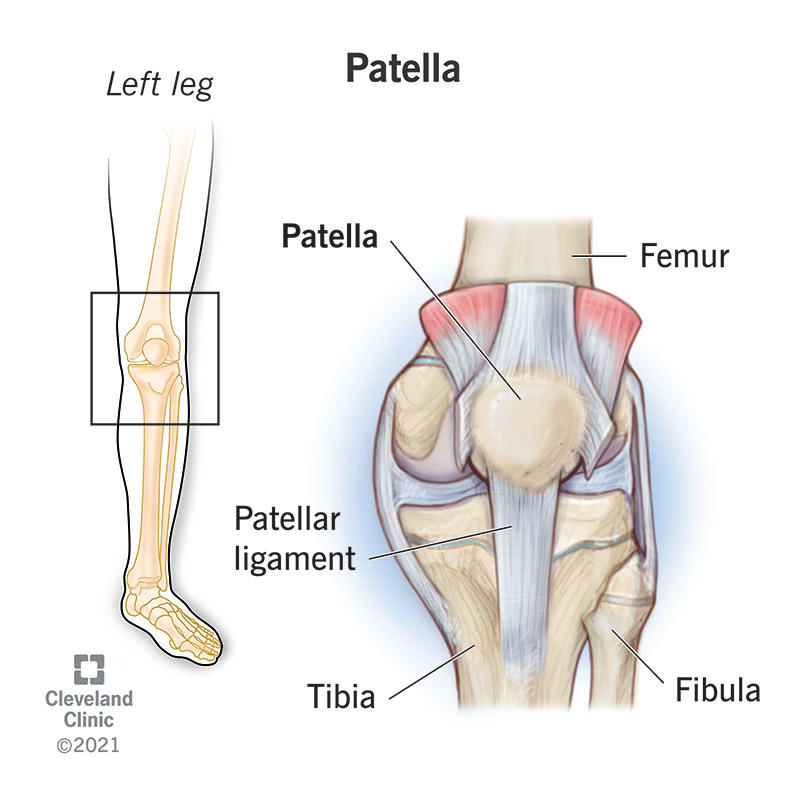
It is also known as the knee cap bone. It is a triangular-shaped sesamoid bone. This bone is located on the anterior side of the knee joint. This bone maintains its position with the help of ligaments and is a moving bone.
Tibia Bone (Tibia Bone):
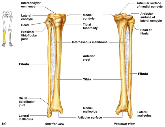
- It is also known as the shin bone. The tibia is a long bone in the lower extremity. It has two extremities and a shaft. This bone is a strong and weight-bearing bone.
- The upper extremity of the tibia bone has two condylar surfaces on its superior surface. The lateral and medial condyles of the femur bone articulate with these surfaces to form the knee joint.
- The anterior surface of the tibia bone has a rough raised area called the tuberosity of the tibia.
- The lateral side of the tibia bone articulates with the fibula bone to form the proximal tibiofibular joint.
- The shaft of the tibia bone is irregularly triangular. Its anterior surface bears a sharp crest known as the crest of the tibia. The part of this crest that lies immediately beneath the skin can be felt by touch.
- The lower extremity of the tibia bone has a smooth elongated process on its medial side. It is called the medial malleolus. This part joins the tarsal bone to form the ankle joint.
- In the lower extremity, the tibia and fibula join to form the distal tibiofibular joint.
Fibula:
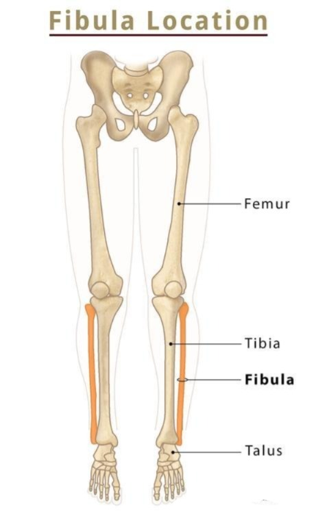
- The fibula bone is a long bone in the leg. It is located on the lateral side of the tibia bone. It has two extremities and a shaft.
- The fibula bone has a quadrangular shaped head at its upper extremity. The narrow part below it is called the neck.
- The fibula bone has an irregular margin on the soft part and a process on the lateral side of the lower extremity called the lateral malleolus.
- The fibula bone is deep in the muscles.
Tarsal Bones:
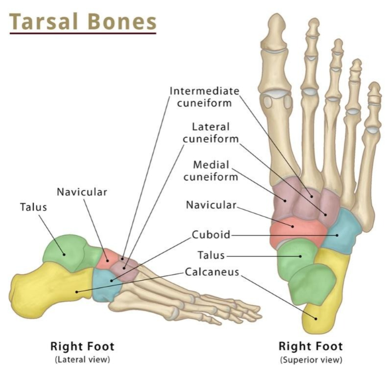
The tarsal bone is also known as the second ankle bone. It consists of 7 different irregularly shaped bones, which are as follows.
- Talus (Talus). 1
- Calcaneus (Calcaneus). 1
- Navicular. 1
- Cuneiform (cuneiform). 3
- Cuboid (Cuboid). 1
All the above tarsal bones join together to form the foot arch.
The talus bone joins the tibia and fibula bones to form the ankle joint.
The calcaneus bone forms the heel part of the foot.
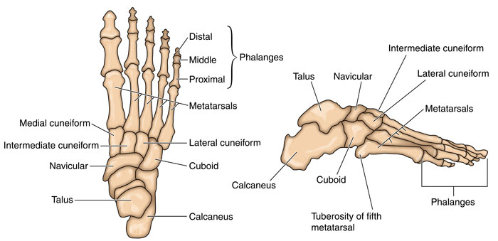
Metatarsals Bones:

Distal to the tarsal bone are the metatarsal bones. They are 5 in number. Below them are the phalanges. They are 14 in number. The toes of the foot have 2 phalanges and the remaining fingers have 3 phalanges. All these phalanges form the interphalangeal joints.