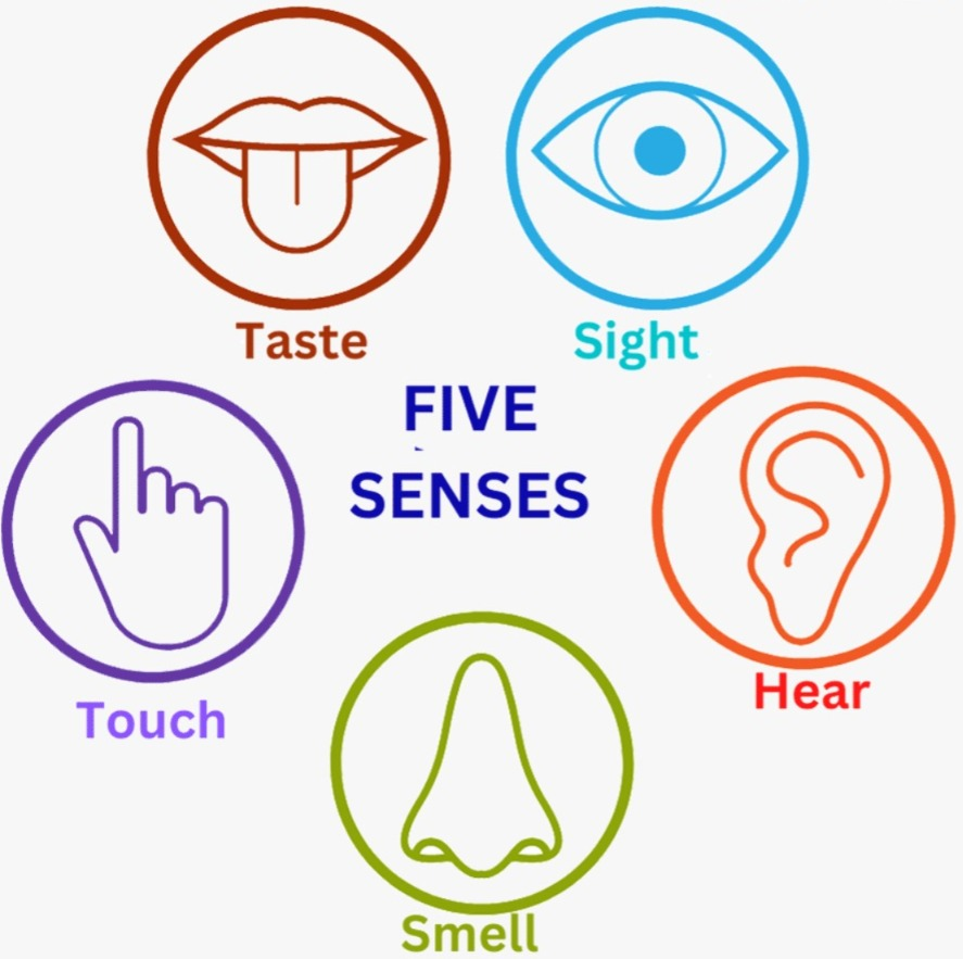
—ENGLISH-ANATOMY UNIT 12. SENSE ORGANS (PART :2). EYE,NOSE AND TOUNGUE
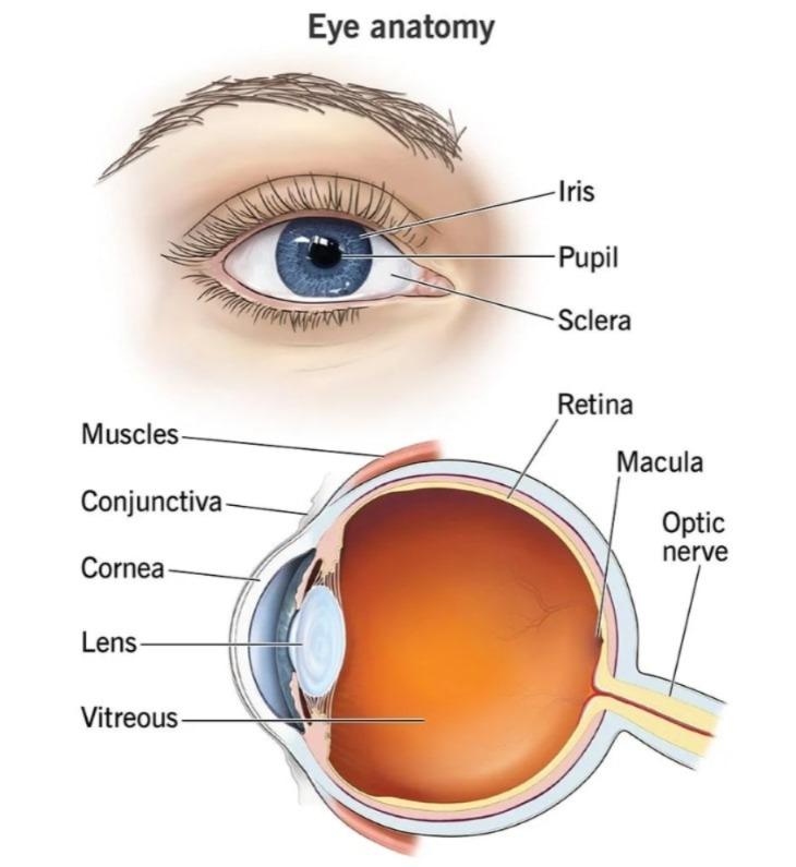
Eye ball:
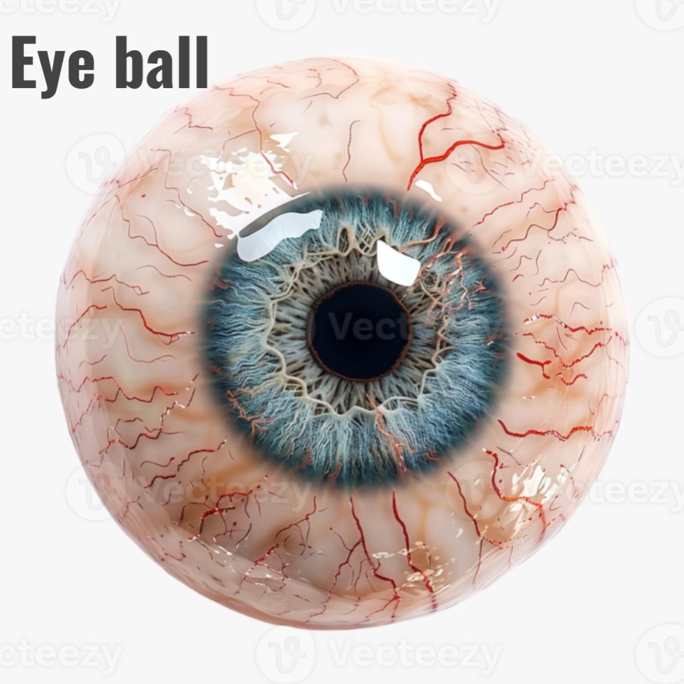
The eye is an important organ for vision, its shape works like a camera. It is a spiracle, its diameter is 2.5 cm. It carries the nerve supply to the brain through the optic nerve and the function of vision is performed.
The eyeball is located in the orbital cavity in the skull. The wall of this cavity has a layer of adipose tissue to protect the eye. The wall around the eyeball is made of bone and this adipose tissue works to protect the eyeball.
Structure:
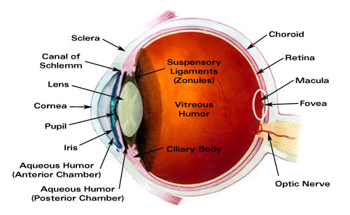
The structure of the eyeball has three layers is.
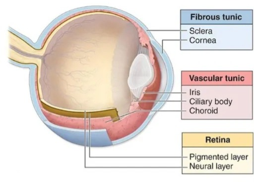
2. The middle layer is a vascular layer containing the choroid, ciliary body, and iris.
3. The inner layer is made of nervous tissue, which contains the retinal.
Sclera (sclera):
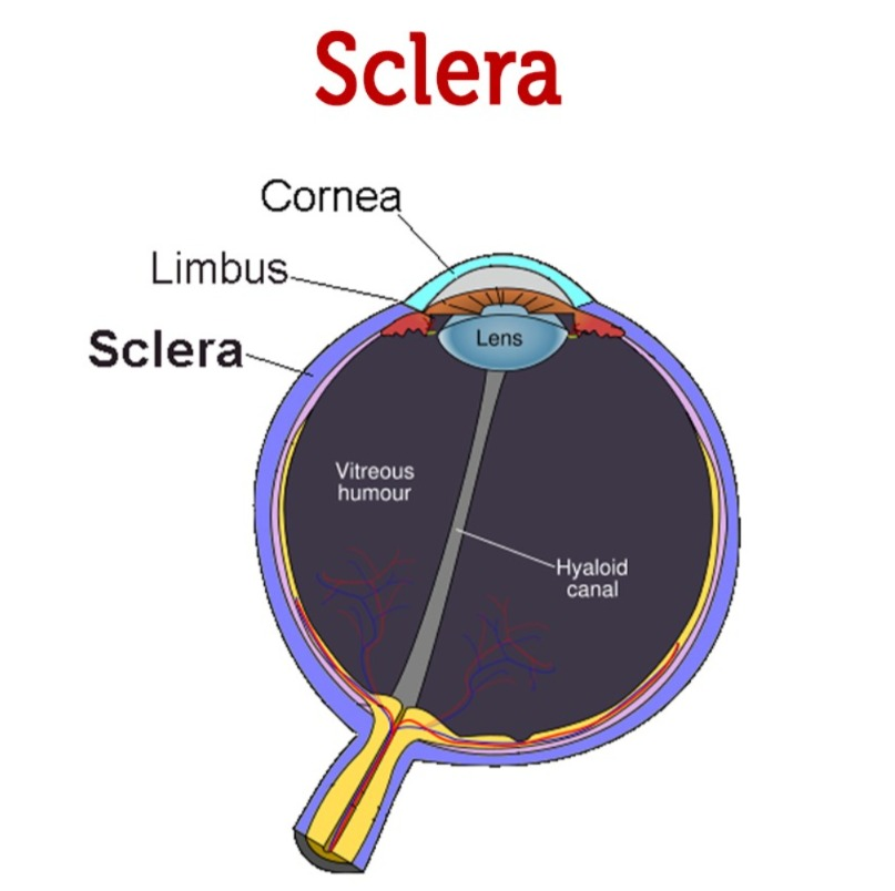
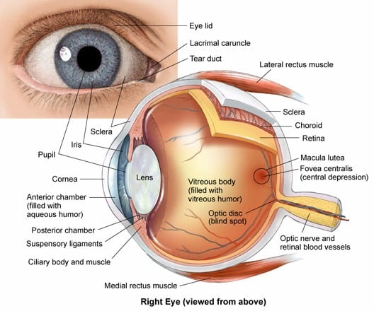
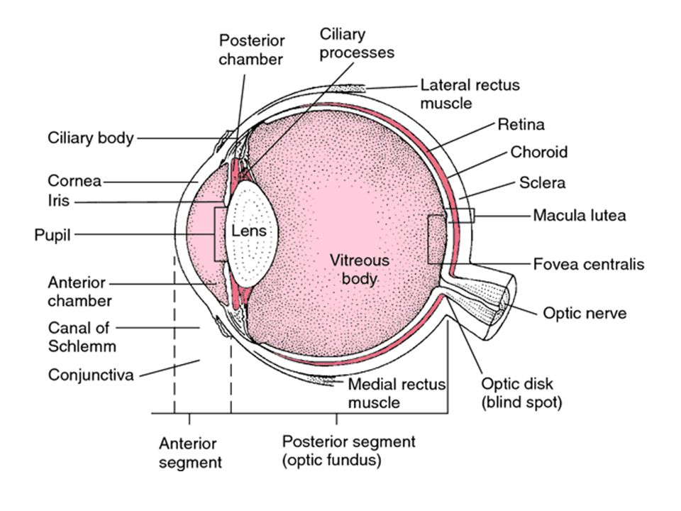
It is the white colored part of the eyeball. Its outer layer is made up of fibrous tissue. It is continuous at the front and connects to the transparent cornea, forming the corneoscleral junction. It functions to protect the delicate structures of the eye and maintain the shape of the eyeball. It provides attachment for the attachment of extrinsic muscles.
Cornea:
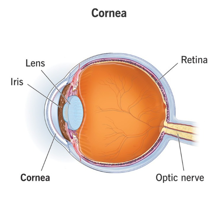
It forms the anterior portion of the eye. It is the colored part of the eye. It is convex and transparent from the front side through which light rays enter and fall on the retina.
Choroid:
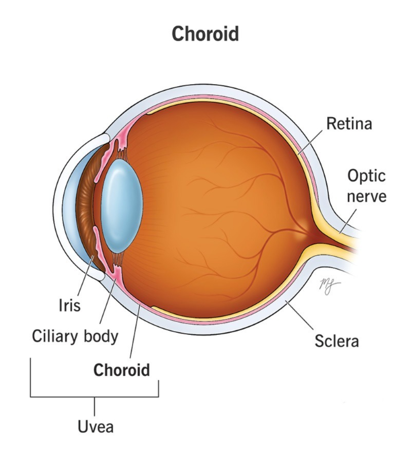
It is the middle layer of the eye and contains blood vessels so it is a vascular layer. It has blue, brown, gray pigmentation and is divided into three structures at the front: the ciliary body, the suspensory ligament and the iris.
Ciliary Body:
It is the thick part of the uveal tract. It is continuous with the iris anteriorly and with the choroid posteriorly. It has a lens cavity in its anterior part which helps in accommodation of the lens for near vision. The ciliary body contains unstriped type ciliary muscles which attach to the suspensory ligament. These ciliary muscles are important in maintaining the shape of the lens during near and distant vision. Thus, due to the change in the shape of the lens, light rays can be focused directly on the retina. These muscles are supplied by the parasympathetic nerves.
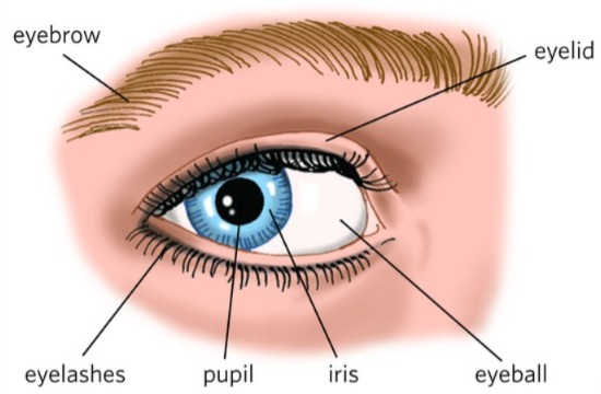
Iris :
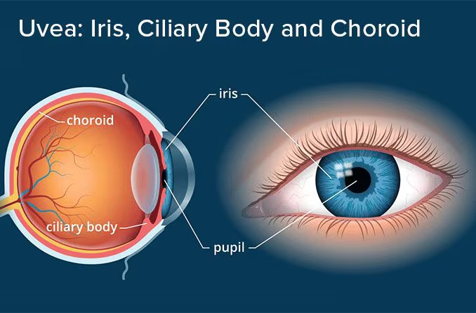
It forms the front colored part of the eye. It is long and flat. It is arranged vertically between the cornea and the lens. It divides the eyeball into anterior and posterior chambers, and these two chambers contain a fluid called aqueous humor. It contains circular and radial smooth muscle fibers and is connected by the ciliary body. The iris has an opening in the middle called the pupil. The amount of light that enters the eye can be controlled by varying the size of the pupil. It is innervated by sympathetic and parasympathetic nerves. The sympathetic dilates the pupil and the parasympathetic constricts the pupil.
Lens:
The lens is the transparent portion of the eye. It has a biconcave structure. The lens is located between the anterior and posterior segments of the eye and behind the posterior chamber of the anterior segment. Its diameter is about 1 cm. Due to the contraction and relaxation of the ciliary muscles, the thickness of the lens changes. If the object is close, the lens becomes thicker and thus the light rays can be easily focused on the retina.
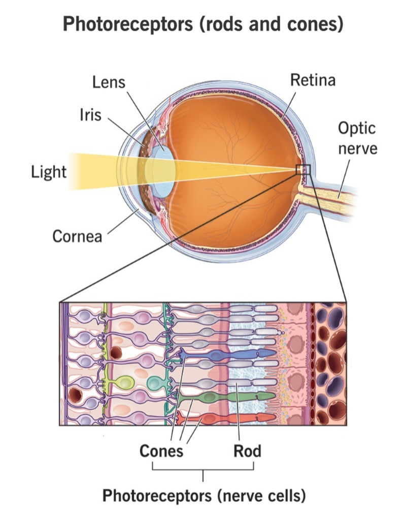
Retina:
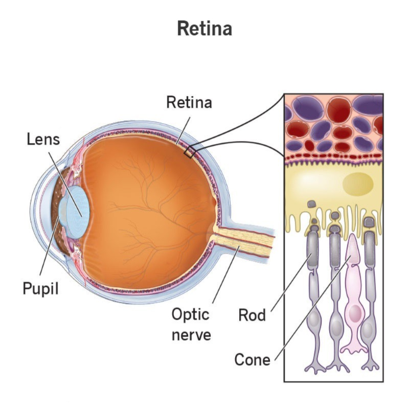
It is the innermost layer of the eyeball. It is made up of nervous tissue. This layer of the retina is continuously connected to the optic nerve at the back. Next to it is a circular area called the optic disc. The retina contains photoreceptors, rod cells and cone cells. Rod cells help in seeing in dim light and cone cells help in bright light and color vision.
At the back of the retina is a depression called the macula lutea. The central part of this is known as the central fovea or fovea centralis, where light rays are focused and help us see. From there, the impulses of vision travel along the optic nerve. It goes to the brain through it and the vision is interpreted. There is a blind spot in the optic nerve, where there are no light-sensitive cells.
Interior of the Eye:
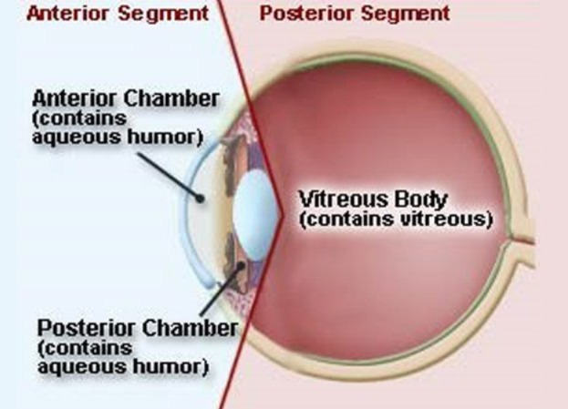
The eyeball is spherical in shape. Inside it is a very large space called a cavity. These cavities are called the anterior and posterior cavities.
The anterior cavity is further divided into two parts, the anterior chamber and the posterior chamber. The anterior chamber is the chamber in front of the iris and forms the back of the cornea. The posterior chamber is formed behind the iris. Both the anterior and posterior chambers contain a fluid called aqueous humor.
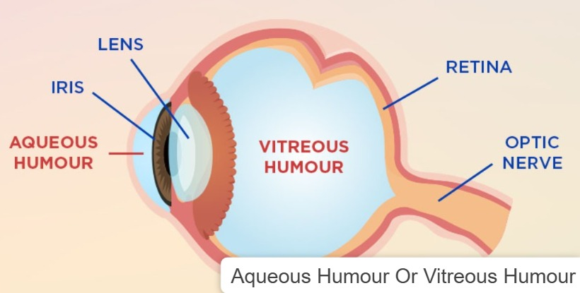
The posterior cavity is the cavity behind the lens and is larger than the anterior cavity. The fluid in this cavity is called vitreous humor. This fluid is mostly water and also contains some salts and proteins. This fluid helps maintain intraocular pressure. Normal intraocular pressure is 16 to 20 mmHg. If this pressure increases, the disease condition is called glaucoma.
Physiology of sight:
Light rays, after returning from an object and entering the eye, pass through the conjunctiva and enter the eye through the cornea. After that, they It reaches the anterior chamber and is focused in the lens and reaches the vitreous body i.e. the posterior chamber of the eye. From there, these light rays are focused at one place on the layer of the retina and through the nerve optic nerve present there, the visual image of these light rays goes to the brain and a clear image is interpreted.
Size of the pupil:
- Bright light pupil constriction
- Dimmed light causes pupil dilation
- The sympathetic nervous system causes pupil dilation
- The parasympathetic nervous system causes pupil constriction.
Accommodation of eye to light:
The eye works like a camera. The process of focusing the eye on any object and seeing it clearly is called accommodation. It has a total of four stages.
1. Refraction Of Light Rays:
Refraction is done by the cornea and lens in which the light rays coming from outside are refracted and reflected due to coming from one medium to another and this bend will be focused on one part of the retina so that a clear image can be seen.
2. Accommodation Of the Lens:
The ciliary muscles attached to the lens, the iris, help the lens to adjust to see near or far objects. The shape of the lens changes to see near objects is called accommodation. To see distant objects, the ciliary muscles relax and the lens becomes flat.
3. Constriction of the Pupil:
The size of the pupil controls the amount of light entering it. If there is bright light, the pupil constricts and if there is dim light, the pupil dilates.
4. Convergence:
Clear vision occurs when the light rays falling on the retina can be focused on a single point.
Muscles of the eye:
Two types of muscles are attached to the eye
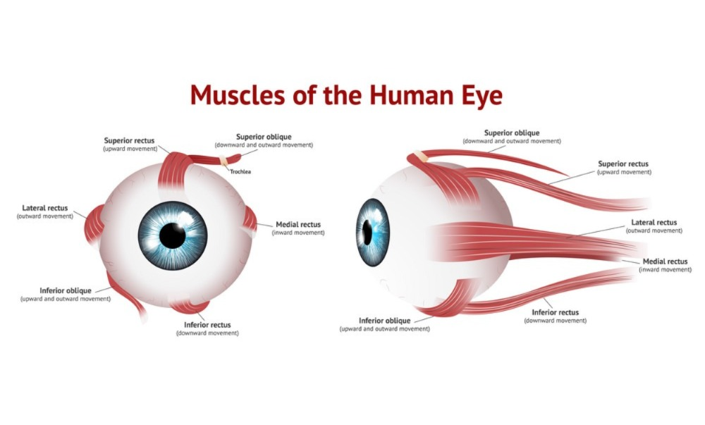
1. Extrinsic:
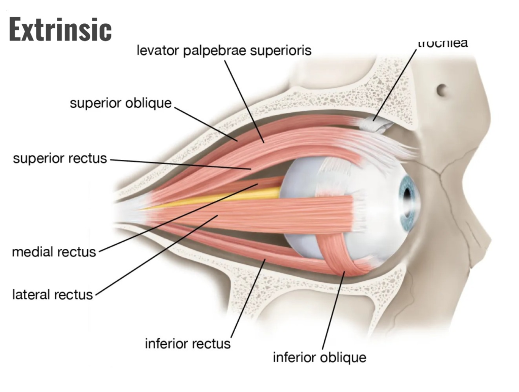
They are also called skeletal muscles. They are attached to the wall of the orbital cavity on the outside and to the sclera of the eyeball on the inside, which These extrinsic muscles are important for moving the eyeball in different directions. They consist of four straight muscles and two oblique muscles, which are as follows.
- Medial Rectus
- Lateral Rectus
- Superior Rectus
- Inferior Rectus
- Superior Oblique
- Inferior Oblique (Inferior Oblique Muscles)
Extrinsic Muscles are voluntary muscles that we can move in different directions at our will and make different movements of the eyeball
2.Intrinsic Muscles (Intrinsic Muscles):
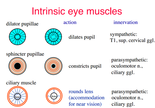
Intrinsic muscles are the muscles inside the eyeball, including the iris and ciliary muscles, which are not under voluntary control. They change size and shape as light from outside enters the eye. This is controlled by the autonomic nervous system.
Accessory Organs of the Eye:
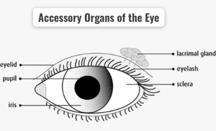
The eye is a very delicate organ and is protected by some accessory structures around it.
1. Eye Lid and Eyelashes
2. Eyebrows (ibros)
3. Lacrimal Apparatus (Lacrimal Apparatus)
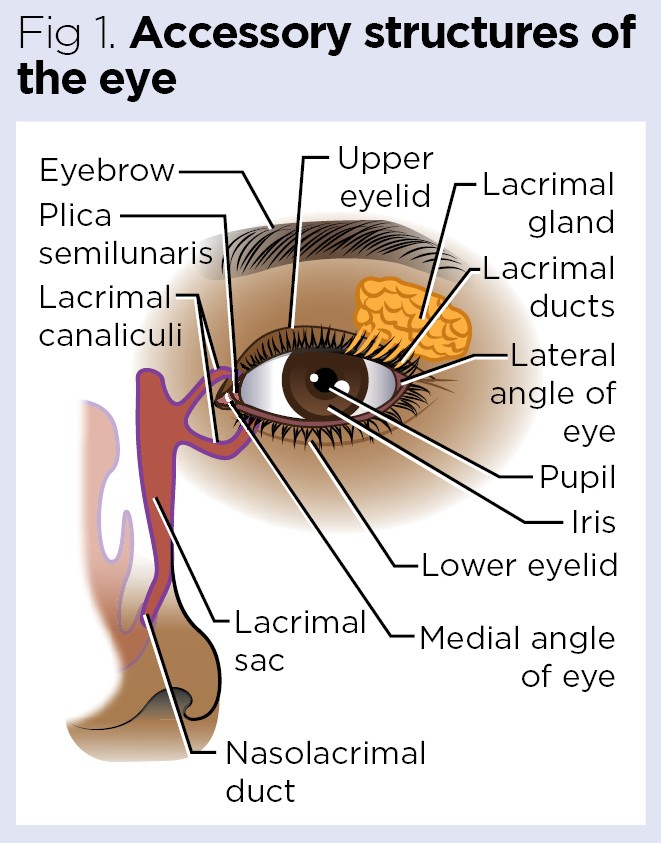
1. Eye Lid and Eyelashes:
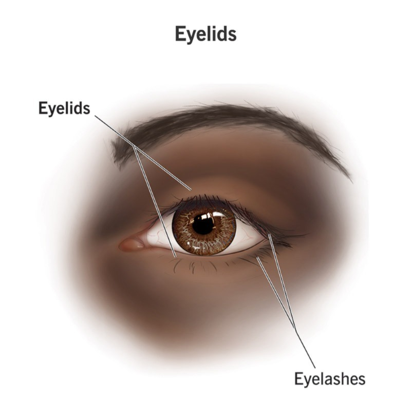
- The eye lids are the upper and lower two, both of which function to protect the eyeball during sleep. The upper eyelid is a movable layer in front of the eyeball that protects the eyeball from any foreign body or object. This eyelid is also called the palpebra. The space between the upper and lower eyelids is called the palpebra fissure. The part where these two eyelids are connected is called the canthus, which is named medial and lateral according to the side of the body.
- The margin of the eyelid has hair-like processes called the eye laces, which protect the eyeball from bright light and prevent foreign bodies. The base of the eye laces, which are located in the margin of the eyelid, has glands that lubricate these hairs. When this hair follicle becomes infected, it is called a stye. This gland is known as the tarsal or meibomian gland.
2. Eyebrows:
Eyebrows are hairs located on the supraorbital margin, the upper margin of the orbital cavity. They prevent sweat from entering the eye area and protect the eyeball from bright light and injury. Works to do. These eyebrows are also known for cosmetic purposes.
Conjuctiva:
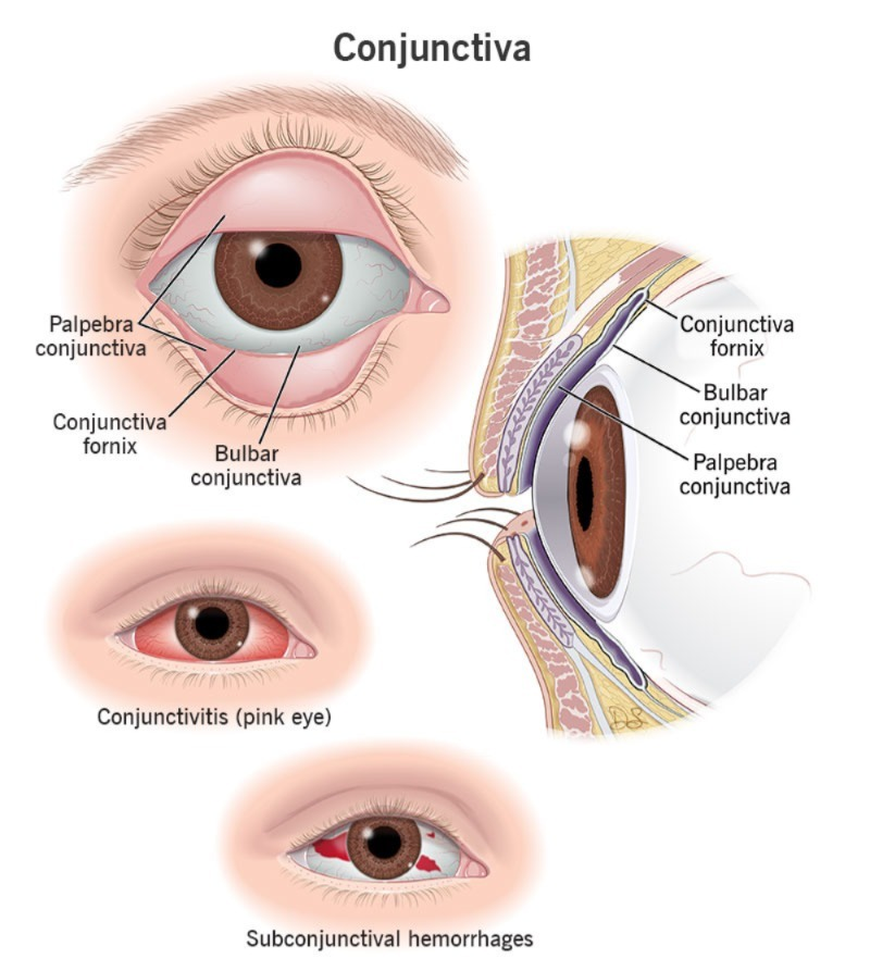
The protective layer on the inside of the eye lid The mucous membrane is called the conjunctiva. It is a membrane made up of columnar epithelium cells. It contains goblet cells that when we close our eyes, they become a close shell and protect the eyeball.
3. Lacrimal Apparatus:
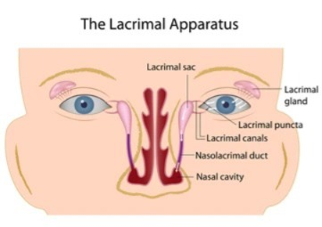
The lacrimal apparatus consists of the following structures which are located around the eyeball and its secretion is the tear which is on the surface of the eyeball Pours on. And the eyeball remains moist. The structure of the lacrimal apparatus is as follows.
1. Lacrimal Gland or its Duct…1
2. Lacrimal Canaliculi…2
3. Lacrimal Sac…1
4. Nasolacrimal Duct…1
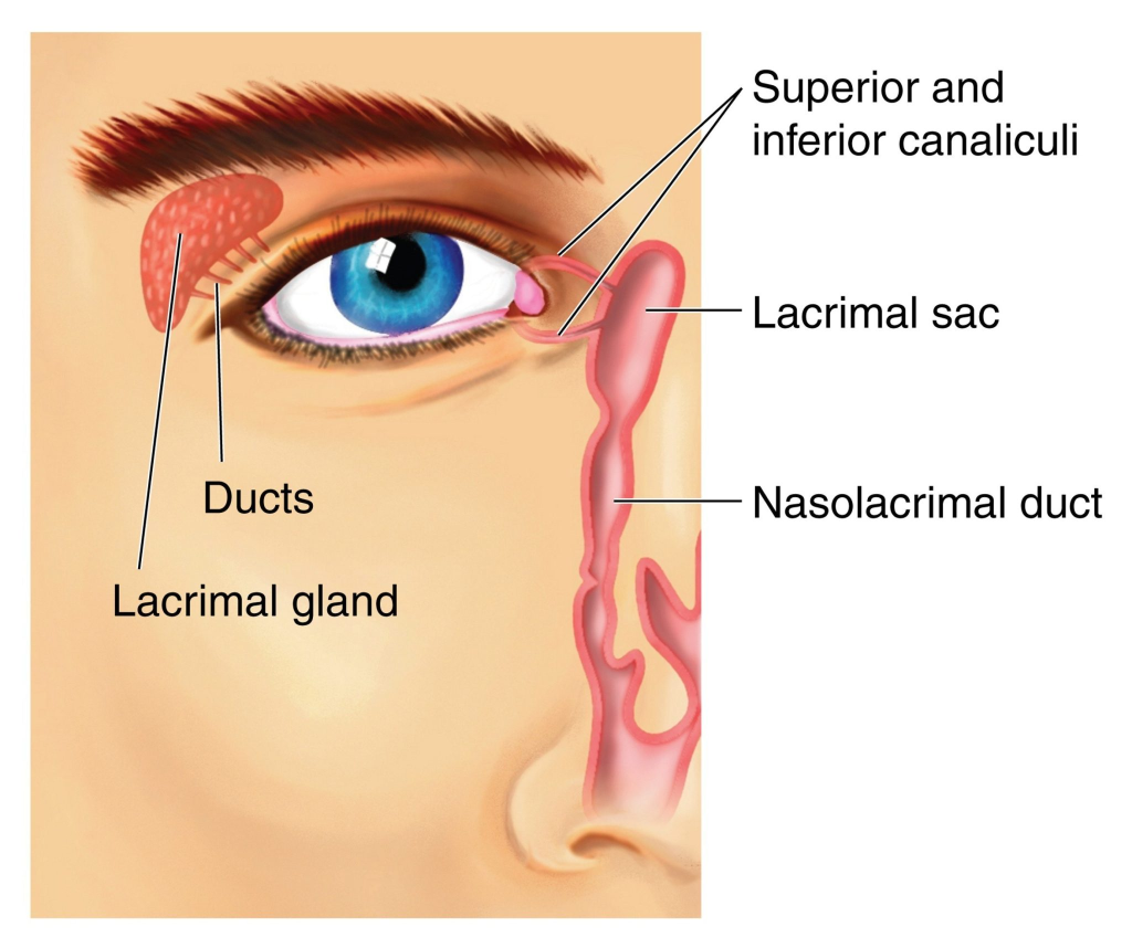
The lacrimal glands are two in number, one on the lateral side of the superior eyelid. They are almond-shaped. It secretes lacrimal fluid, the fluid called tears. This fluid drains through the lacrimal duct and spreads over the surface of the conjunctiva and the eyeball, keeping the eyeball moist.
Composition of Tears.
- Water (water)
- Mineral Salt (mineral salt) Salt)
- Antibodies and Lysozymes which contain bacteriocidal enzymes
The secretion of this lacrimal gland is collected in the lacrimal sac through the superior and inferior canaliculi leading to the middle canthus of the eyeball and from there opens into the nasal cavity through the nasolacrimal duct.
Functions of the Tear of the tear):

- Provides nourishment to the cornea
- It works to flush out irritating substances and dust that fall on the eyeball
- Prevents infection by microorganisms due to its bactericidal properties
- Acts as a lubricant due to its oily nature
- It prevents the eyes from drying out
Structure of Nose and it’s function:
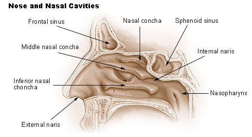
- The nose is an organ at the beginning of the respiratory system. It is located at the front of the face.
- When viewed from the outside, the nose has two openings. These are called external nostrils or nasal striations. There is a partition between these two openings, which has a cartilage-like septum on the front side and a vomer bone on the back side, which is the structure separating the two openings.
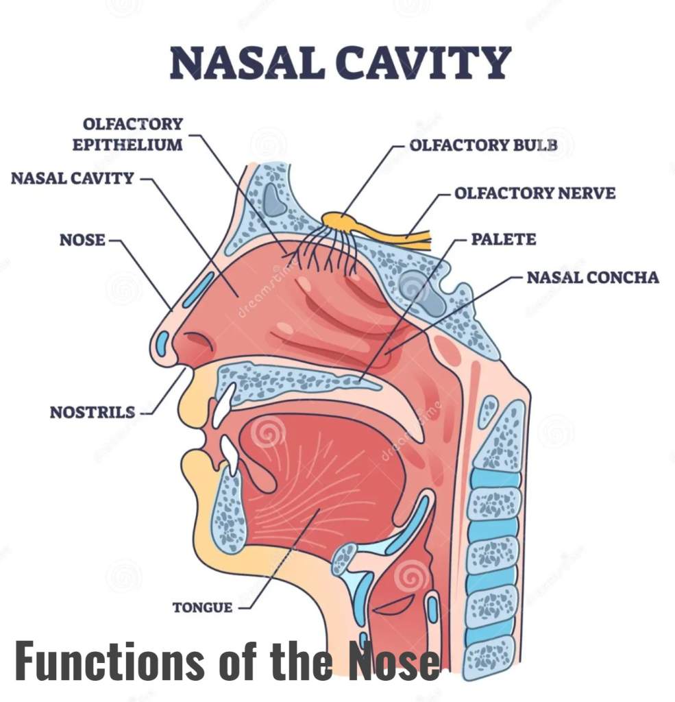
- The external nose is covered with skin on the outside and the inner lining is made of mucous membrane.
- It contains pseudo stratified ciliated epithelium cells. Here, goblet cells are present, which secrete mucus. Due to which the inner lining remains moist.
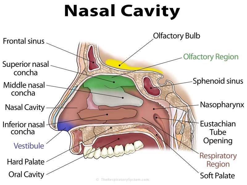
- The nasal cavity is a cavity located in the face, which is arranged above the oral cavity and the cribiform plate of the ethmoid bone of the cranial cavity is present on the upper side of the nasal cavity. In addition, the frontal bone and the sphenoid bone are also found in this part.
- The medial wall of the nasal cavity is formed by cartilage and the vomer bone. This wall is known as the nasal septum.
- The floor of the nose is made up of the soft and hard palates.
- The lateral wall of the nose contains the nasal bones. In addition, a bony structure called the inferior conchae also forms the lateral wall.
- Openings at the back of the nasal cavity, called the posterior nares, are connected to the pharyngeal fossa.
- Hairs are located on the inside and anterior side of the nasal cavity, which warm the air, filter it, and prevent small foreign particles from entering.
Openings into the Nasal cavity (Opening of the nasal cavity):
- There are 2 openings in the front of the nasal cavity, called the anterior nares is called. 2 such openings are located on the posterior side of the nasal cavity, which open into the part of the phalanges. They are called posterior nares.
- In addition, the openings of the sinuses of the bones surrounding it open into the part of the nasal cavity. It is called paranasal sinuses.
Functions of the Nose:
- The nose is an external organ of the respiratory system. Its functions are as follows.
- It performs the act of respiration. In which oxygen-rich air from the external environment enters the lungs through the nose and the air containing carbon dioxide waste from the body exits through the nose.
- The hairs in the inner lining of the nose clean the air. So that no foreign particles enter the respiratory track.
- Due to the hair and vascular mucus membrane in the inner lining of the nasal cavity, the air entering inside becomes warm and reaches the lungs as warm as the body temperature, so that the lung tissue does not get irritated or damaged.
- Due to the presence of goblet epithelium cells in the inner membrane of the nose, it is a moist membrane. Due to the passage of air through it, it becomes moist. So that the lining of the inner mucus membrane does not get damaged or irritated. Thus, it also performs the function of humidification.
- The vestibule structure at the beginning of the nose, which is covered with cells and hair processes, is also found in it. It functions to filter the air entering inside. It prevents dust particles and foreign materials from entering the respiratory tract.
- The nose senses smell, that is, it also performs the function of distinguishing odors. The mucous membrane of the nose contains receptors of the olfactory nerve. When that air enters the nasal cavity, it comes into contact with the odorous or foul-smelling chemicals present in it, which stimulate those receptors, and their impulses travel to the brain via the olfactory nerve, resulting in the sensation of smell. Thus, it also performs the function of smell detection.
Physiology of Smell:
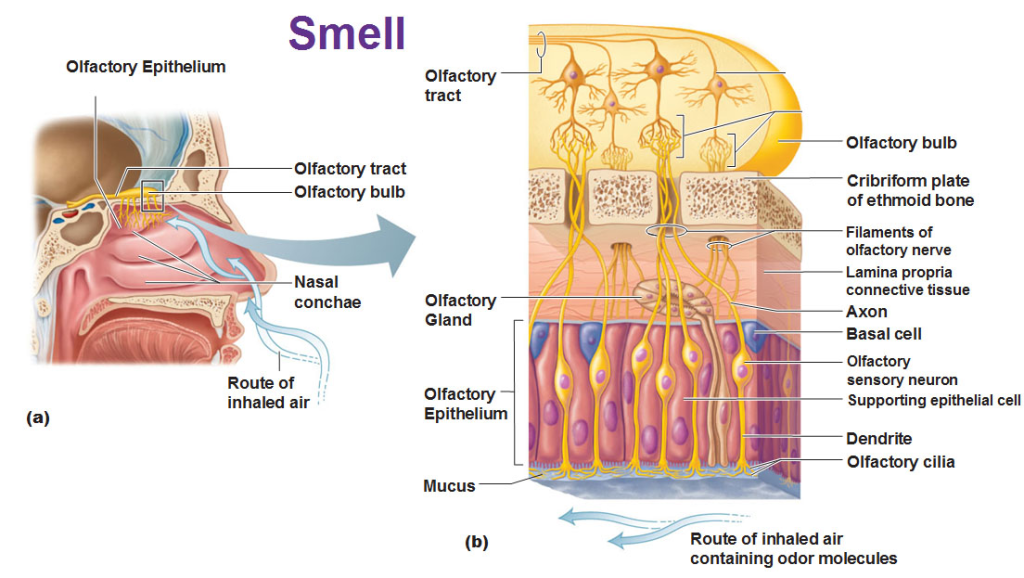
1. Olfactory Receptors:
These receptors are located in the olfactory epithelium in the superior nasal cavity inside the nose.
Functions:
These receptors identify and bind to odor molecules in the external environment and It finally generates Electrical signals. These receptors are special types of G-protein coupled receptors (GPCRs).
2. Olfactory Nerve:
The Olfactory Nerve is the “Cranial Nerve I”.
Impulses generated from the Olfactory receptors reach the Olfactory bulb through the Olfactory nerve.
3.Olfactory Bulb and Tract:
The olfactory bulb is the primary processing center where impulses are first analyzed.
Impulses from the bulb travel through the olfactory tract to the olfactory cortex.
4.Central Processing:
Primary Olfactory Cortex:
Here odors are identified and final perception occurs.
Amygdala and Hypothalamus:
These structures are responsible for emotional and autonomic responses.
Hippocampus:
Smell related memories are stored here.
5.Physiological Process:
1.Stimulus:
Odorant molecules enter the nose.
2.Transduction:
Receptors recognize these molecules and generate signals.
3.Conduction:
Signals are carried to the olfactory bulb by the olfactory nerve. reaches.
4.Perception:
Odor can be recognized and experienced as smell in the olfactory cortex of the brain.
6.Special Features:
Olfactory neurons are the only neurons that regenerate at regular intervals.
Smell is directly associated with emotional memory.
Olfaction is a chemical sense: it detects volatile substances.
The physiological system of smell begins with impulses from olfactory receptors reaching the olfactory cortex.
This system is not only important for odor detection, but is also important for emotional and behavioral responses, as well as memory recall.
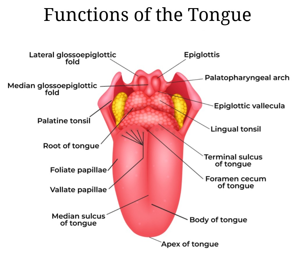
Tongue:
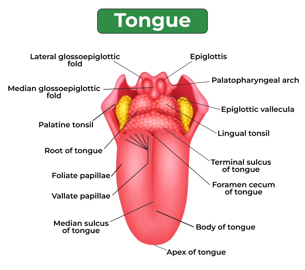
- The tongue is made up of skeletal muscles covered by a layer of mucous membrane. It forms the floor of the oral cavity. It divides the oral cavity into two parts in the middle. The tongue is attached inferiorly to the hyoid bone at the back. The tongue is attached to a fold of mucous membrane at the base of the oral cavity called the frenulum.
- The superior surface of the tongue is made up of stratified squamous epithelium cells. Papillae are located on the upper and lateral sides of the tongue. These papillae are connected to nerve endings and are also known as taste buds, which are responsible for the sensation of taste. These papilla are of three types.
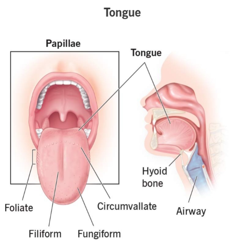
Filiform papilla.. Papilla)
- These papillae are located in the anterior two-thirds of the tongue and do not contain taste buds.
Fungiform papillae..
- They are located on the tip of the tongue and on its sides. They are like small dots and are known as taste buds for most birds.
Valet papilla..
- They are arranged in a V-shape on the posterior side of the tongue and are the largest of all the papillae.
Physiology of Taste (Physiology of Taste):
Introduction (Introduction):
Taste is a specific sensory function, which in medical terms is called gustation. Taste is experienced through taste receptors and taste buds, which are mainly located on the tongue and other parts of the mouth.
Primary Taste Sensations:
There are five main types of taste in humans to identify different types of chemical substances:
1. Sweet
2. Salty (Salty)
3. Sour (Sour)
4. Bitter (Bitter)
5. Umami (Umami) – which is created from substances such as amino acids and monosodium glutamate.
Taste Receptors and Taste Buds:
Taste buds are sensory organs, which are present on various papillae. There are mainly three types of papillae on the tongue, in which the taste buds are found:
Fungiform Papillae (Fungiform Papillae – Fungiform Papillae)
Folate Papillae (Foliate Papillae – Foliate Papillae)
Circumvallate Papillae (Circumvallate Papillae – Circumvallate Papillae)
Filiform Papillae (Filiform Papillae – Filiform Papillae) responsible for the taste No.
Each taste bud contains about 50 to 150 sensory cells.
Mechanism of Taste Sensation:
1. When chemical molecules of food come into contact with the taste buds, they bind to the taste receptors.
2.This causes depolarization and generates an action potential.
3.These impulses reach the brain through the sensory nerve fibers.
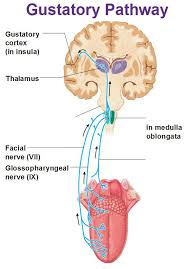
Sensory Pathway:
The sense of taste is carried to the brain through the following cranial nerves: Reaches:
1. Facial Nerve (CN VII):
Conveys information from the front 2/3 of the tongue.
2. Glossopharyngeal Nerve (CN IX):
Conveys taste from the back 1/3 of the tongue.
3. Vagus Nerve (CN X):
Conveys taste sensations from other parts of the mouth.
All these nerves go to the Solitary Nucleus (which is in the Medulla Oblongata) → then to the Thalamus → then to the Gustatory Cortex (which is in the Insula and Frontal Operculum).
Associated Factors in the Perception of Taste:
Olfaction (Smell) Senses)
Trigeminal Nerve (for identifying spicy or pungent substances)
Functions of the Tongue:
- It plays an important role in the act of speaking and speech.
- It performs the function of detecting taste due to the presence of taste buds on its surface.
- It performs the act of mastication, i.e. chewing. Both the tongue and teeth complete the act of chewing. The tongue is important for the moment of food in the mouth.
- During the act of swallowing, the tongue helps in the act of pushing the food backwards.