—ENGLISH-ANATOMY UNIT 12. SENSE ORGANS (PART :1) EAR
THE SPECIAL SENSES :
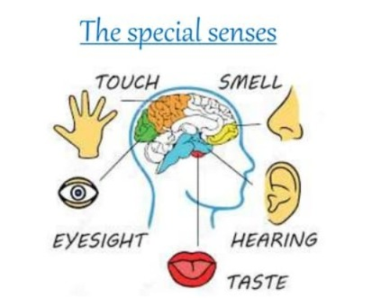
There are many senses in the body, some of which are general senses For example, touch, temperature, pain, and some are known as special senses, for example, vision, hearing, balance, taste, and smell. To recognize these senses, the body has certain sense organs such as the ears, nose, eyes, and mouth. Let us see about this sense organ…
EAR:
Ears are located on either side of the head. It is an organ involved in hearing and maintaining body balance.
Structure:
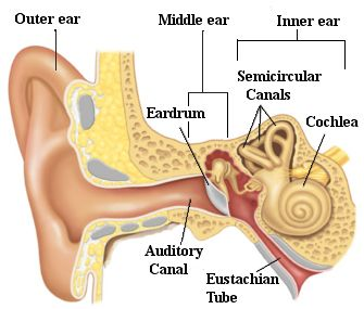
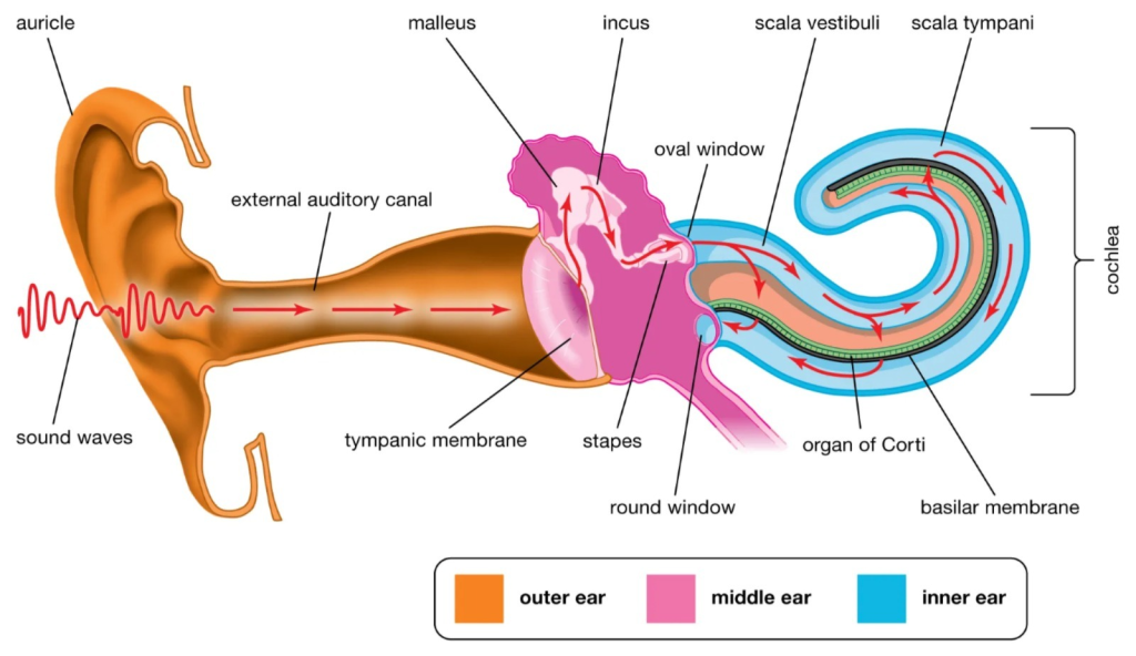
Structure-wise, the ear is anatomically divided into three parts
- External or Outer Ear
- Middle Ear
- Inner Ear
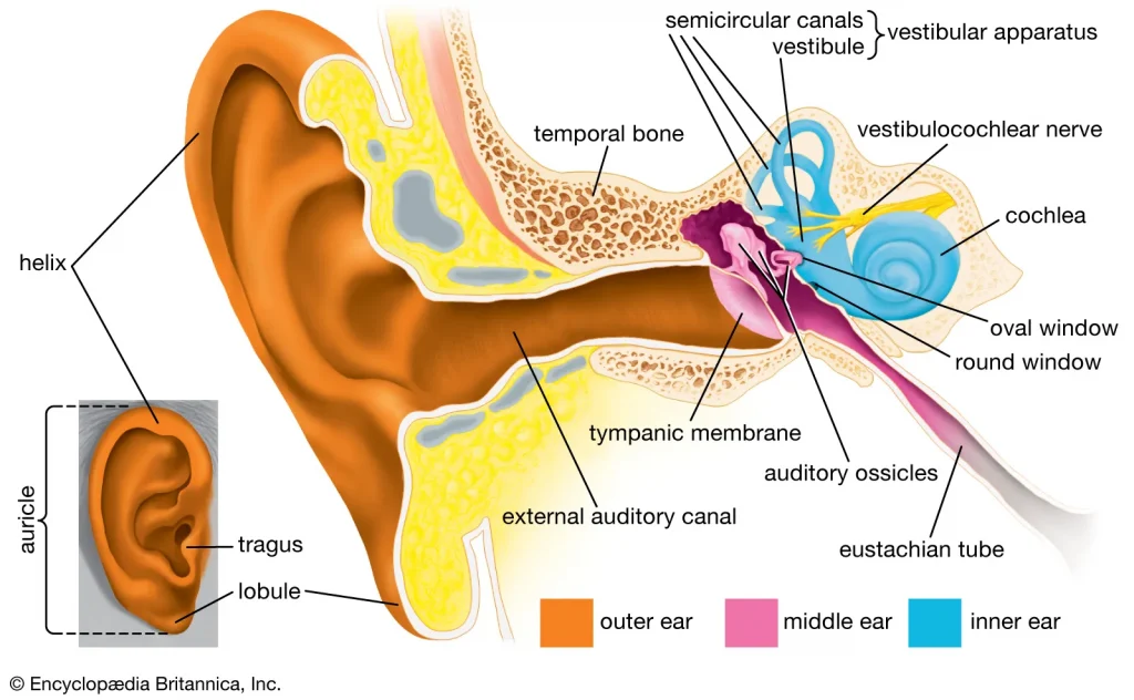
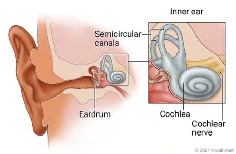
1.External or Outer Ear:
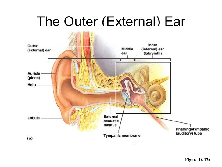
The external ear collects sound waves and transfers them to the inside. The external ear consists of the following parts.
Auricle or pinna (Auricle or Pinna):
External acoustic meatus i.e. auditory canal
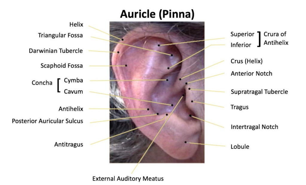
Auricle:
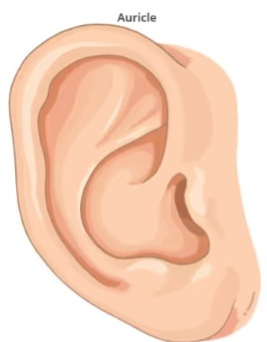
It is a flap-like part on both sides of the head, made of elastic cartilage and covered with skin on the outside. It collects sound waves coming from the outside environment and transmits them to the inner hole. Its uppermost part is called the helix and the soft part hanging below is called the lobule.
External acoustic meatus:

Also known as the auditory canal, it is a curved tube in the shape of an S. Its length It is about 2.5 cm long and is an elongated canal on the inside and the glands in its wall secrete a wax-like viscous fluid, also known as cerumen. Some hairs are also present in this canal which, combined with the viscous fluid, prevent dust and foreign bodies from reaching the inner wall, i.e. the tympanic membrane.
Tympanic membrane (tympanic membrane):
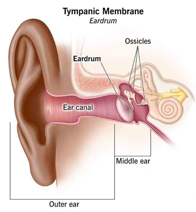
It is a thin membrane that separates the external ear and the middle ear and is oval in shape. This membrane is made up of epithelium tissue and connective tissue, which contains fibroblast cells.
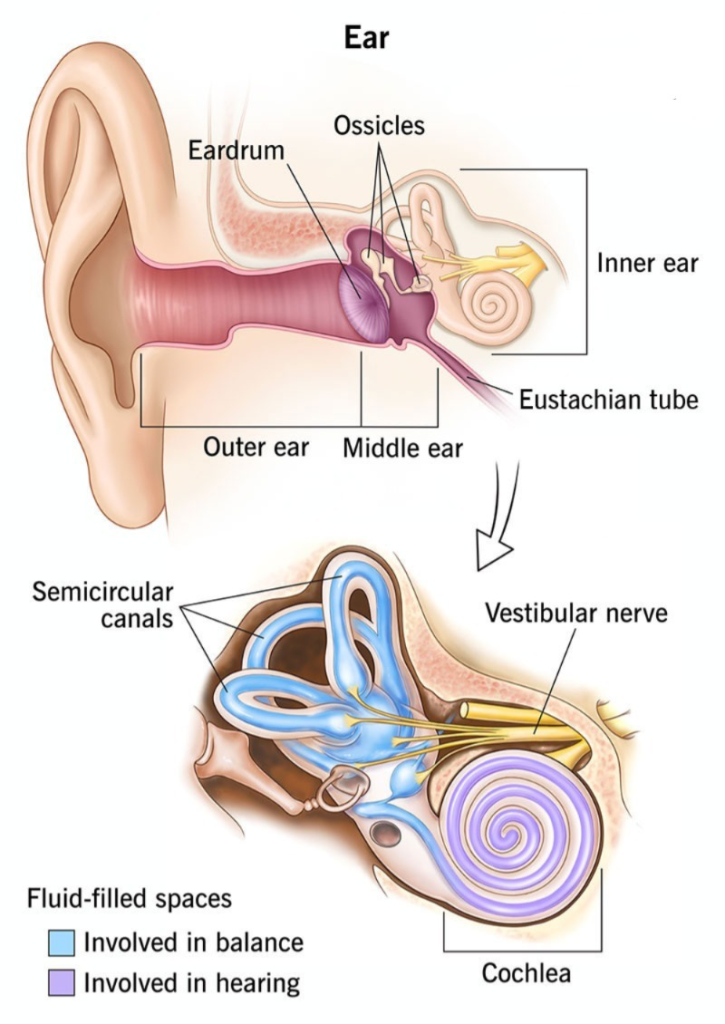
2. Middle ear:
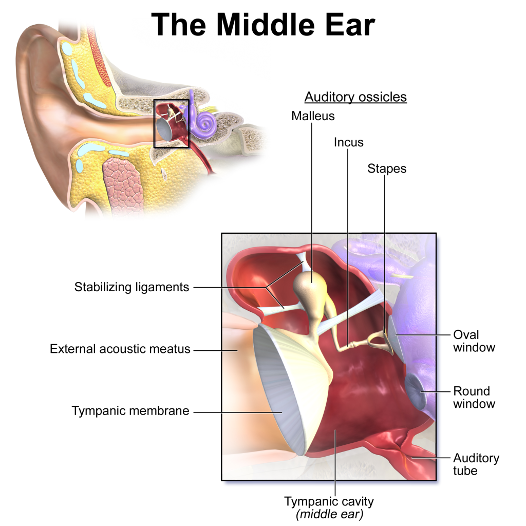
It is a small air-filled cavity located in the temporal bone. The lining of this cavity is made of epithelium tissue and it is called the tympanic membrane. The outer ear is separated from the middle ear by the membrane, or ear drum, and the inner ear is separated from the middle ear by the oval and round windows.
Its anterior wall has an opening called the Eustachian tube. This tube connects the middle ear to the structures of the nasal passages and maintains pressure within the tube cavity, which prevents the tympanic membrane from rupturing during yawning and swallowing.
Auditory ossicles:
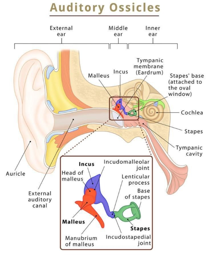
The bones in the middle ear are called auditory ossicles, which include the malleus, incus, and stapes bones. Their number is one in each ear, meaning there are a total of 6 auditory ossicles in the body.
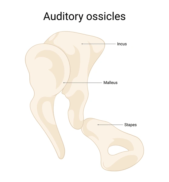
Malleus (Malus):
It is hammer-shaped and its handle is attached to the wall of the tympanic membrane. The head of the malleus is attached to the incus bone.
Incus (Incus):
It is the middle bone which is in the shape of an anvil, it is connected to the malleus bone and its head is connected to the stapes bone. The joint part has fibrous tissue.
Stapes (stapes):
It is a stirrup-shaped bone whose head is connected to the incus bone and its foot plate is attached to the oval window, which has a round window at its bottom.
Thus, from the tympanic membrane The structure between the oval window and the round window is called the middle ear.
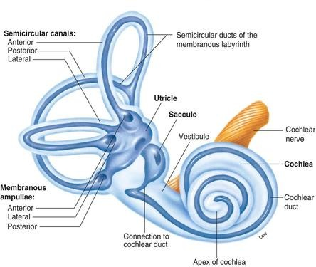
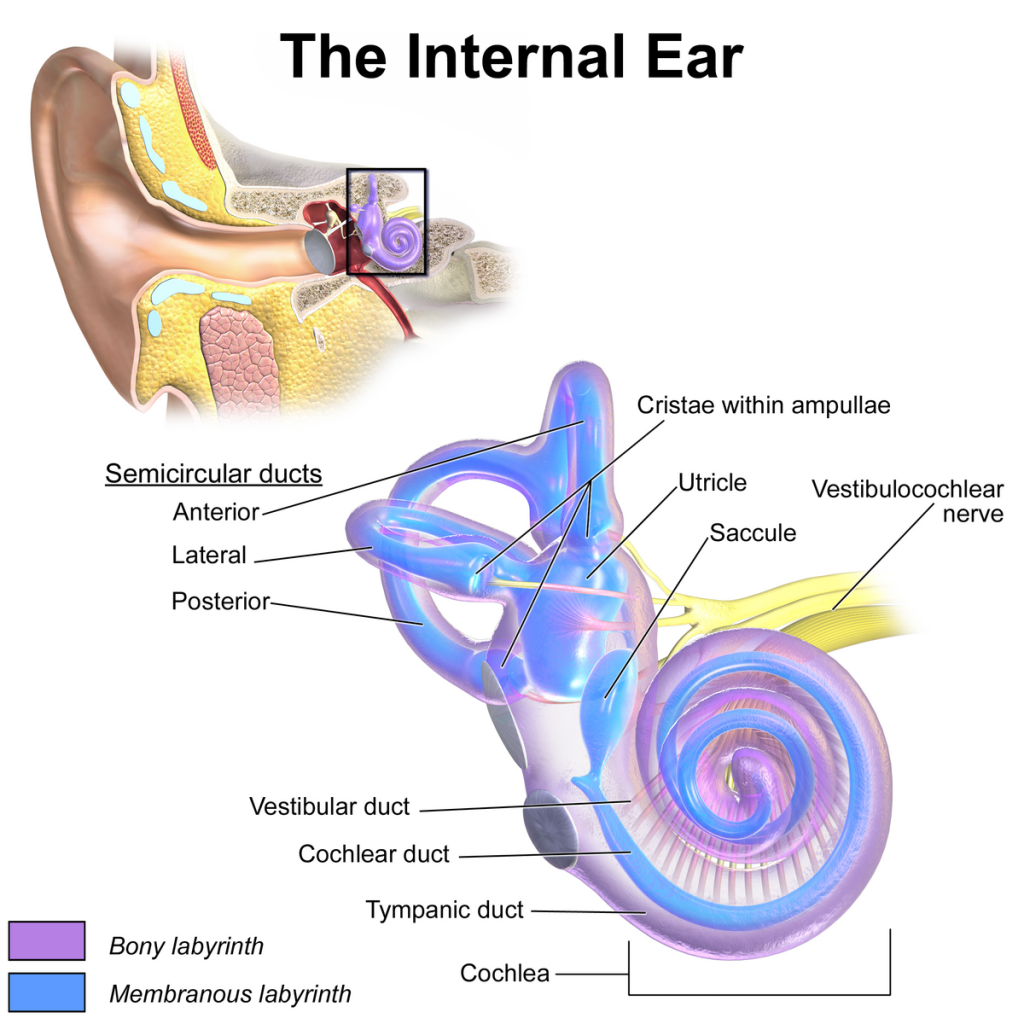
3.Inner ear:
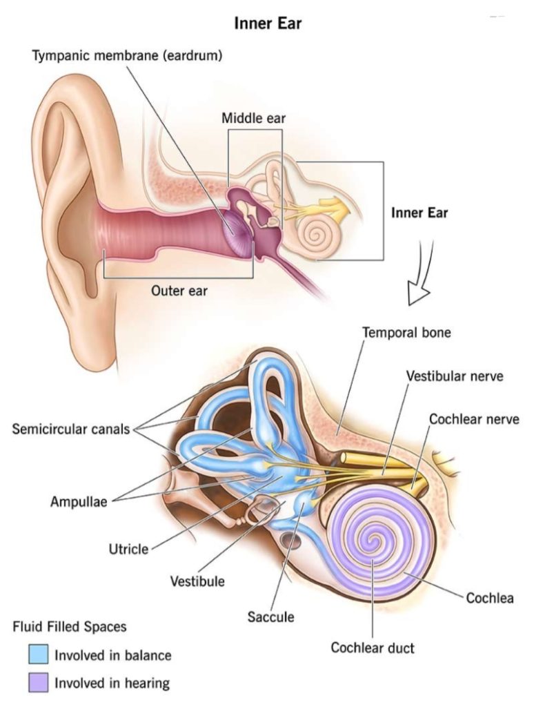
It is also known as a labyrinth. It has a complex structure and is involved in hearing and balance.
It has two parts: the bony labyrinth and the membranous labyrinth.
Bony Labyrinth:
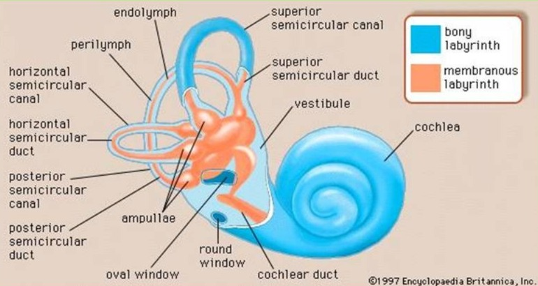
It is a cavity located in the petrous portion of the temporal bone, whose wall is made of a layer of periosteum. Inside it is a fluid called perilymph. It has three parts. A vestibule, a cochlea and three semicircular canals.
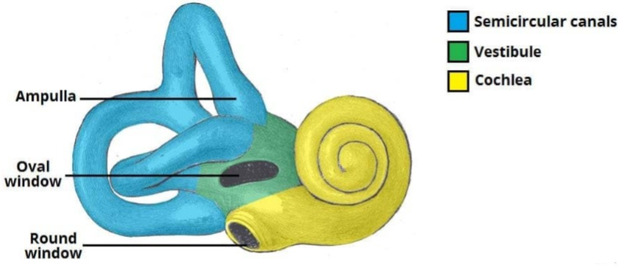
Vestibule:
It is the middle oval-shaped portion of the bony labyrinth, which contains the utricle and saccular structures, and its lateral walls have oval and round windows.
Cochlea:
This part of the bony labyrinth is associated with hearing, and is a ball-like structure also known as the Snail’s cell.
Semi Circular Canal:
The structure of this canal is associated with balance. The three tubes of the semicircular canal are arranged at right angles to each other.
Membreneous Labyrinth:
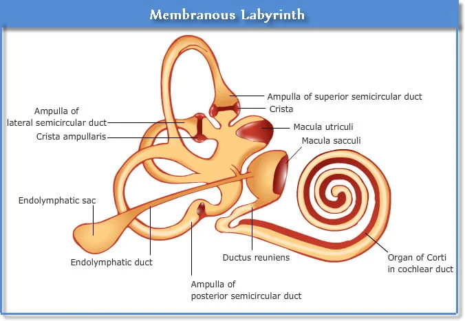
Inside the bony labyrinth is a membranous tube called the membranous labyrinth, which contains a fluid called endolymph. The properties of this fluid are similar to CSF.
1. Vestibule which contains the utricle and saccule.
2. Vestibule and semicircular canals.
- The vestibule and semicircular canals in the inner ear work to maintain the body’s balance.
- In which the semicircular canals, the cochlea, and the utricle help to maintain the body’s dynamic equilibrium. The semicircular canal is a tube-like structure that lies behind and above the vestibule and the canals are arranged at an angle to each other. The semicircular canal and the cochlea, whose outer wall is the bony labyrinth and inside it is the perilymph fluid, have another tube inside it called It is called the membranous labyrinth and contains endolymph.
- The utricle is a membranous sac that is part of the vestibule and all its ducts open into a dilated portion called the ampulla. The saccule is part of the vestibule and communicates with the utricle and cochlea.
- The walls of the utricle, saccule and ampulla contain small hair-like projections of specialized epithelial cells, which contain sensory nerve endings, forming the vestibular part, through which the vestibulocochlear nerve passes.
3. Cochlea
The cochlea, also known as the snail, is a coiled structure that resembles a snail. Inside the cochlea is a membrane called the cochlear duct, which is mainly involved in the process of hearing. The cochlea’s cross The following parts are found in the section.
- Scala Vestibuli (Scala vestibuli)
- Scala Media (or Cochlear Duct)
- Scala Tympani (Scala Tympani)
The cochlear duct is a triangular-shaped tube. The bony part of the cochlea is divided into two parts, upper and lower, in which the upper part is called the scala vestibuli and the lower part is called the scala tympani. In the middle part is the cochlear duct and its roof membrane is called the basilar membrane. The organ of Corti, also known as the hearing organ, is located on the basilar membrane.
Physiology of Hearing:

The physiology of hearing is the act of hearing. The wavelength of the waves for hearing is 20 to 20,000 Hz. The human ear has a capacity for frequencies between 500 and 5,000 Hz. The frequency at which sound waves vibrate is known as pitch. As the vibration increases, the pitch increases.
Every sound produces sound waves that strike the outer part of the auricle and from there enter through the external auditory canal. These sound waves vibrate the tympanic membrane, i.e. the ear drum, which is the junction between the external ear and the middle ear.
The tympanic membrane is connected to the melas bone. These waves go to the incus with the melas bone and the incus with the incus to the stapes and this stapes bone is further connected to the oval window. From the oval window, these sound waves reach the perilymph fluid which goes to the cochlea and from there to the endolymph and when the round window vibrates, it goes to the cerebrum through the vestibule cochlear nerve and the sound is recognized.
Balance and Ear…
Vestibule And Semi Circular Canal:
The vestibule and semicircular canals in the inner ear work to maintain the body’s balance. The semicircular canals, sacculus, and utricle help maintain the body’s dynamic equilibrium. The semicircular canals are tube-like structures that lie behind and above the vestibule, and these canals are arranged at an angle to each other. The semicircular canals and cochlea, whose outer wall is the bony labyrinth and inside it is the perilymph fluid, have another tube called the membranous labyrinth, which contains the endolymph fluid.
(Physiology of Equilibrium) Physiology of Equilibrium:
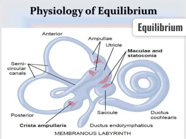
Introduction:
Equilibrium is the ability of the body to maintain balance. For this, the coordinated actions of specific sense organs and the nervous system in the body are responsible. There are two types of equilibrium:
1. Static Equilibrium: Maintaining position when the body is stationary.
2. Dynamic Equilibrium: Maintaining balance when the body is in motion.
Both of these processes involve the Vestibular Apparatus, Visual Input and is regulated by Proprioceptive Feedback.
Vestibular Apparatus:
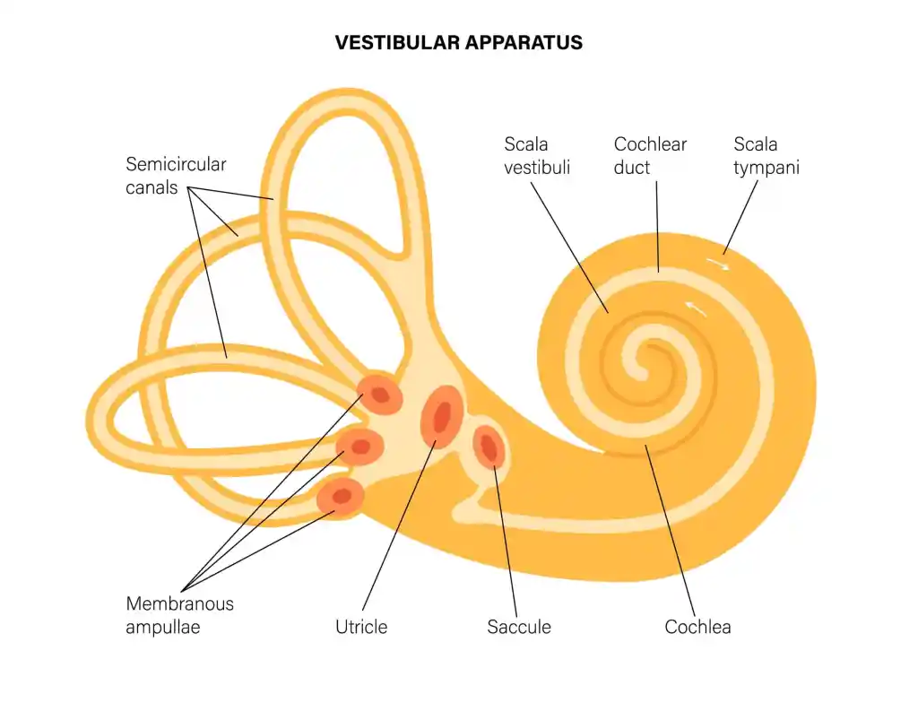
The vestibular apparatus is located in the internal part of the ear, and its main function is to maintain the body’s equilibrium. It is divided into the following components:
1. Semicircular Canals:
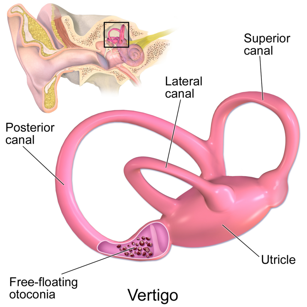
- A tube-like structure with three axes – Anterior, Posterior, and Lateral.
- Each canal begins with an ampulla containing a crista ampullaris.
- The crista ampullaris contains hair cells.
- The movement of endolymph occurs when the head moves. It gives pressure to the hair cells.
- This information reaches the brain through the Vestibulocochlear Nerve (CN VIII).
- These components are responsible for dynamic equilibrium.
2. Utricle and Saccule:
- Both have a sensory area called the macula.
- The otolith membrane above the hair cells (Otolith Membrane) which contains Otoliths (Otoliths – Calcium Carbonate Crystals).
- When the head is in movement, the change in the position of the otoliths puts pressure on the hair cells.
- This pressure informs the brain about static position and linear motion.
- The utricle and saccule are responsible for static equilibrium.
Visual Input:
- Visual information from the eyes is also important in maintaining equilibrium.
- If vision is blocked, it becomes difficult to maintain the body’s balance, which shows that the visual system also participates in equilibrium.
Proprioceptive Feedback:
- Sensory information from muscles, tendons, and joints reaches the brain through proprioceptors.
- This information helps the brain know the position of the body’s organs.
Nerve Pathways and Brain Integration:
- Vestibular information travels to the brainstem via the Vestibulocochlear Nerve.
- It then reaches the Cerebellum, Medulla Oblongata, and Cerebral Cortex.
- This coordination allows the body’s position and Motion is controlled.
The physiology of equilibrium is a very complicated but coordinated process in which information received from the vestibular apparatus, eyes, and proprioceptive system reaches the brain, is processed by the brain, and the body’s balance is maintained by obtaining the appropriate motor response.