—ENGLISH-ANATOMY UNIT 11. NERVOUS SYSTEM (PART : 1) CENTRAL NERVOUS SYSTEM
INTRODUCTION (Introduction):
- The nervous system and the endocrine system work together to perform important functions in our body.
- It works to maintain homeostasis in our body and allows us to survive.
- The nervous system recognizes changes in our body and responds to those changes.
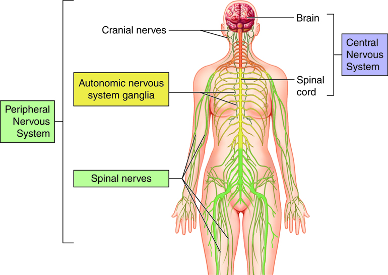
ORGANIZATION OF NERVOUS SYSTEM
- NERVOUS SYSTEM (nervous system)
CENTRAL NERVOUS SYSTEM
- BRAIN AND SPINAL CORD
PERIPHERAL NERVOUS SYSTEM
- AFFERENT ( SENSORY ) NERVE
- EFFERENT ( MOTOR ) NERVE
- SOMATIC ( MOTOR ) NERVE
AUTONOMIC NERVOUS SYSTEM
SYMPATHETIC (Sympathetic)
PARASYMPATHETIC (parasympathetic)
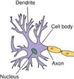
NEURONE (neurons):
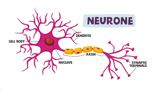
The brain contains a large number (100 billion) of neurons is.
Each neuron has the following characteristics
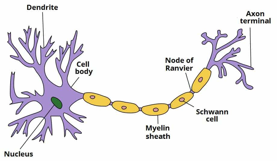
Cell Body And Its Process
Axone
Dendrites (Dendrites)
Some thread-like nerve fibers may also be present.
- Neurons cannot divide and require constant oxygen for their survival. It gets its energy from glucose.
- Neurons have the properties of conductivity and excitability, which allow them to respond to stimuli from the external environment, which can be mechanical, electrical, and chemical.
- These stimuli travel from the dendrites of the nerve cell to the cell and axon, which is called forward conduction.
Cell Body:
- Nerve cells vary in size and shape that can be seen with the naked eye. No.
- The part of the body of a nerve cell that forms gray matter is located in the periphery of the brain and the middle of the spinal cord.
- The cell body, like any other cell, contains a nucleus, cytoplasm, and other organelles.
- The body of a nerve cell is made up of nuclei in the central nervous system and ganglia in the peripheral nervous system.
Axone and Dendrites :-
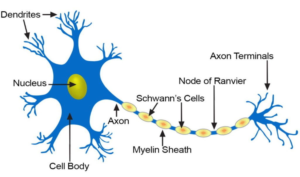
- They are processes extending from the cell body. Each neuron has an axon and a large number of dendrites called nerve fibers. Each nerve contains bundles of sensory and motor nerve fibers.
- axons and dendrites make up white matter. Which is in the central part of the brain and in the peripheral part of the spinal cord.
Structure of Axon:

- An axon is a process extending from the cell body. A neuron has one axon. An axon is a thin, cylindrical process. Its length can be 100 cm.
- The mass of an axon is called a track.
- Axons also have organelles similar to the organelles of the cell. The liquid part inside it is called axoplasm and the membrane surrounding it is called axolymph.
- Schwann Cells are present in the peripheral nervous system and surround the axon.
- The small gap between Schwann Cells is called the Node of Ranvier. These nodes and myelin sheaths are essential for proper nerve conduction along nerve fibers.
- The membrane surrounding the schwann cell is called the neurilemma.
- Axons of neurons are surrounded by a multilayered sheath of lipids and proteins called the myelin sheath.
- A neuron that has such a sheath around its axon is called a myelinated neuron and one that does not have a sheath is called non-myelinated or Unmyelinated neuron is called.
- Unmyelinated neurons have poor nerve transmission.
Dendrites:
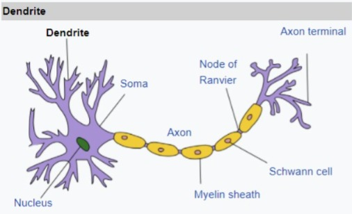
- They are branches extending from the cell body. Which carry impulses towards the cell body of the neuron. Dendrites are not myelinated.
SYNAPSE AND NEUROTRANSMITTERS:
- The bulb-shaped structure at the end of the axon of one neuron is called a synaptic knob. The raised area in the dendrites of the cell body of another neuron is called a vesicle.
- Both of the above structures have a sac-like structure called synaptic vesicles which contain chemical substances called neurotransmitters. Here the transmission of impulses takes place which is called synapse.
- Noradrenaline, gamma amino butyric acid (GABA), acetyl choline, dopamine, serotonin etc. act as neurotransmitters.
- The above Neurotransmitters help in the conduction of nerve impulses.
NERVES (nerves):
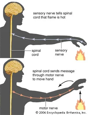
Sensory or afferent Nerves:-

These carry sensory impulses from the skin, sense organs, muscles, joints and visceral organs to the central nervous system via the spinal cord. In which the following areas are found.
1. Somatic cutaneous which conveys impulses of sensations such as pain, temperature, touch, vibration etc.
2. Special senses which convey impulses such as taste, smell etc.
3. Proprioceptors are special senses such as vision, hearing, balance, etc. that transmit impulses through cranial nerves.
MOTOR OR EFFERENT NERVES:-
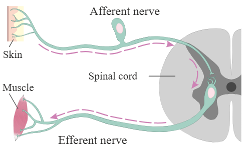
Which conveys impulses from the central nervous system to the effector organ, muscle or gland.
1. Somatic nerves provide impulses to control the contraction of skeletal muscles.
2. Autonomic nerves (sympathetic and parasympathetic) which control the contraction of smooth muscles, cardiac muscles, glands through cranial and spinal nerves.
MIXED NERVES (Mixed Nerves) :-
The spinal cord contains sensory and motor nerves, while elsewhere in the body they are separated by connective tissue. It is called mixed nerve.
NEUROGLIA (Neuroglia):
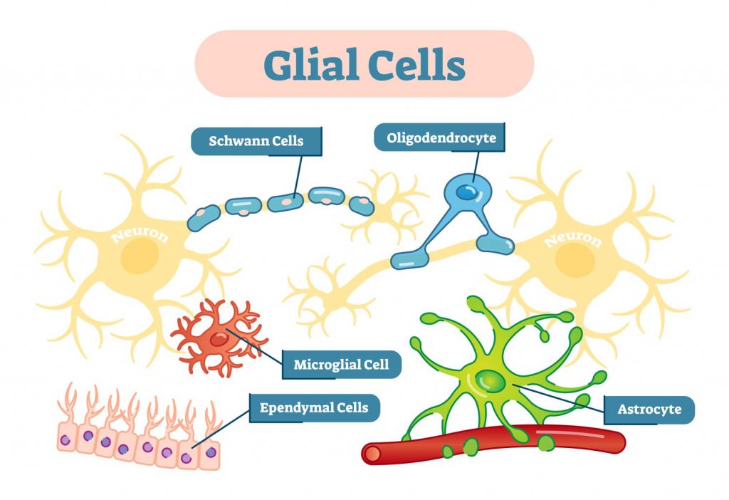
They are the support cells of the nervous system. They are generally smaller than neurons. They have the capacity to multiply and divide in the mature nervous system. They are supported by four types of non-excitable cells: astrocytes, oligodendrocytes, microglia, and ependymal cells. They perform different functions in the nervous system.
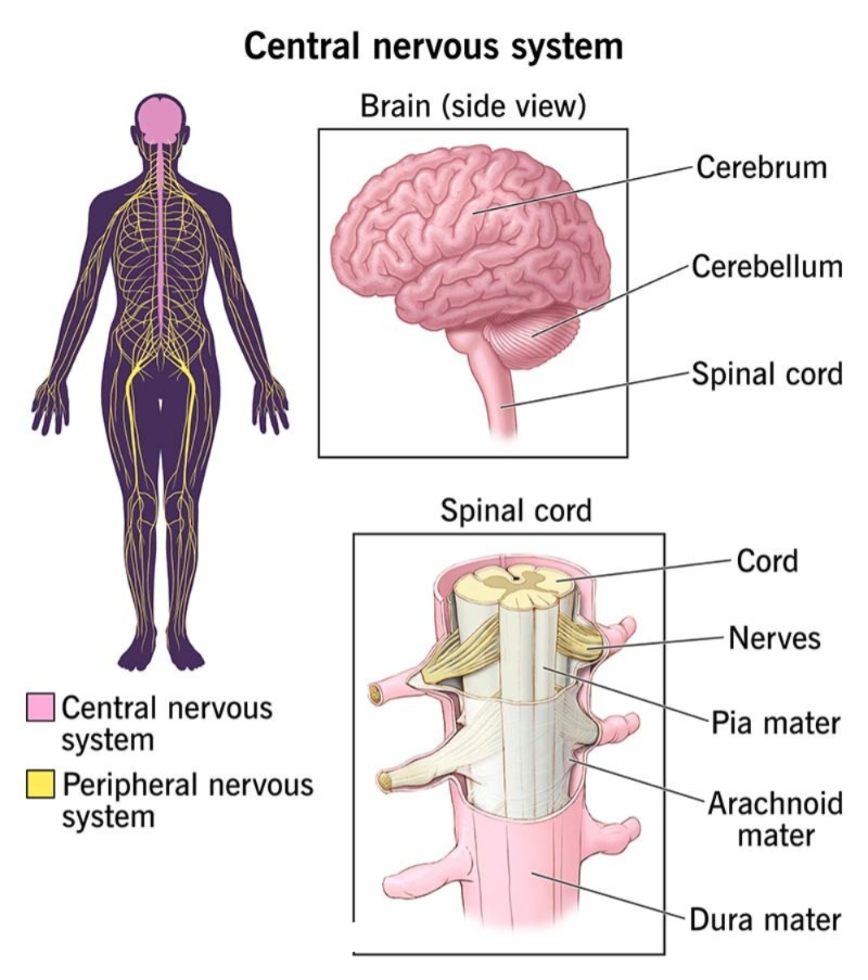
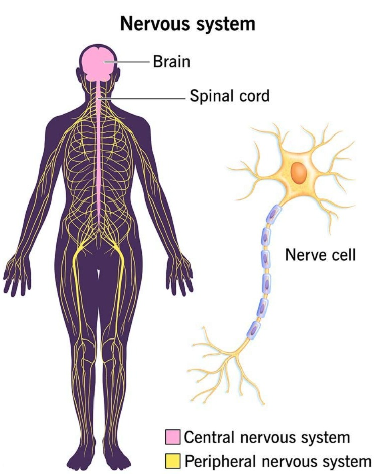
CENTRAL NERVOUS SYSTEM:

•The central nervous system includes the brain and spinal cord.
MENINGIES (Meninges):
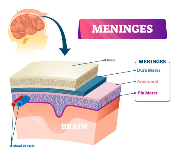
The brain and spinal cord are wrapped in a layer called the meninges, which works to protect the delicate nerve structures. There are three layers of meninges
1. Dura mater
2. Arachnoid mater
3.Piamater
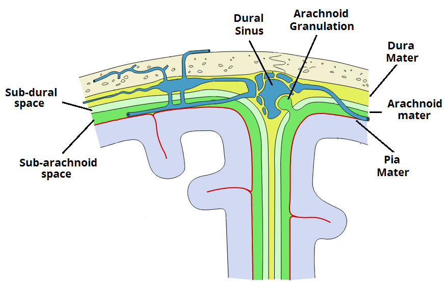
Dura mater:
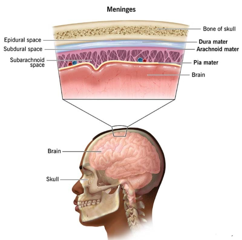
- The dura mater is the outermost layer of the brain and spinal cord. The lateral layer is the dura mater. The dura mater is dense and tough It is arranged in a double layer. The outer layer forms the outer lining. The inner layer is attached to it except where the dura mater forms partitions in some places.
- Such as the fovea cerebri which is located between the two cerebral hemispheres which is located on the upper side which is the superior longitudinal fissure where the sagittal sinus is located which receives the venous blood of the brain. Tentorium cerebelli which separates the cerebrum and cerebellum.
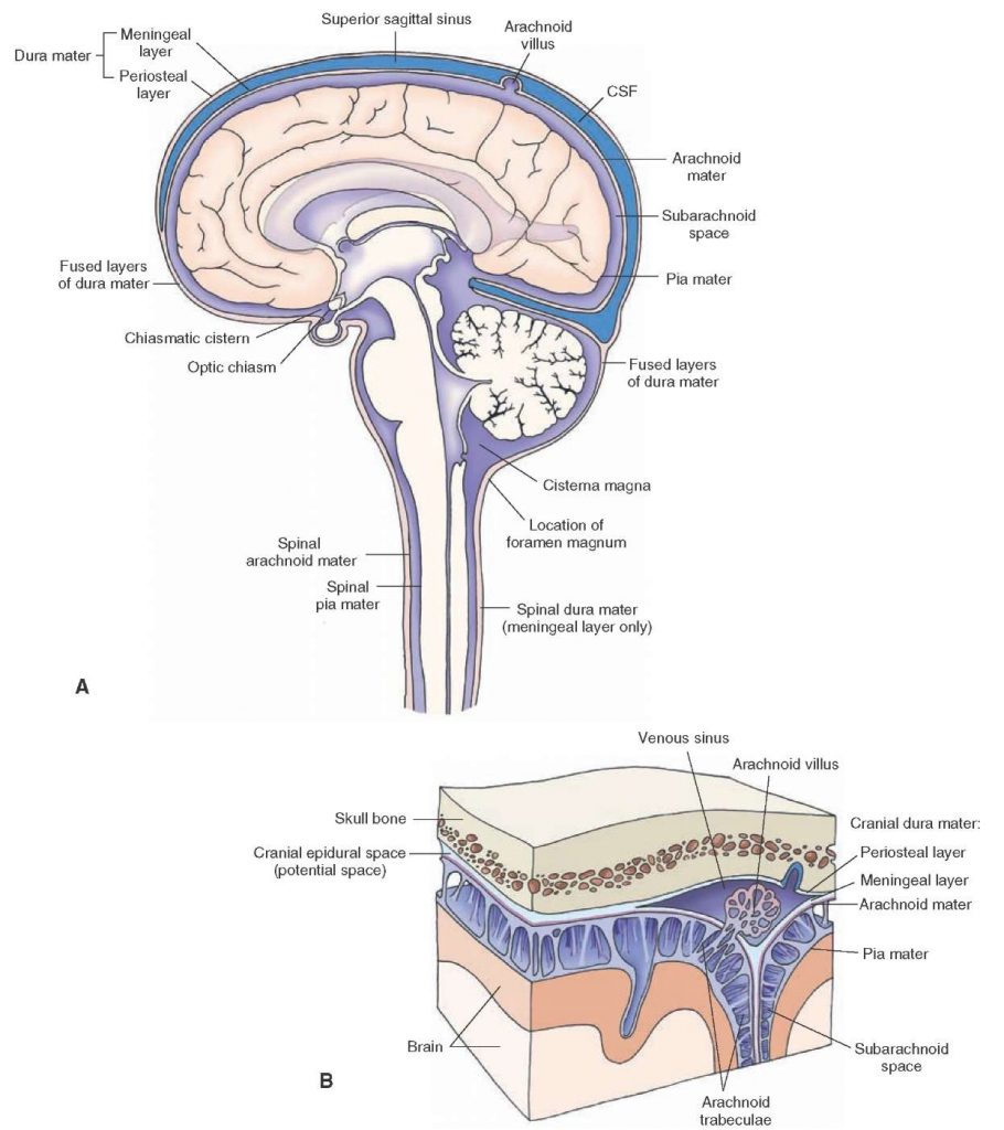
Arachnoid mater:
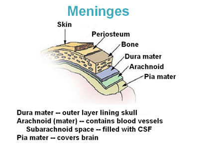
- The delicate serous membrane that lies between the dura mater and the pia mater is Contains collagen fibers and elastic fibers. The space between the skull and the dura mater is called the epidural space.
- The space below the dura mater is called the subdural space.
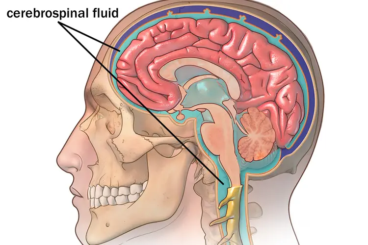
- The space between the arachnoid mater and the pia mater is called the subarachnoid space where the cerebrospinal fluid is located.
Basic Matter/Pia Mater (Pia Mater):
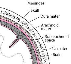
- It is the innermost layer. The thin and vascular membrane that surrounds the spinal cord contains a large number of collagen fibers and fine elastic fibers. It also contains many blood vessels that supply nutrition and oxygen to the spinal cord. Its end is called the filum terminale.
VENTRICLES OF THE BRAIN:
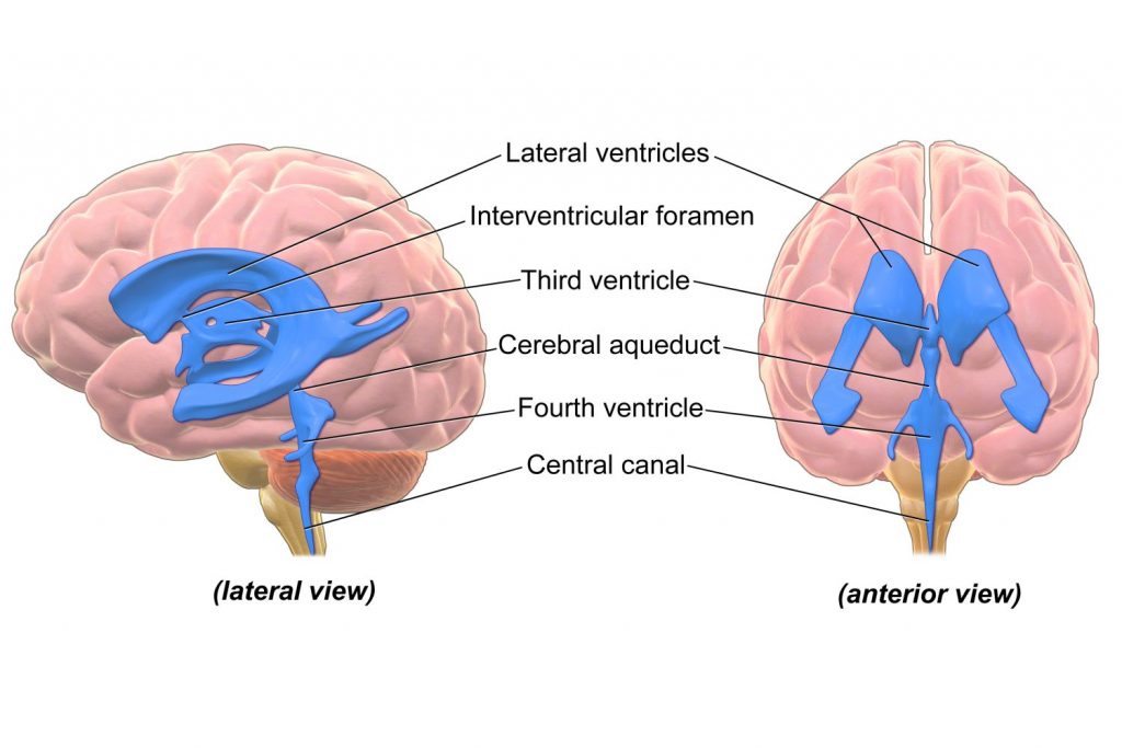
The cavities inside the brain are called ventricles, in which fluid is produced and circulates around the brain and spinal cord. These ventricles are as follows.
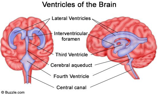
1.Right And Left Ventricles/Lateral Ventricles
2.Third Ventricles
3.Fourth Ventricles
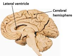
1.Lateral Ventricles:
This cavity is located below the corpus callosum of both cerebral hemispheres. There is a septum lucidum that separates it. Its lining is made of epithelial tissue. Its wall contains a network of capillaries called the choroid plexus from which the cerebrospinal fluid is produced. It connects to the third ventricle through the interventricular foramen.
2.Third Ventricles:
The narrow cavity below the right and left lateral ventricles is called the third ventricle. It connects to the fourth ventricle via the cerebral aqueduct
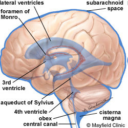
3.Fourth Ventricles:
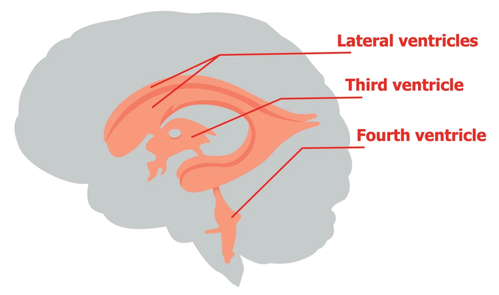
It is diamond-shaped, located below the third ventricle, and is the center of the spinal cord. Continuous with the canal, the foramen (Lashka and Magendi) on its roof are connected to the subarachnoid space.
CEREBRO SPINAL FLUID (Cerebro Spinal Fluid):
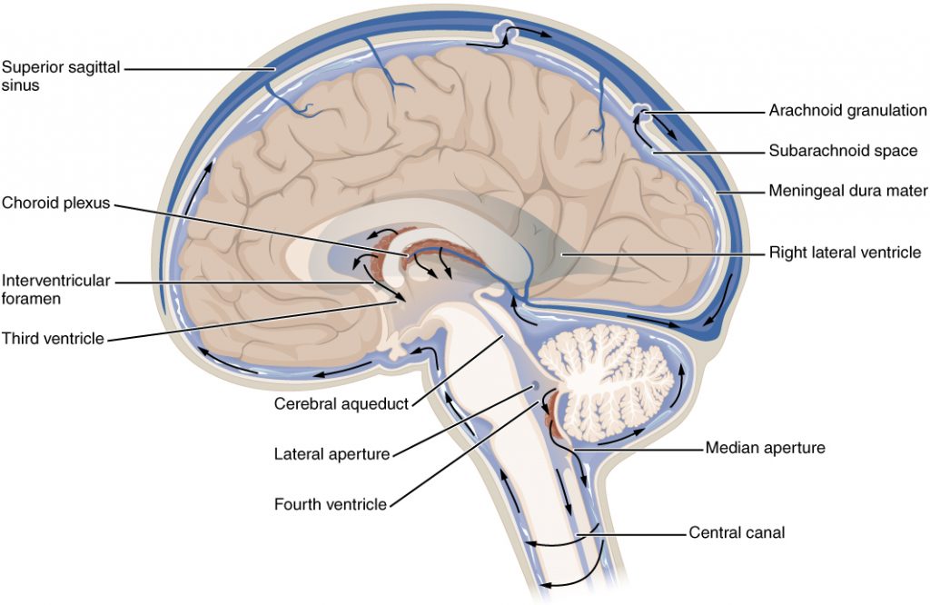
Cerebrospinal fluid is produced by the choroid plexus in the wall of the ventricle. The choroid plexus is a network of capillaries located in the wall of the lateral ventricle. Cerebrospinal fluid from this lateral ventricle passes through the interventricular foramen of Munro into the third ventricle and from there into the fourth ventricle through the cerebral aqueduct. From there, some fluid passes into the central canal of the spinal cord and some fluid passes through the foramen of Lascaux and Magendie and circulates in the subarachnoid space.
The fluid circulating in the subarachnoid space is absorbed into the blood through the arachnoid membrane. CSF is formed at a rate of 20 ml per hour, i.e. 480 ml per day, and is also absorbed at the same rate.
- CSF pressure can be measured using a lumbar puncture needle which is 10 cm h2o long position and 30 cm h2o in the sitting position.
- CSF is a clear fluid that is slightly alkaline and has a specific gravity of 1.005
- Its composition includes water, mineral salts, glucose, plasma proteins, some albumin and globulin, creatinine, urea, and some leukocytes.
FUNCTIONS OF CSF (CSF Functions):
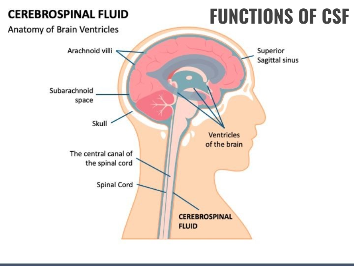
1. Mechanical protection in which it acts as a shock absorbing medium and provides protection to the delicate structures of the brain.
2. Chemical protection in which CSF provides an optimal chemical environment for neural signals.
3. Circulation in which CSF serves as a medium for the exchange of nutrients and waste products between nervous tissue and blood.
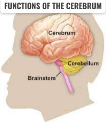
BRAIN:
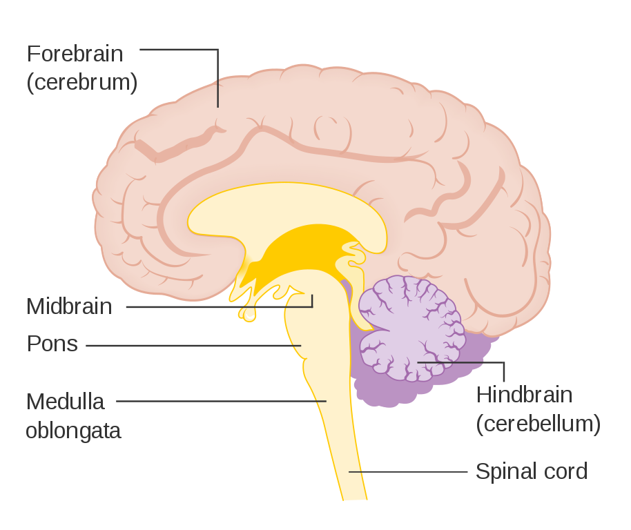
- The normal weight of the brain is 1200 to 1400 grams
- It is located in the cranial cavity
- Its parts are as follows
- Cerebrum
- Midbrain
- Ponse Veroli
- Medulla oblongata
- Cerebellum
- The blood supply to the brain is provided by anastomoses of many arteries of the Circle of Wills occurs from.
CEREBRUM :
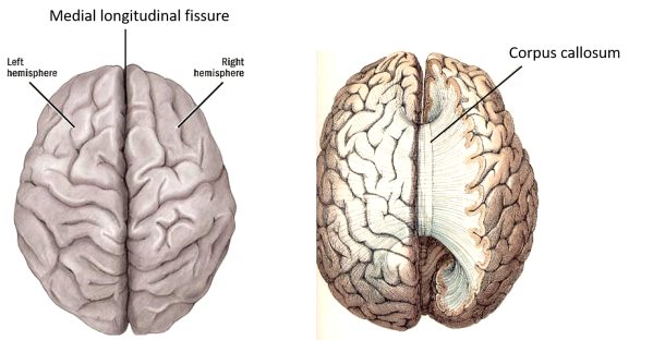
- The cerebrum is made up of the right and left cerebral hemispheres.
- The two cerebral hemispheres are connected by the corpus callosum. Each hemisphere has a cavity called the lateral ventricle.
- The superficial part of the cerebrum is made up of gray matter, which is called the cerebral cortex. White matter is found in the deep part of the cerebrum.
- The cerebral cortex contains raised areas called gyri or convolutions and is separated by grooves called fissures or sulci.
- The deepest of these fissures is the longitudinal fissure, which separates the right and left hemispheres.
The hemispheres of the cerebrum are divided into different lobes, which are as follows.
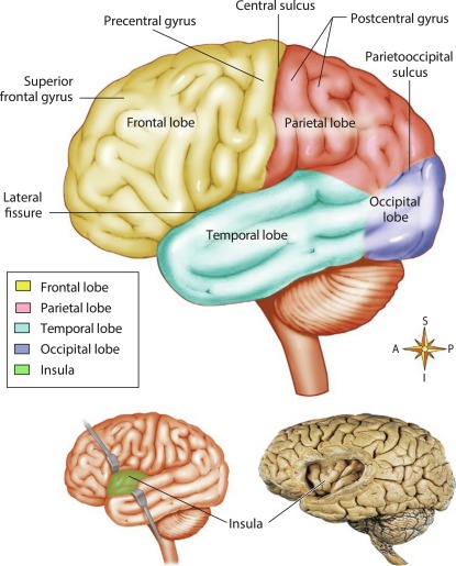
•Frontal Lobe
•Parietal Lobe
•Temporal Lobe
•Occipital Lob.
•Important fissure on the side of the cerebrum;
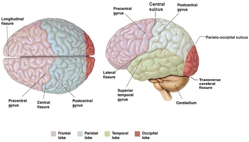
•Longitudinal sulcus or fissure which is the deepest and divides the cerebrum into two hemispheres
•Central sulcus which is located between the frontal and parietal lobes.
•Lateral sulcus which is a deep groove and separates the temporal lobe from the frontal and parietal lobes.
•parieto-occipital sulcus which separates the occipital lobe from the two parietal lobes….
•The inner part of the cerebrum is lined with nerve fiber tracks such as
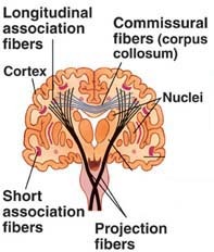
•Association fibers
•Commissural Fibers
•Projection Fibers
These fibers are connected to each other and connect one area to another and help in the transmission of impulses.
FUNCTIONS OF THE CEREBRUM:
1. The cerebrum controls intelligence, memory, reasoning, thinking, speaking, reading, writing, etc.
2. Sensory perception such as pain, temperature, touch, sight, There is no control for the perception of hearing, taste, smell etc.
3. Control for the contraction of skeletal muscles is found in this area.
Functional areas of the Cerebrum (areas of the Cerebrum):
There are many areas in the hemisphere of the cerebrum which are as follows.
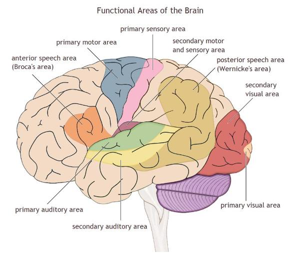
Motor Area:
Which is mainly located in the front part of the cerebral hemisphere. In which the following areas are found.
1. Primary motor area which is located in the frontal lobe and controls voluntary muscle contraction.
2. Motor speech area which is also called Broca’s area which is located in the frontal lobe.
Sensory Areas of Cerebrum:
• Primary somatosensory area or general The area located in the parietal lobe behind the central sulcus is the sensory area for touch, pain, and temperature.
•The primary visual area located in the occipital lobe interprets vision.
•The primary auditory area located in the temporal lobe and is associated with hearing.
•The primary gustatory area located in the parietal cortex behind the central sulcus is associated with taste.
•The primary The olfactory area is located in the temporal lobe. The area associated with smell.
OTHER AREAS OF CEREBRUM:
There are some special areas in the cerebrum which are involved in the transmission of impulses and other important functions which are as follows.
1.Basal ganglia. It is an area in each cerebral hemisphere which works to coordinate muscle tone, posture and voluntary muscle movement.
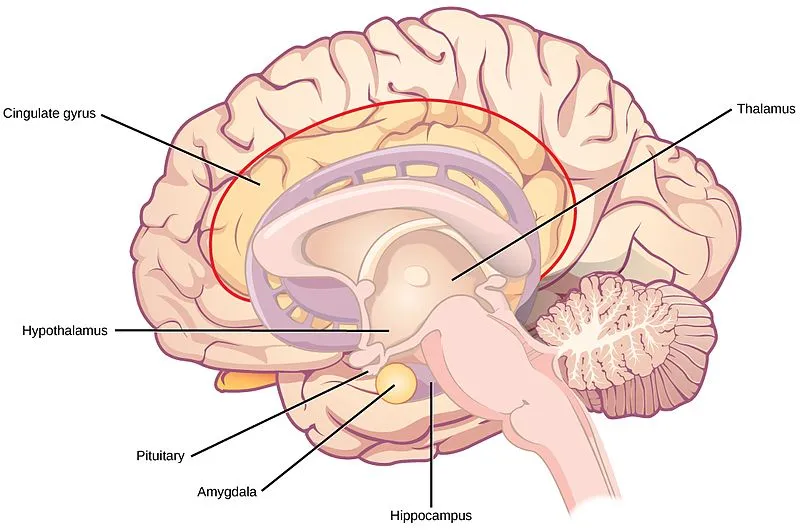
2. Thalamus. It is located in the upper part of the brain stem in the middle of the brain. It acts as a relay station for sensory impulses coming towards the sensory areas of the cerebral cortex, which allows the interpretation of proper sensations and some movement control.
3. Hypothalamus. The hypothalamus is made up of many groups of nerve cells located below the thalamus, whose functions are as follows.
•The hypothalamus controls the following functions.
•Body temperature
•Hunger and thirst
•Emotional reactions
•Autonomic nervous system
•Sexual Behavior
•Biological Clock or Circadian Rhythm
•Secretion of Certain Hormones
All of the above are done by the hypothalamus.
BRAIN STEM (Brain Stem):
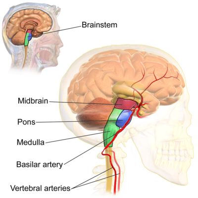
The following parts are included in the brain stem
1.Mid Brain (Mid Brain)
2. Pons Veroli
3.Medulla oblongata
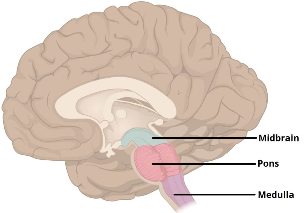
It forms the lowest part of the brain stem and connects the pons Veroli to the spinal cord
It is approximately 2.5 cm long and contains the following centers.
1.Respiratory center
2.Cardiovascular center
3.Vasomotor center
4.Reflex centers for vomiting, coughing, and swallowing.
Some special functions are found in the medulla oblongata which are as follows
1. The descending motor pathway crosses from the medulla and passes into the spinal cord which mainly provides impulses to the skeletal muscles.
2. Like the motor pathway, the sensory pathway also crosses from the medulla and passes towards the brain.
3. The medulla contains the cardiovascular center which controls the rate and force of the heart. Sympathetic stimulation increases the heart rate and force while parasympathetic stimulation decreases the heart rate and force.
4. The medulla contains the respiratory center which controls the rate and depth of respiration, in which inspiration and expiration occur when nerve impulses reach the intercostal muscles and diaphragm.
5. The medulla contains the vasomotor center which controls the diameter of blood vessels, causing vasoconstriction and vasodilation.
6. The reflex center in the medulla controls vomiting, coughing and hiccups, which is also a protective response.
CEREBELLUM (Cerebellum):
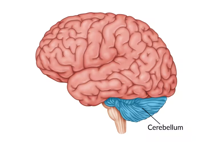
It is the second largest area of the brain, located posterior to the medulla and pons veroli
It is separated from the cerebrum by the transverse fissure, where the inner layer of the dura mater intrudes to form the tentorium cerebelli.
•The cerebellum performs the following functions
1. It regulates posture and postural activity
2. It controls the movement of the muscles Plays an important role in coordination
3. Plays an important role in maintaining body balance.
SPINAL CORD (Spinal Cord):

- It starts from the medulla.
- It passes through the foramen magnum and is continuous to the first and second lumbar vertebrae.
- It is about 45 cm long is.
- The spinal cord acts as a connection between the brain and body parts.
- Sensory impulses are transmitted to the brain and motor impulses are transmitted to the spinal cord From the cord to different parts of the body.
- Some activities are individualized through the spinal cord which are not required for the functioning of the brain. After the completion of the function, the brain becomes aware of them. Which action is completed through spinal reflexes.
- For the action of spinal reflex, sensory and motor neurons in the spinal cord are connected through a connecting neuron which is found in different levels of the cord.
- The spinal cord is incompletely divided into two equal parts in which the anterior median fissure is seen in the front and the posterior median septum is seen in the back. is found.
- In the spinal cord, gray matter is found in the middle part and white matter is found in the periphery.
- The spinal cord also has layers of meninges, just like the brain.
Gray Mater (Gray Matter):
- Spinal H-shaped gray matter is found in the middle of the cord. This H-shaped gray matter has two posterior, two anterior and two lateral columns.
- The central canal, which originates from the fourth ventricle and contains cerebrospinal fluid, is also arranged in between.
- The gray matter contains sensory neurons, motor neurons and connecting neurons.
White Matter:
- The white matter in the spinal cord is located in the periphery
- Which is divided into anterior, lateral and posterior columns. These columns also form tracks through sensory neurons, motor neurons, and connecting neurons.
FUNCTIONS (Functions):
1. The spinal cord transmits sensory impulses to the brain where they are interpreted.
2. Motor impulses coming from the brain pass through the spinal cord to different parts of the body.
3. Reflex arches are formed by the spinal cord, which allows for instant action, which reduces the workload of the brain.
Reflex action:
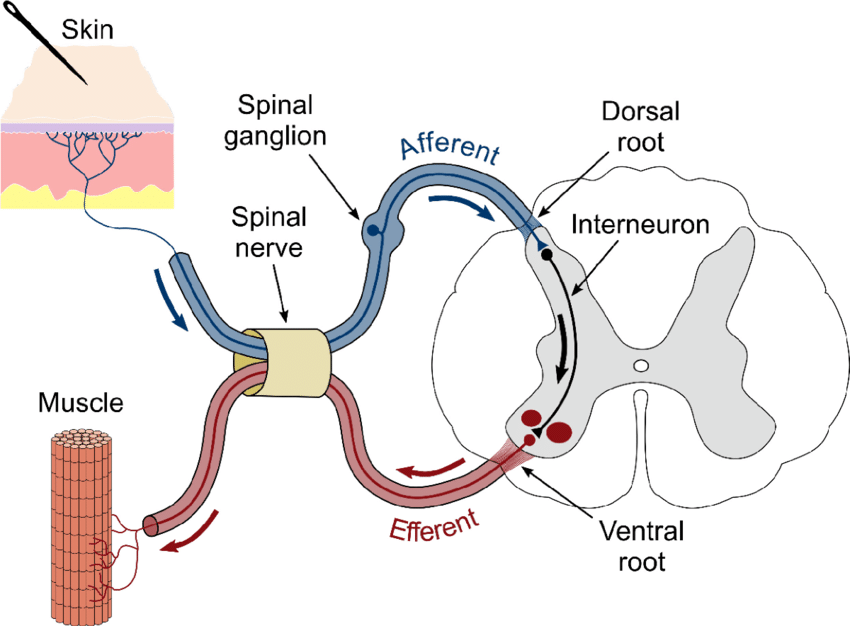
- The spinal cord works as a connection between the brain and the body parts. The length of the spinal cord is about 45 cm.
- It conveys sensory impulses to the brain and motor impulses go from the spinal cord to different parts of the body.
- Some activities are individualized through the spinal cord which do not require the brain to function. After the function is completed, the brain becomes aware of them.
- Which action is completed through spinal reflexes. This is called a reflex action.
- For the action of spinal reflex, sensory and motor neurons in the spinal cord are connected by connecting neurons which are found at different levels in the cord.
- The spinal cord forms a reflex arch which allows for rapid action which reduces the workload of the brain.