—ENGLISH-ANATOMY UNIT 7. DIGESTIVE SYSTEM. ( PART :2 ) Pancreas, Liver, Gall bladder
Pancreas :
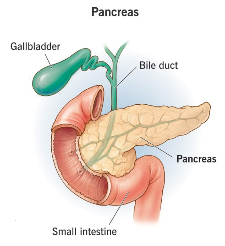
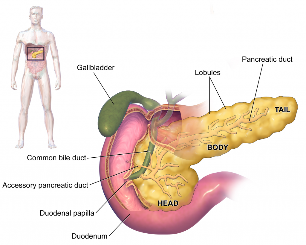
The pancreas is a gray and pink colored gland. It is 6 to 9 inches long. It weighs about 60 grams. It is located in the abdominal cavity. Its shape is fish-shaped, in which its head and neck are found in the C-shaped curved part of the duodenum. Its body part extends horizontally to the supine and its tail part touches the supine part. The abdominal aorta and inferior vena cava are found posterior to the pancreas.
The pancreas gland is an exocrine gland that functions both as an exocrine and endocrine gland.
Exocrine pancreas:
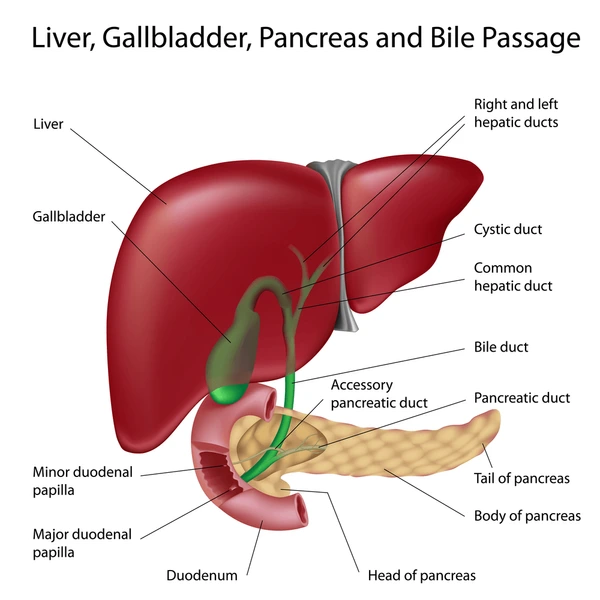
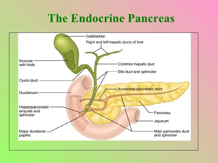
- This part of the exocrine pancreas is made up of many lobules. Secretory cells are located inside these lobules. These secretory cells secrete their secretions into small ducts and these small ducts join to form a large duct called the pancreatic duct. It carries the secretions of the pancreas to the small intestine.
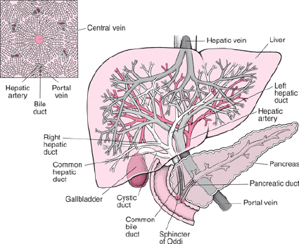
- This pancreatic duct connects to the common bile duct coming from the liver and gall bladder and opens into the duodenum of the small intestine through the hepatopancreatic ampulla and discharges the secretions there. This hepatopancreatic ampulla has sphincter muscles that control the flow of secretions. These sphincter muscles are also known as the sphincter of Oddi.
- The exocrine part of the pancreas secretes pancreatic juice which helps in the digestion and absorption of carbohydrates, fats and proteins.
Endocrine pancreas:
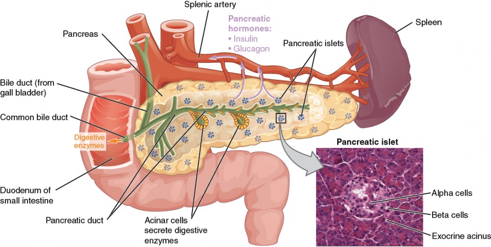
- One percent of the total cells in the pancreas function as the endocrine pancreas. These cells are arranged in clusters and these clusters are also known as pancreatic islets or islets of Langerhans, which are called alpha, beta and delta cells.
- These cells in the pancreas secrete hormones that secrete glucagon, insulin and somatostatin hormones respectively. This part of the pancreas functions as the endocrine pancreas. This hormone maintains the blood glucose level in the body.
- The pancreas secretes hormones that perform endocrine functions.
- The pancreas secretes juices that help in the digestion of proteins, fats, and carbohydrates.
- Thus, the pancreas performs both endocrine and exocrine functions.
Liver:
The liver is the largest gland in the body. It is located in the right quadrant of the abdominal cavity, below the diaphragm. It weighs approximately 1.4 kilograms and is found inside an adult. It is located under the ribs of the chest. The ribs protect it. Some of it is also located in the region of the left abdominal cavity.
The upper surface of the lever is smooth. This part is attached to the diaphragm and has an irregular surface and margin behind and below the liver.
Organs surrounding the liver:
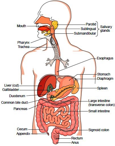
- The organs such as the diaphragm, anterior abdominal wall muscles, stomach, duodenum, kidneys, inferior vena cava, gallbladder, etc. are arranged around the liver.
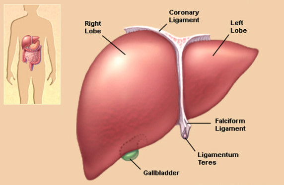
- The liver is mainly divided into two lobes, the right lobe and the left lobe.
- The right lobe is larger than the left lobe and the two lobes are separated by the falciform ligament.
- The quadrate lobe is seen on the posterior side of the liver and the quadrate lobe is seen on its inferior side.
- Thus, anatomically four lobes of the liver can be seen.
Portal Fissure (Portal Fisher):
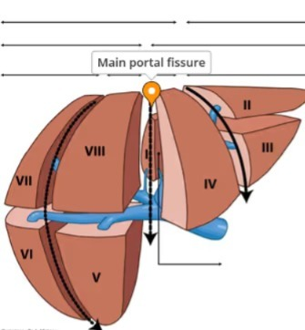
- The groove on the posterior surface of the liver is called the portal fissure. Some structures enter the liver through this fissure and some exit the liver through it, such as..
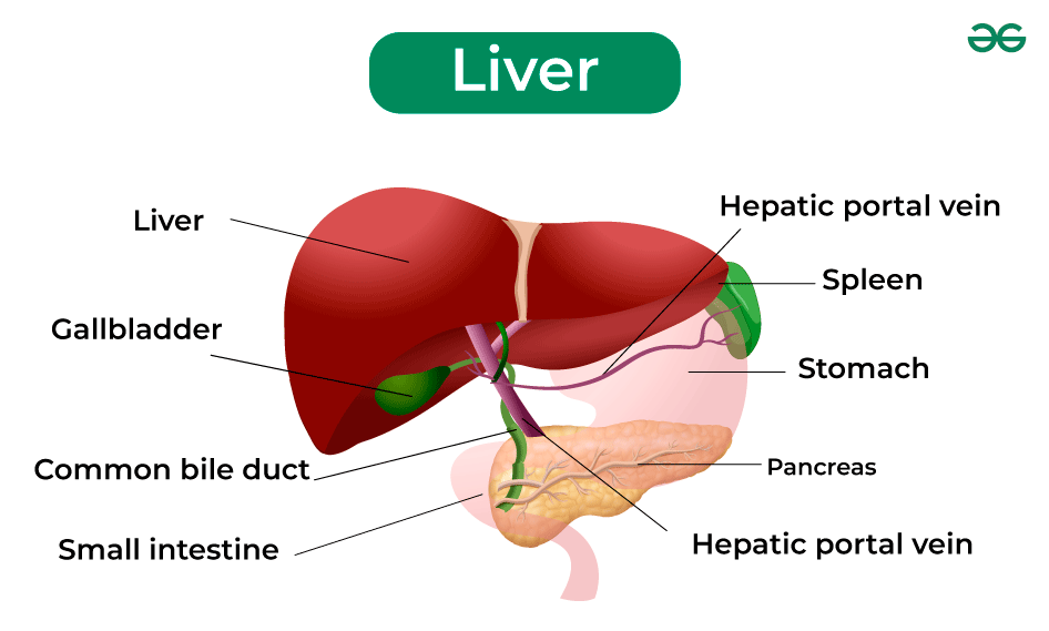
- The portal vein carries deoxygenated and nutrient-rich blood from the intestine and enters the liver through this fissure.
- The hepatic artery carries oxygenated blood and enters the liver through this fissure.
- The nerve fibers of the sympathetic and parasympathetic nerves enter the liver through this fissure.
- The left and The right hepatic duct exits this fissure to carry bile from the liver to the gallbladder.
- The hepatic vein exits this fissure to carry deoxygenated blood from the liver.
Structure of the liver:
- The liver is an organ located in the right quadrant of the abdominal cavity. It is mainly made up of 2 lobes. These lobes are made up of many lobules. These lobules are made up of special types of epithelium cells and cells called hepatocytes. These cells are found in a hexagonal pattern in the liver.
- After the hepatic artery and portal vein enter the liver, a capillary network of arteries and veins is formed. This capillary network is called sinusoids. Kupffer cells are found in the walls of these sinusoids. Kupffer cells work to protect the liver from bacteria, foreign material or toxins and perform a protective function in the liver.
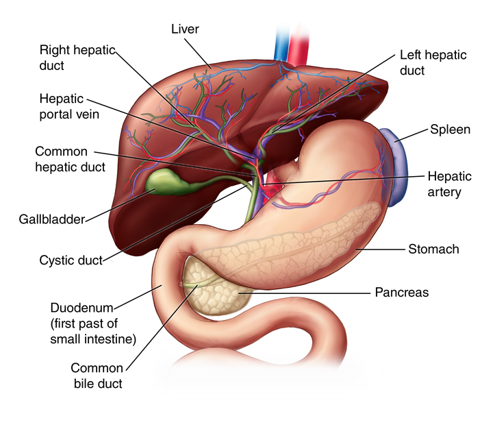
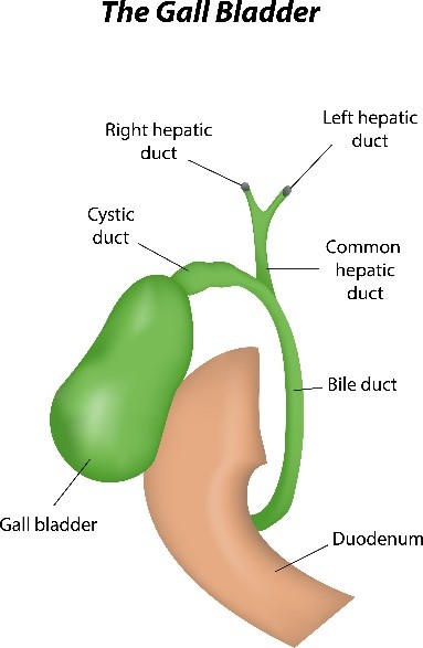
- Hepatocytes cells found inside the liver secrete bile. This bile enters the bile canaliculi. These bile canaliculi bring the bile into the small bile ducts. These small bile ducts join to form the right and left hepatic ducts. Which drains bile from the liver through the common hepatic duct. Further, this common hepatic duct joins with the cystic duct coming from the gallbladder to form the common bile duct. Bile is drained into the small intestine and plays an important role in the digestion of fat.
Blood Supply Of The Liver (Blood Supply of the Liver):
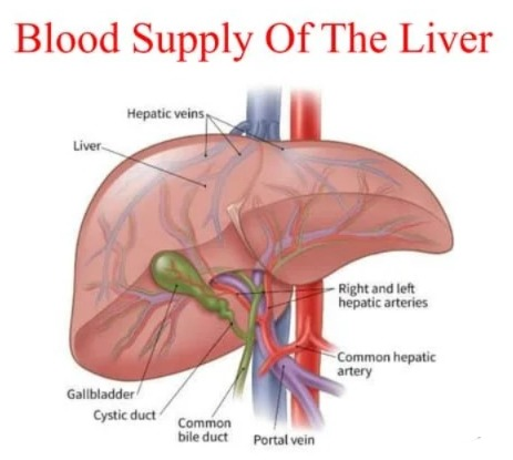
- The liver is supplied with blood by the hepatic artery and the portal vein takes the nutritious blood and enters the liver. is.
- The hepatic vein carries deoxygenated blood from the liver and then joins the inferior vena cava.
Functions of the liver:
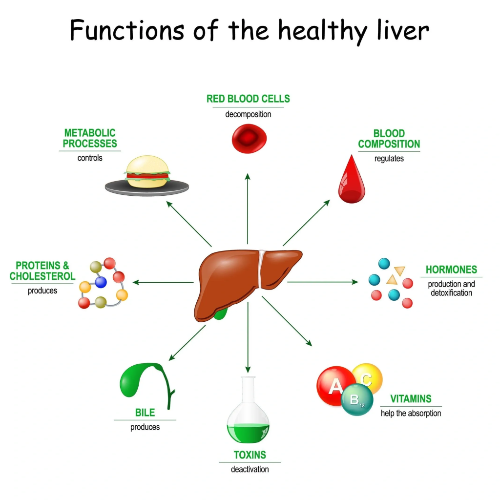
- The liver is a very important organ of our body. It is associated with many important functions. These functions are as follows.
- The liver performs the function of carbohydrate metabolism.
- The liver functions to maintain normal carbohydrate i.e. blood glucose levels. When the blood glucose level decreases, it creates glucose from glycogen through the action of glycogenolysis and maintains the blood glucose level.
- When the blood glucose level increases, it converts glucose into glycogen through the action of glycogenesis.
- The liver converts fat into fatty acids so that it can be used in the body. This is known as fat desaturation, thus it helps in lipid metabolism.
- The liver helps in protein metabolism so that amino acids are used in the body. Synthesis occurs.
- The liver converts ammonia and forms urea so that this waste product can be excreted through urine.
- The death of red blood cells releases bilirubin, which is modified by the liver and helps to remove excess bilirubin from the body.
- The liver helps to detoxify toxic substances, alcohol, drugs, etc. that have entered the body and remove them from the body.
- The liver is a Synthesizes which is necessary for the emulsification of fats and hence the absorption of lipids and cholesterol.
- Helps maintain the level of vitamin D in the body.
- Since Kupffer cells are present inside the liver, they perform a protective function by protecting the liver from harmful substances and performing the action of phagocytosis.
- The liver functions to store vitamins and minerals and releases these vitamins and minerals when needed in the body.
- The liver performs the function of heat production in the body.
Composition of bile:
- The liver performs the function of secreting bile. It secretes approximately 500ml of bile during the day.
- The composition of this bile includes components such as water, mineral salts, mucus, bile pigments, bile salts, cholesterol, etc.
- Bile is a yellow colored liquid. It also has brown or olive green characteristics. Its P. H. It has an alkaline pH of 7.6 to 8.6.
- Bile helps in the emulsification of fats, i.e. in converting its large molecules into small molecules.
- Bilirubin present in bile pigment is its main component and due to the breakdown of bilirubin, stercobilin is formed which is brown in color and it is excreted through stool.
GALL BLADDER (Goalblader):

The gallbladder is a pear-shaped sac. It is located in the abdominal cavity on the posterior side of the liver. It is 7 to 10 cm long and 3 cm wide. It has the capacity to store 30 to 50 ml of bile. It is connected to the areolar connective tissue.
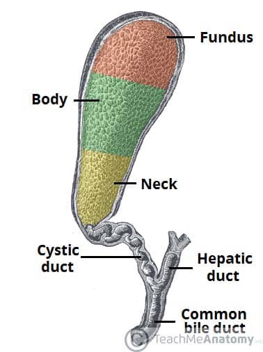
Structure of the gallbladder (Structure of the gallbladder):
The gallbladder is divided into three parts.
- Fundus
- Body
- And Neck.
- It is surrounded by three tissue layers.
- On the outermost side There is a serous peritoneal layer.
- The middle layer is made up of unstriped type muscles with oblique muscle fibers.
- The innermost layer is a layer of mucous membrane. This layer is also continuous with the inner lining of the ducts leading from the gallbladder.
- The inner lining of this mucous membrane is made up of columnar epithelium cells. Which secretes mucin and absorbs water and electrolytes rapidly but does not absorb bile salts and bile pigments, so that bile is concentrated and bile becomes scarce.
- The cystic duct, which carries bile from the gallbladder, is 4 cm long. Which joins the common hepatic duct coming from the liver and forms the common bile duct which carries bile to the duodenum of the small intestine.
- The blood supply to the gallbladder is through the hepatic and biliary arteries and the venous return is through its own venous branch.
- It is supplied with nerve by sympathetic and parasympathetic nerve fibers.
Functions of the gallbladder (Functions of the Gallbladder):
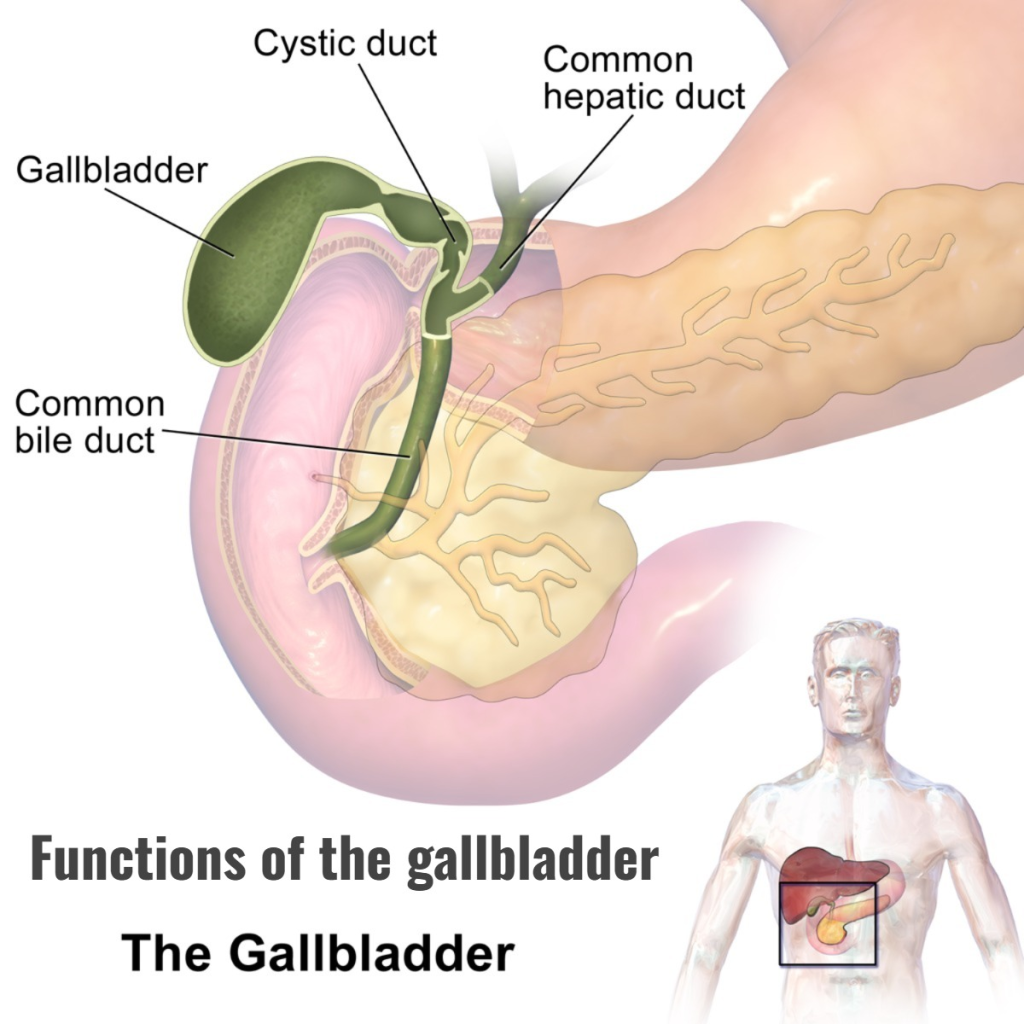
The gallbladder acts as a bag to store bile.
The gallbladder releases bile into small interstitial spaces as needed, which helps in digestion.
Metabolism:
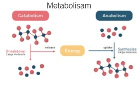
Metabolism is the process of metabolism. It is a chemical reaction in the body in which ATP is transported in living cells in the body.
Metabolism includes two types of processes.
1. Catabolism which is the chemical process of converting complex molecules into simple molecules. In which energy is released in the form of ATP. For example, converting glycogen into glucose. Converting proteins into amino acids.
2. Anabolism is a type of chemical reaction in which simple and small molecules combine to form complex molecules or larger molecules. This process requires ATP. Like the formation of glycogen from glucose.