—ENGLISH-ANATOMY UNIT 7. (PART :1) DIGESTIVE SYSTEM.
DIGESTIVE SYSTEM:

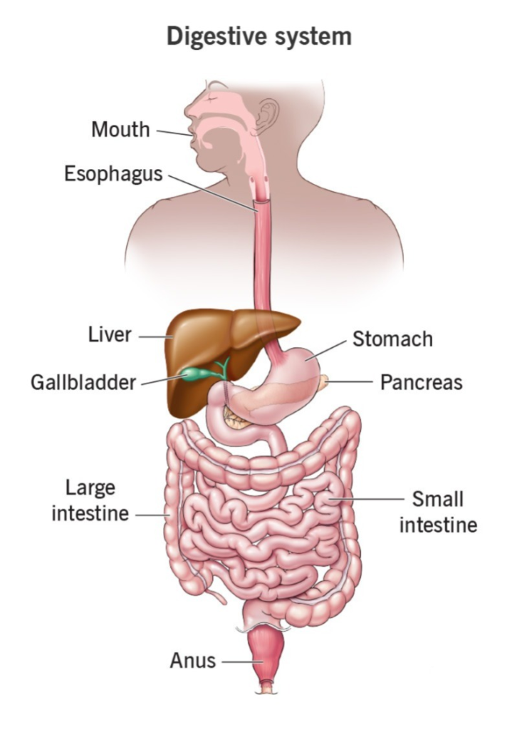
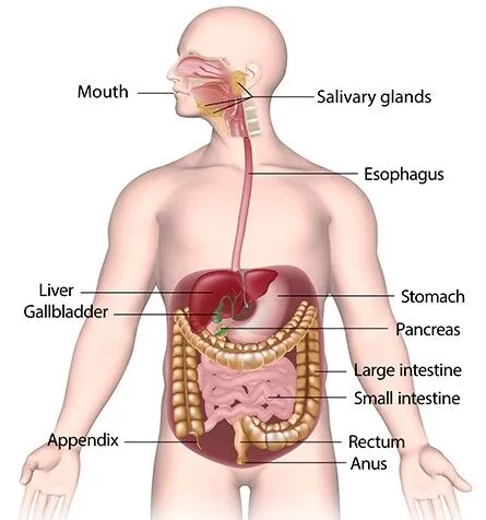
Digestive The Digestive System is also known as the Alimentary Canal. Food contains a large amount of nutrients that work in the body for body building, growth and cell repair. This food material also provides energy to the body. The Alimentary Canal starts from the Mouth and ends at the Anus.

We take food from the mouth and this food is digested and absorbed and the waste material comes out through the anus in the form of stool.
Digestion:
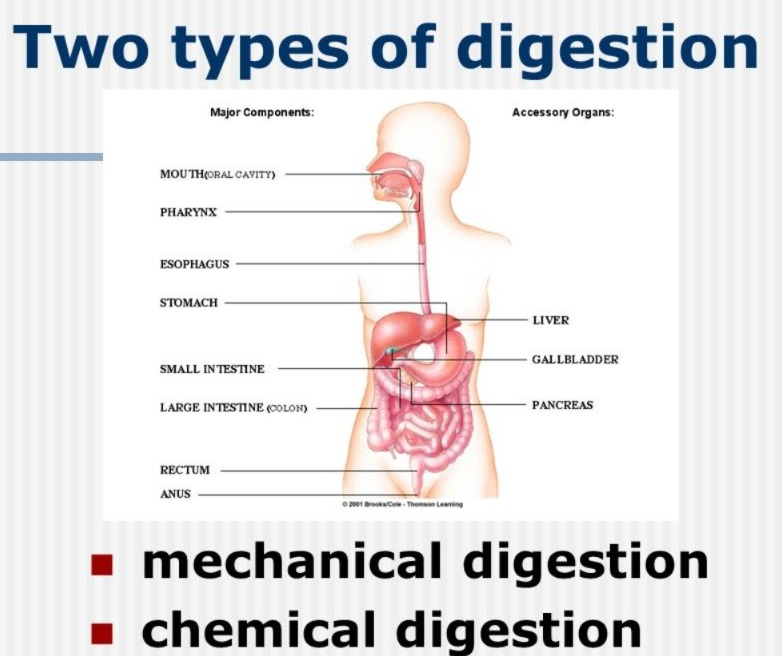
Digestion is the conversion of large food molecules into small molecules, which occurs in two ways.
1.Mechanical Digetion (Mechanical digestion)
2.Chemical digestion
1.Mechanical Digestion (Mechanical Digestion):
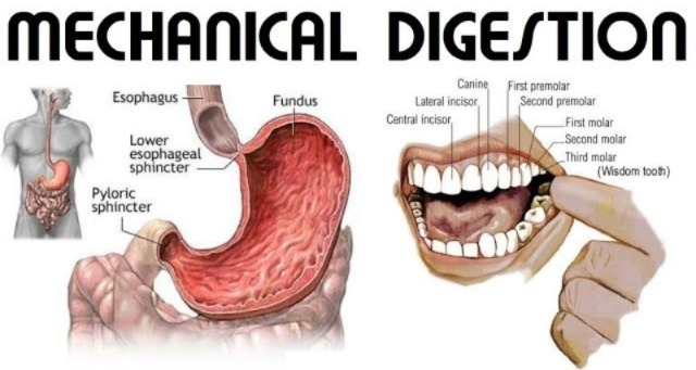
It works to mechanically break down food. This work is mainly done through the act of chewing food, in which large food molecules are broken down into smaller molecules. Mechanical digestion also occurs to some extent in the stomach.
2.Chemical digestion:
In this digestion, chemicals and enzymes are mixed with food molecules which help in digestion. In this chemical digestion, food mixes with saliva and different enzymes. Food mixes with different chemicals at different places in the digestive tract and is digested. In chemical digestion, secretions from the main organs and accessory organs of the digestive tract also help in digestion.
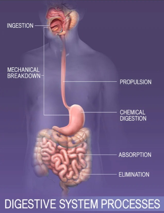
Process of digestion:
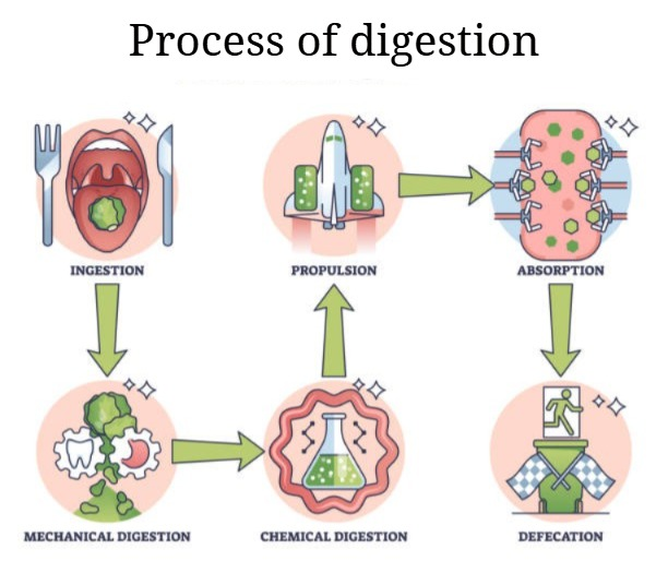
Ingestion:
It is the first step in the design process. In which food is placed in the mouth is called Ingestion.
Propulsion:
It is the second step in the process of digestion. According to this step, the food placed in the mouth slowly moves forward into the alimentary canal which is called Propulsion.
Digestion (Digestion):
This Digestion (Digestion) is mainly seen in two types: mechanical digestion and chemical digestion. It occurs in the mouth, stomach, and intestines.
Absorption:
The small digested molecules are absorbed into the body as nutrients through the mucous membrane of the digestive tract. It occurs through all the organs of the digestive tract.
Elimination:
The residual material that is excreted after the digested nutrient materials are absorbed in the digestive tract The waste that is called feces or stools that exits the body through the rectum and anus through the act of defecation is called Elimination .
Organs of the Digestive System (Organs of the Digestive System):
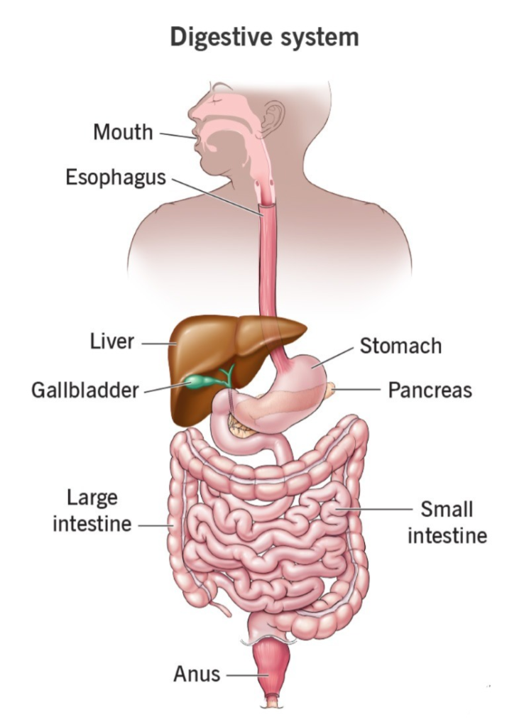
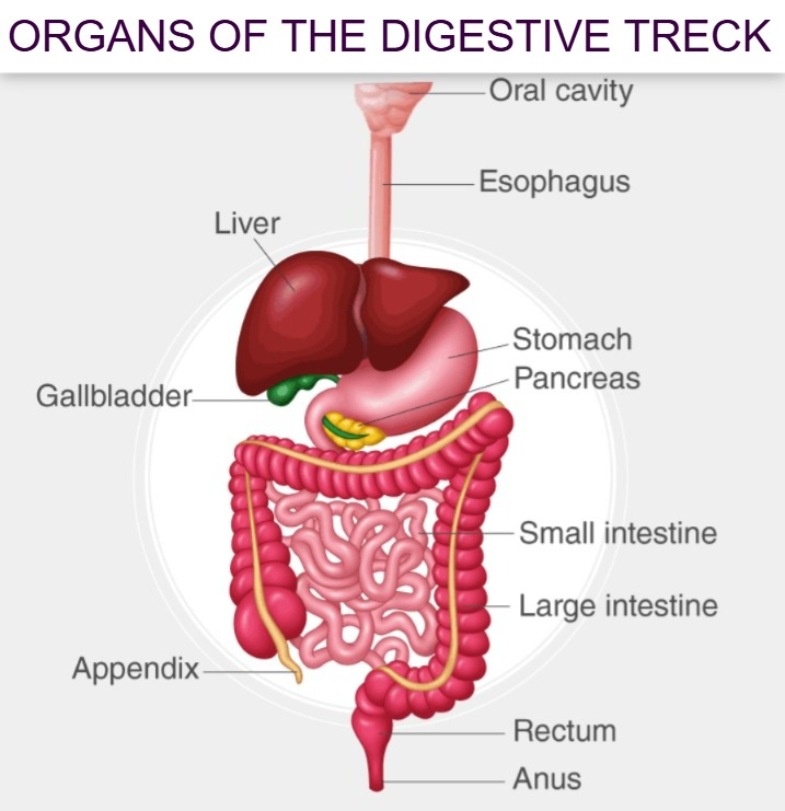
The Digestive Track is also called the Alimentary Track which is a hollow tube that starts from the mouth and extends to the anus and its organs are as follows…
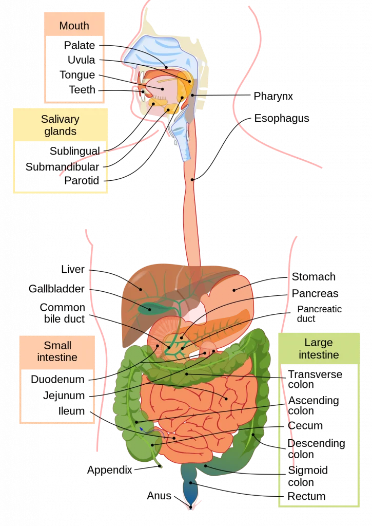
- Mouth
- Pharynx
- Esophagus
- Stomach
- Small Intestine Consists of the Duodenum, Jejunum, and Ileum.
- Large Intestine which includes the Cecum, Ascending Colon, Transverse Colon, Descending Colon, Sigmoid Colon, Rectum and Anal Canal include.
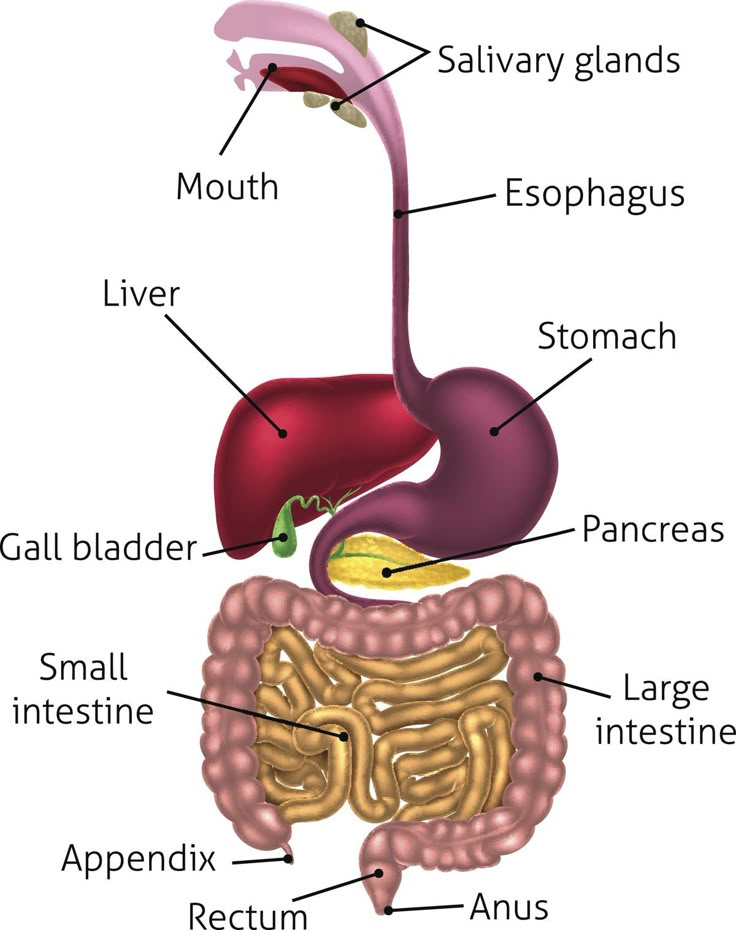
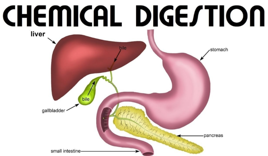
Accessory Organs of the Digestive Tract:
These organs do not come in the main track of the digestive tract but are located on the side which pour their secretions into the alimentary canal and help in the process of digestion hence they are called accessory organs. These help in digestion with the special secretions of their glands which organs are as follows.
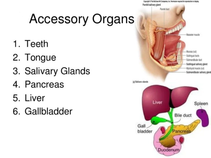
- Teeth
- Tongue
- 3 Pairs Of Salivary Gland
- Liver And The Biliary Track
- Gall Bladder
- Pancreas
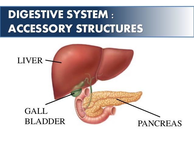
The above organ is an accessory organ which is involved in digestion. Helps.
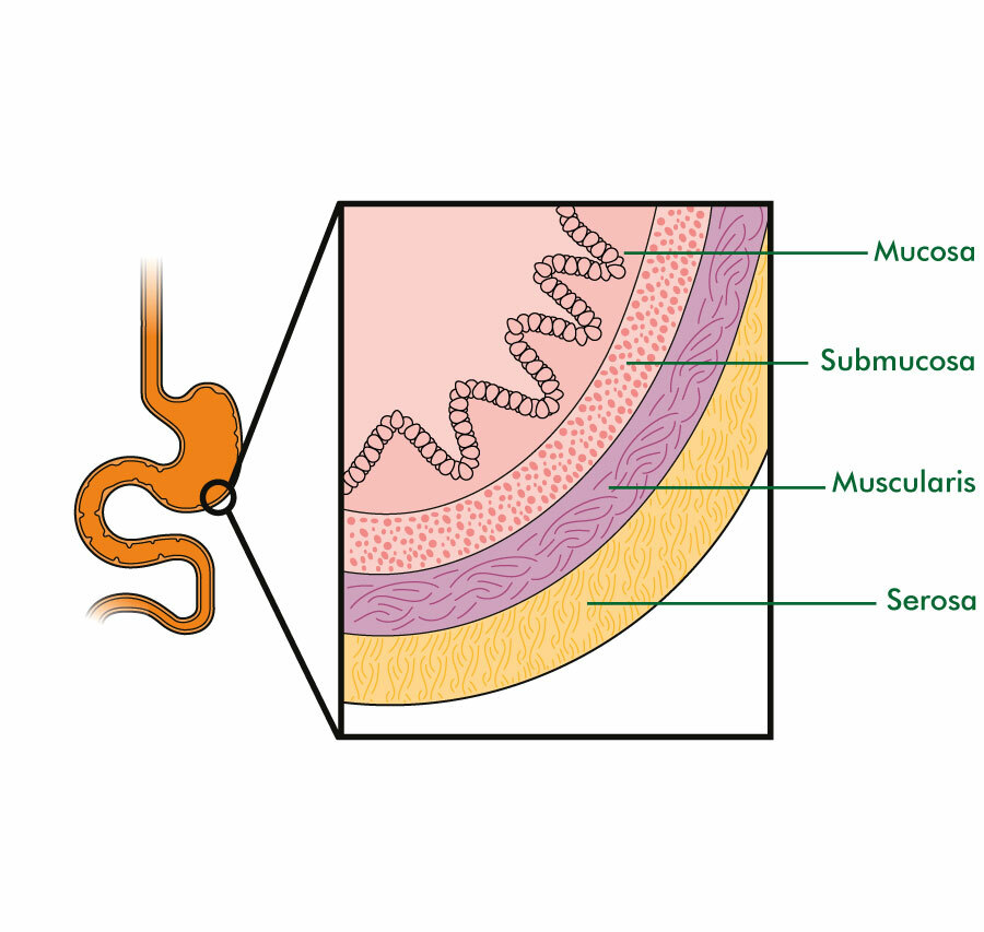
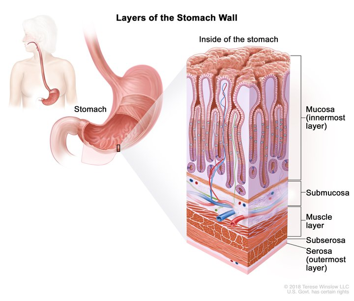
Basic Structure of the Alimentary Canal:
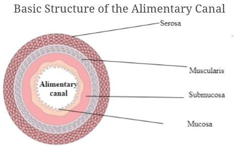
The alimentary canal is composed of four continuous tissue layers from the esophagus to the anus. The tissue layers are arranged from the inside to the outside, as follows:
- Mucosa (Mucosa/Inner Layer)
- Submucosa
- Muscular Layer (Muscular Layer/Muscularis)
- Serosa or Adventitia / Outer Covering
The above tissue layers are arranged from the inside to the outside.
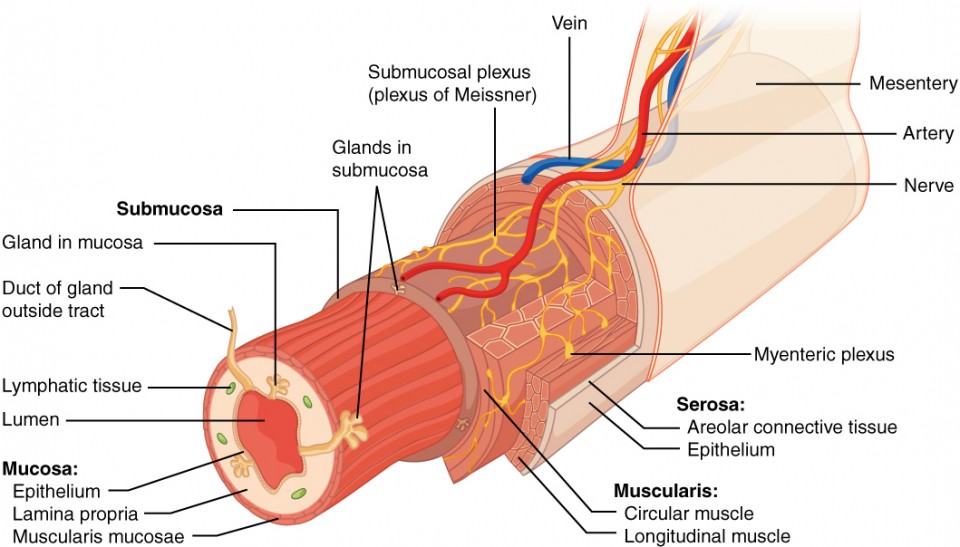
Mucosa layer:
- It is the innermost layer of the alimentary canal. It is in direct contact with food. Its wall is made up of columnar epithelium tissue. Its wall also contains goblet epithelium cells which secrete mucus. This mucus acts as a lubricant for the food. Since mucus is a viscous liquid, the food does not rub against the inner wall of the alimentary tract and acts as a protector for its wall. It is a layer associated with the functions of protection, secretion and absorption. Different glands secrete their secretions on this mucus layer, such as saliva in the oral cavity and gastric juice in the stomach. All these liquids help in the chemical digestion that is in contact with the mucosal layer.
Submucosal layer:
- This layer is made up of areolar connective tissue. It is the vascular layer that contains blood vessels and nerves. These blood vessels and nerves control the functions of the digestive tract. The nerve supply consists of sympathetic and parasympathetic nerve supplies that control the movement of the mucosal layer.
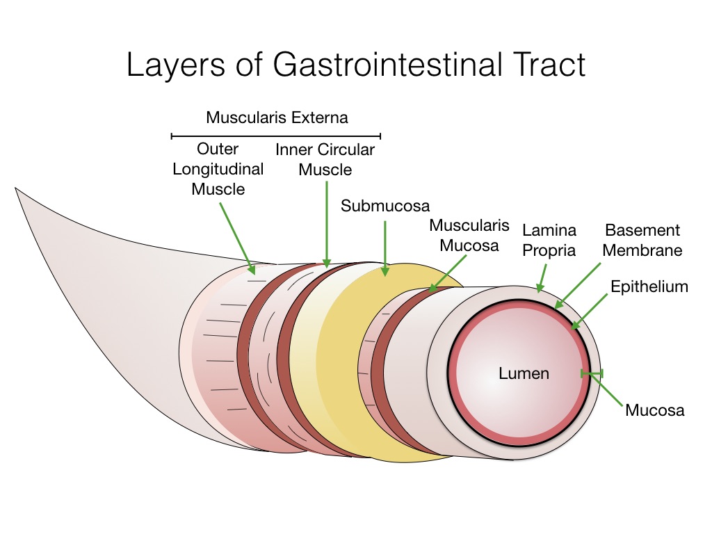
Muscular layer:
- The muscular layer is found in a double layer of smooth muscles in which the outer layer is made up of longitudinal muscles and the inner layer is made up of circular muscles. These muscles have involuntary control. The contraction of these muscles causes the breakdown of food. The contraction and relaxation of these muscles propels the food along the track. This movement is called peristalsis movement. This movement propels the food forward and the sphincter valve prevents the food from moving backwards.
Adventasia:
- This is the outer covering of the digestive tract, i.e. the superficial layer. It consists of loose fibrous tissue. This serous membrane covers the organs of the abdominal cavity called the peritoneum.
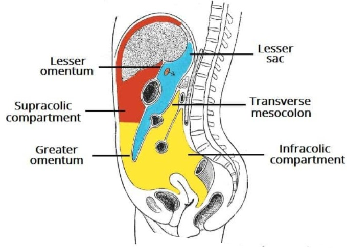
Peritoneum:
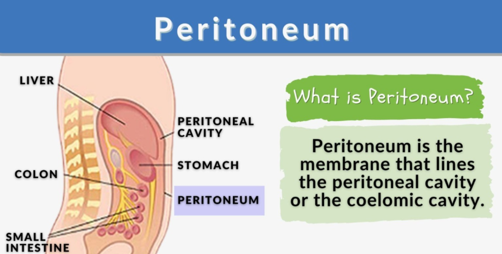
- It is a covering on the organs of the abdominal cavity. It is a very large membrane. It is divided into two parts and forms a cavity in the middle called the peritoneal cavity. This cavity contains a fluid called serous fluid. The outer layer of this cavity is called the parietal peritoneum and the inner layer is called the visceral peritoneum. The serous fluid is present between them.
- The important folds of the peritoneum include the falciform ligament, the lesser omentum, and the greater omentum.
- The falciform ligament attaches the liver to the anterior abdominal wall and diaphragm.
- The lesser omentum is a fold of peritoneum that attaches to the stomach and duodenum, while the greater omentum is a large fold of peritoneum that attaches to the transverse colon and small intestine.
ORGANS OF THE DIGESTIVE TRACK:
Mouth:
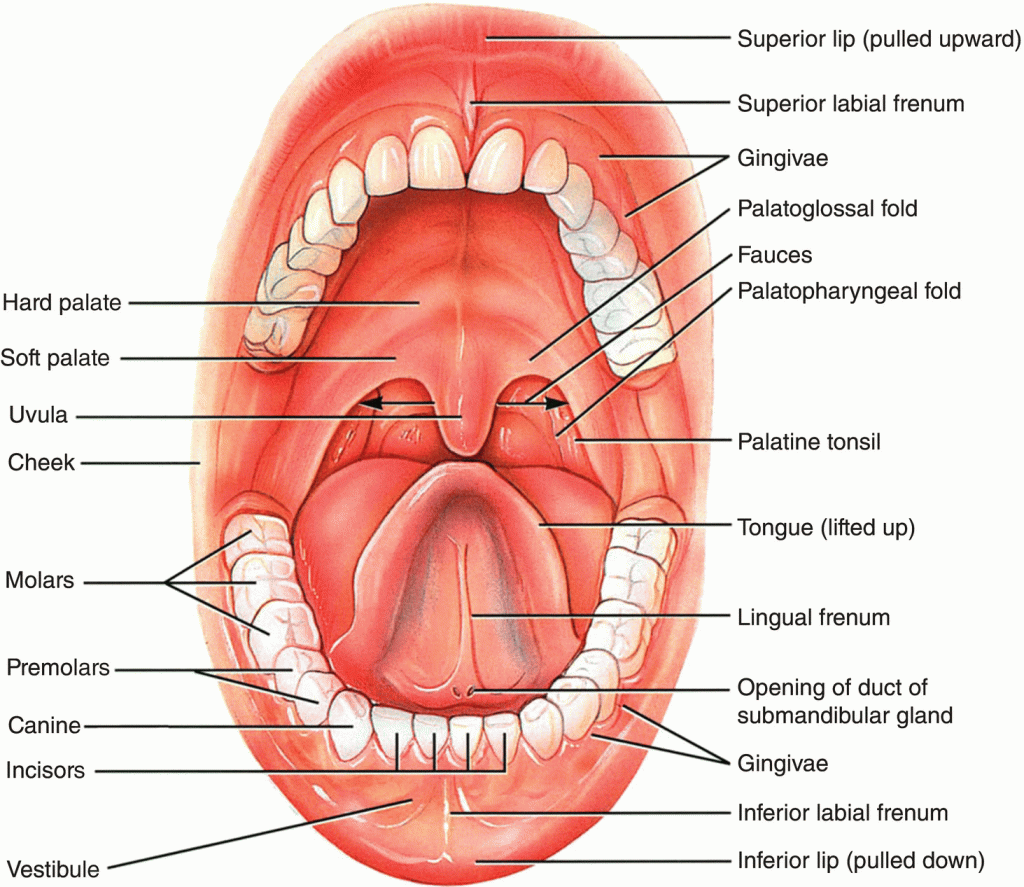
- The mouth is also called the oral or buccal cavity. The mouth is a cavity made of bones and muscles. The structure around it is as follows.
- The lips and the opening of the mouth cavity are located in its front part.
- The mouth is connected to the oropharynx at the back.
- Its side walls are made of cheek muscles.
- Its upper part is made of hard palate made of bones and its back part is made of soft palate made of muscles.
- The tongue is located in the lower part of the mouth. The lining of the cavity inside the mouth is known as the mucous membrane. It consists of stratified squamous epithelium cells.
- In the back middle part of the oral cavity, there is an arch-like structure called the uvula. On both the lateral sides of this uvula, there are folds on both sides, in which the fold on the anterior side is called the palatoglossal arch and the fold on the posterior side is called the palatopharyngeal arch. On either side of this arch are collections of lymphoid tissue called the palatine tonsils.
Tongue:
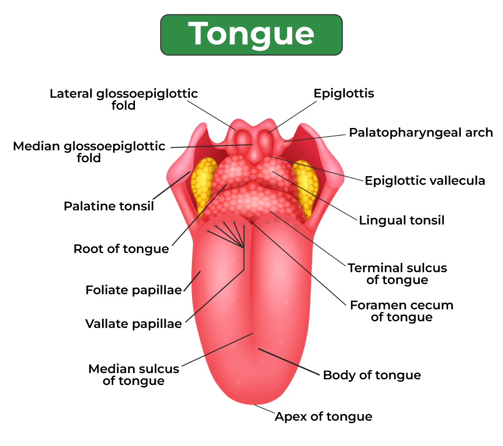
- The tongue is made up of skeletal muscles and is covered by a layer of mucous membrane. It forms the floor of the oral cavity. It divides the oral cavity into two parts in the middle. The tongue is attached inferiorly to the hyoid bone at the back. The tongue is attached to a fold of mucous membrane at the base of the oral cavity called the frenulum.
- The superior surface of the tongue is made up of stratified squamous epithelium cells. The papillae are located on the upper and lateral sides of the tongue. These papillae are connected to nerve endings and are also known as taste buds, which are responsible for the sensation of taste. These papilla are of three types.
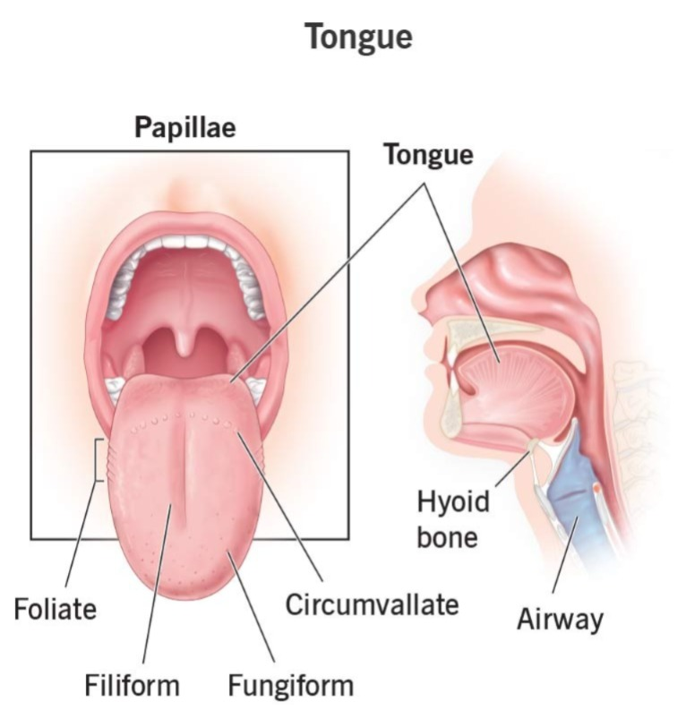
Filiform papilla.. Papillae)
- These papillae are located in the anterior two-thirds of the tongue and do not contain taste buds.
Fungiform papillae..
- They are located on the tip of the tongue and on its sides. They are like small dots and are known as taste buds for most birds.
Valet papilla..
- They are arranged in a V-shape on the posterior side of the tongue and are the largest of all the papillae.
Functions of the Tongue:
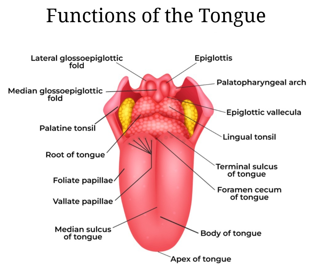
- It plays an important role in the act of speaking and speech.
- It performs the function of testing due to the presence of test birds on its surface.
- It performs the act of mastication, that is, chewing. Both the tongue and teeth together complete the act of chewing. The tongue is important for the moment of food in the mouth.
- During the act of swallowing, the tongue helps in the act of pushing food backwards.
Teeth (Teeth):
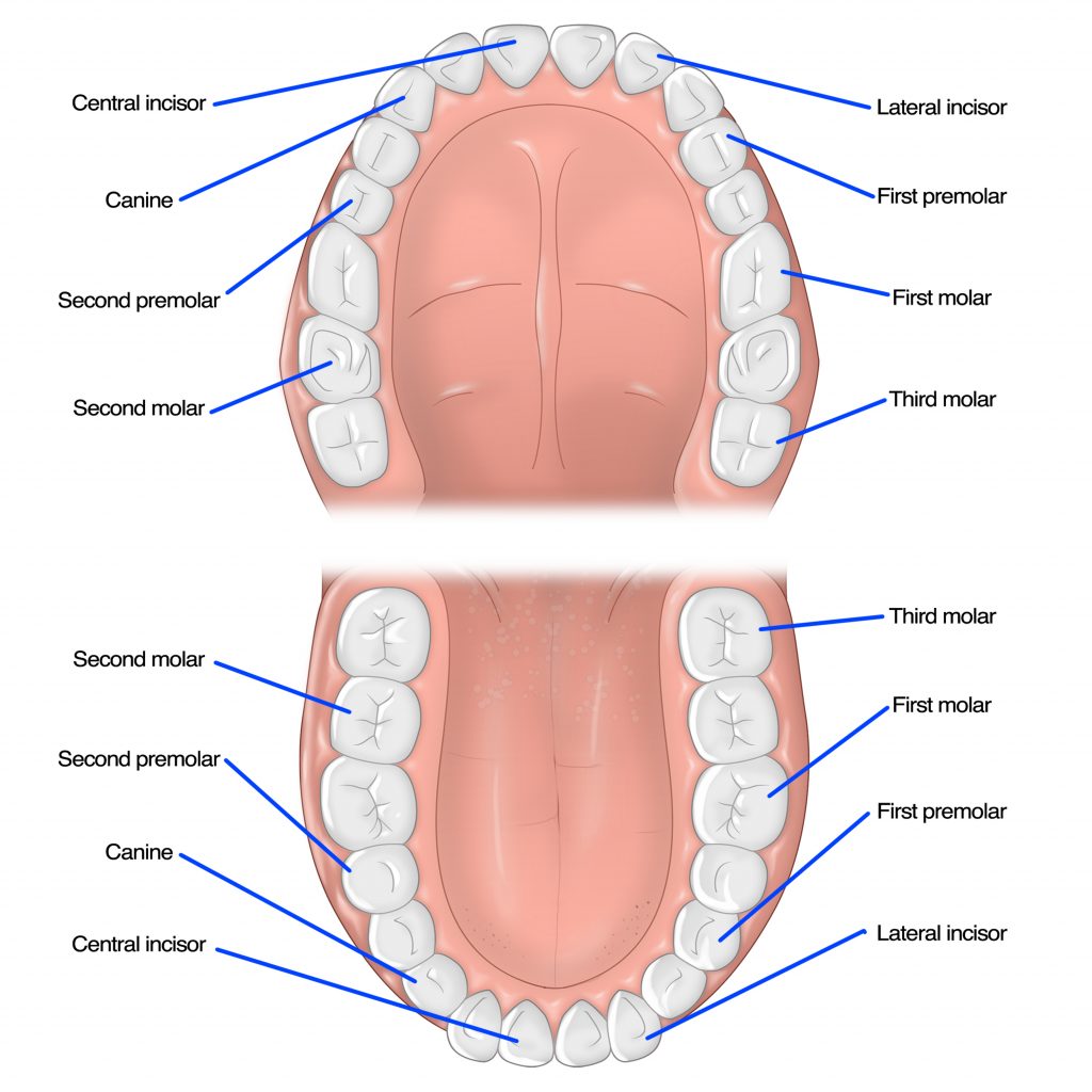
- Teeth are the parts used for chewing. They are fixed to the jaw. In humans, teeth are replaced only once.
- The first set of teeth to appear in childhood are called milky teeth or deciduous teeth, and the second set is called permanent teeth.
- The number of deciduous teeth is 20, with 4 incisors, 2 canines, and 4 molars in each jaw.
- Permanent Teeth There are 32 teeth, including 4 incisors, 2 canines, 2 premolars and 3 molars.
Structure of a tooth:
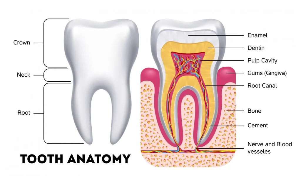
Each tooth has three parts.
- Crown: Which is visible above the gum.
- Root: Which is pressed into the jaw below the gum.
- Neck: Which is located between the crown and the root.
Other Structure of Tooth:
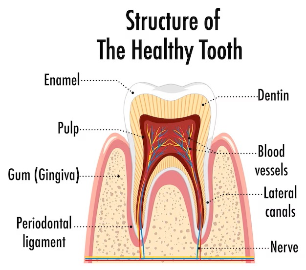
Other structures of the tooth include the following parts.
1. Pulp:
- It is a cavity of loose fibrous tissue in the middle of the tooth that contains blood vessels and nerves.
2. Dentine:
- The structure surrounding the pulp is called dentine and is made of calcified material.
3. Enamel:
- The outer part of the tooth, i.e. the crown or the part covering the dentin, is called enamel and forms the hard part of the tooth.
4. Cementum:
- The upper part of the dentin at the root of the tooth is called cementum and has a bone-like structure.
5. Periodontal Membrane:
- This membrane is useful for holding the root of the tooth in its socket.
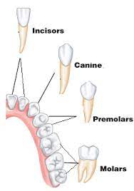
Function of Teeth:
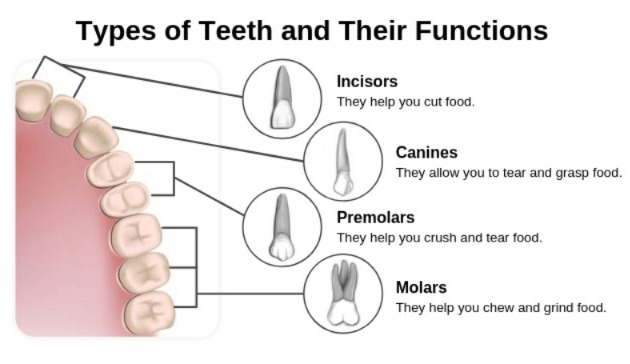
- The functions of teeth are determined by their shape. Different shaped teeth perform different functions such as…
- Incisor and canine teeth are sharp and important for cutting and tearing.
- Premolar and molar teeth have flat surfaces and are important for grinding.
Salivary glands:
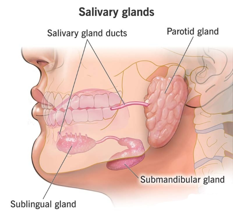
Salivary glands are accessory structures. They are located on the side of the wall of the mouth. They secrete a fluid called saliva. These salivary glands are in pairs of three which are as follows…
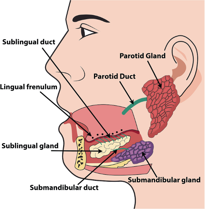
1.Perotid Gland:
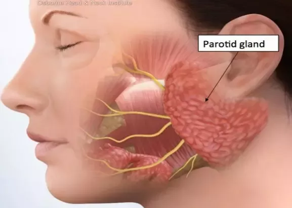
- It is a pyramid-shaped gland. It is a gland located between the skin and muscles at the bottom of the external ear. Each gland has a parotid duct and this duct opens in the upper jaw at the second upper molar tooth. This gland produces a fluid called saliva that contains enzymes but no mucus.
2. Submandibular Gland:
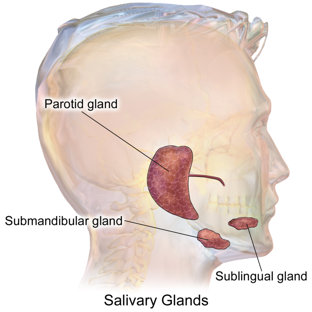
- This gland is located below the angle of the mandible. It secretes both mucus and enzymes. Its submandibular duct, i.e. Wharton’s Duct, opens at the lingual frenulum.
3. Sublingual Gland:
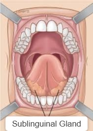
- This is a small gland that is located on the opposite side of the submandibular gland. It opens into the floor of the mouth through the sublingual duct, i.e. the Duct of Rivinus.
All the above glands have a fibrous capsule on their outer side. It has a large number of lobules and these lobules are made up of secretory cells which secrete their secretions into small ducts and all those small ducts join and form a large duct which opens at the mouth.
The composition of saliva :
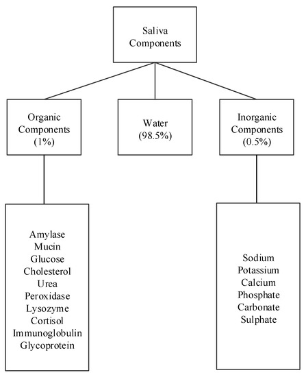
- Saliva is secreted by the salivary gland. This gland is located in the oral cavity. The fluid that it secretes is called saliva. Its P. H. is slightly acidic. It is 6.35 to 6.85. An estimated one and a half liters of saliva is produced daily. Its composition includes water, mineral salts including sodium, potassium, chloride and some enzymes including salivary amylase and mucus organic substances such as urea, uric acid, lysosomes and some blood clotting factors.
- Saliva secretion It is controlled by the autonomic nervous system, in which the parasympathetic nervous system increases saliva production and the sympathetic nervous system decreases saliva production. The parotid gland secretes salivary amylase.
Function of the saliva:
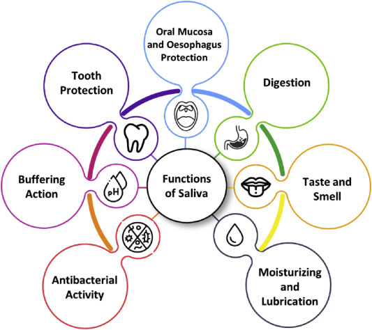
- When food is placed inside the mouth, saliva mixes with the food and lubricates it so that the lining of the mucus membrane is not damaged.
- The water in the composition of saliva keeps the mouth clean and the mucus keeps the mouth lubricated so that the tissues of the mouth remain soft and moist.
- The salivary amylase present in saliva is a polysaccharide chemical Helps in digestion and digests it into disaccharides or monosaccharides.
- Saliva helps in destroying microorganisms due to its composition of lysosomes, immunoglobulins and clotting factors.
- Food comes into contact with water by mixing with saliva and it dissolves, which is detected by the test.
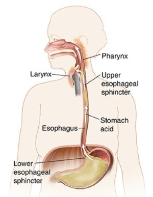
Pharynx (pharyngitis) :
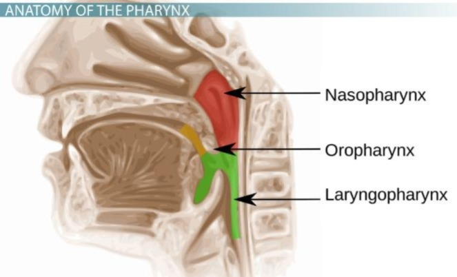
The fairings are wide muscular tubes that are located behind the nose and mouth. The pharynx is mainly divided into three parts.
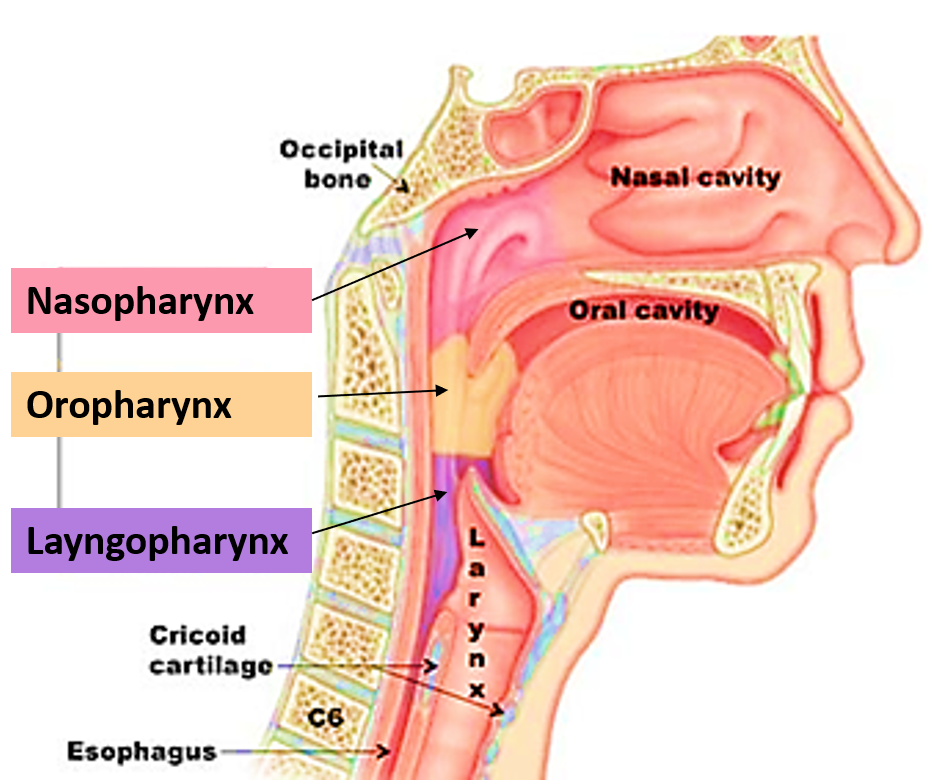
1.Nasopharynx (Nasopharyngs)
2. Oropharynx (Oropharynx)
3. Laryngopharynx (Laryngopharynx)
- The nasopharynx serves as a passage for the act of respiration.
- The oropharynx and laryngopharynx are the parts of the digestive system. Passage as well as the respiratory system.
- Food passes from the oral cavity into the esophagus through the pharynx, so the pharynx acts as both a food passage and an air passage.
- The structure of the pharynx has layers similar to the basic structure of the digestive tract, in which the mucosal layer is the innermost. Above it is the submucosal layer and above it is the muscular layer. Which is discussed in detail in the basic structure.
Esophagus:
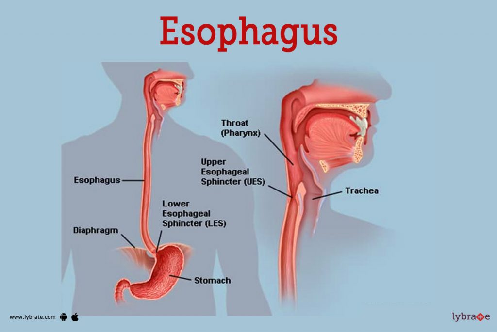
Esophagus is thin The esophagus is a muscular tube that lies between the pharynx and the stomach and functions as a food passage. It extends from the lower part of the neck to the stomach. It is 25 centimeters long and two centimeters wide. The lumen of this tube is a collapsible tube and widens during the act of swallowing food.
The upper and lower ends of the esophagus are the sphincter muscles. The upper part is the Cricopharyngeal Sphincter and the lower part is the Lower Oesophageal Sphincter or Cardiac Sphincter which prevents the gastric contents of the stomach from coming up i.e. preventing reflux.
- The trachea is located in the front of the esophagus, the vertebral column is located in the back, and the lungs are located on the right and left sides.
- The structure of the esophagus also has tissue layers similar to the basic structure of the digestive tract, in which the innermost part is the mucosal layer, above it the submucosal layer, above it the muscular layer, and on the outermost side the adventitia layer. The esophagus acts as a food passage.
Stomach :

- It is an organ of the digestive system. It is a J-shaped organ. It forms the widest part of the alimentary canal. It is mainly located in the epigastric region of the stomach and some part of it is located in the left hypochondriac region.
- It has some part of the liver in its front part, and the abdominal aorta, pancreas and spleen in its back part. The diaphragm is located at the top and the intestines are located at the bottom.
Structure:
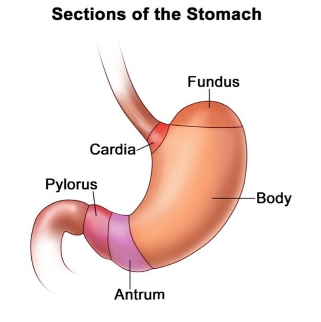
The structure of the stomach is divided into three parts
- Fundus
- Body
- Pylorus
1.Fundus:

It forms the uppermost part of the stomach. It is dome-shaped. It is located above the level where the esophagus joins the stomach. It is sometimes filled with gas. Here, the esophagus connects to the stomach, and between them are the cardiac sphincter muscles.
2.Body:
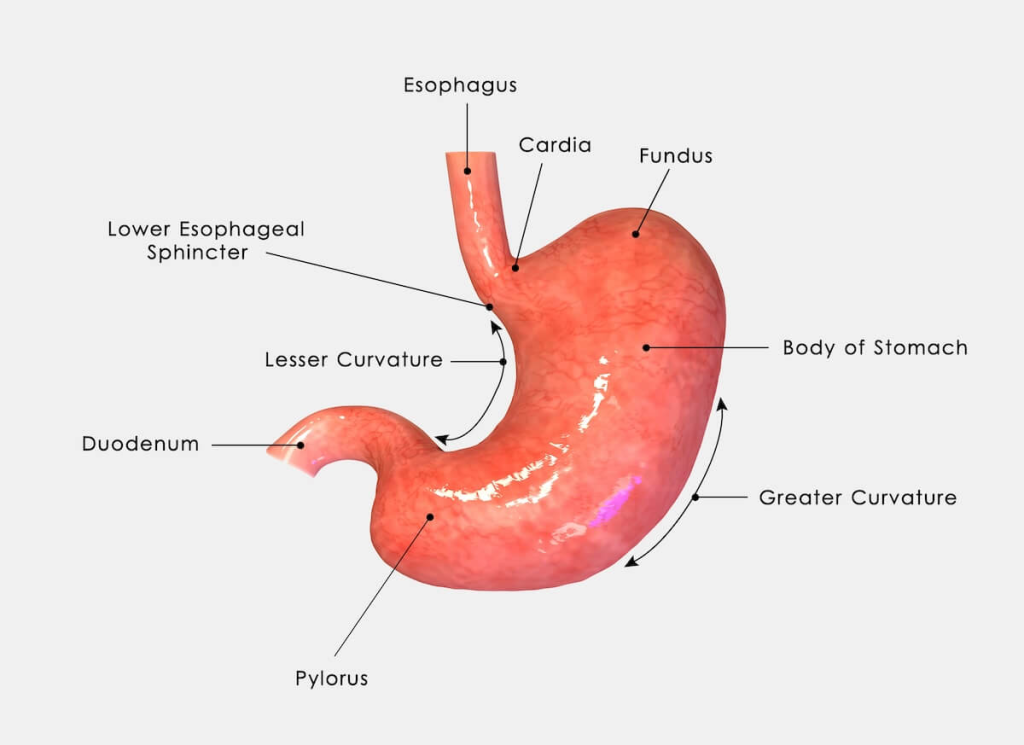
The part below the fundus of the stomach is called the body, which is located at the bottom of the stomach, i.e. the pylorus.
3. Pylorus:

The stomach has a part that comes down after the body, in which the initial part is called the pyloric antrum, which is the narrow part after the body, from there, going towards the duodenum, there is a canal-like structure called the pyloric canal, which opens into the duodenum and in between there are sphincter muscles called the pyloric sphincter.
In addition, two curvatures can be seen on the body portion of the stomach. There is a lesser curvature that forms the right border of the stomach and a greater curvature that forms the left border of the stomach. Creates a left border.
Layers of the stomach:
- The stomach wall, like the basic structure of the digestive tract, is made up of four tissue layers, with the outermost layer being the peritoneum layer, also called the adventitia layer, which forms the serous lining.
- Below it is a layer of muscles. In the stomach, this muscular layer is made up of three layers, one of which is longitudinal muscle fibers that are arranged superficially, the other is circular muscle fibers that are arranged in a circular shape, which lie below the superficial layer and these fibers are also important for forming sphincter muscles. Three oblique muscle fibers are mainly found in this layer of the stomach, which is not found in the basic structure. It is especially found in the wall of the stomach, which is responsible for the movement of food into the stomach. It is a very important layer for movement and mechanical digestion.
- Below the muscular layer is the submucosal layer which contains a network of blood vessels and lymph vessels.
- The innermost layer of the stomach wall is called the mucosal layer. This layer is made up of columnar epithelium tissue. This layer secretes mucus and also acts as a lubricant. This layer contains folds of mucous membrane which are especially visible when the stomach is empty, which is called flatulence. This flatulence disappears when the stomach is full of food.
Functions of the Stomach:
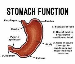
The stomach performs the following functions:
- The stomach functions to store food for a short time. Before the food moves into the intestinal tract, it is partially mechanically digested.
- The stomach functions to secrete gastric juice. This gastric juice has an acidic content and the enzymes present in it help in the digestion of food.
- The muscular layer in the wall of the stomach is specially arranged, so its contraction causes a churning movement, which helps in the mechanical digestion of food and breaks down food into smaller pieces. and then the food moves on to the duodenum.
- The inner wall of the stomach secretes intrinsic factor, which is necessary for the absorption of vitamin B12.
- Some amount of water, alcohol and some drugs are also absorbed from the wall of the stomach.
- It also works to destroy bacteria or viruses that are inside the stomach. In which these bacteria reach the stomach through mucus and oral cavity, they are destroyed in acidic secretion and It stops the progress of the intestinal tract, thus it also works as a protection for the body.
Digestion in the stomach:
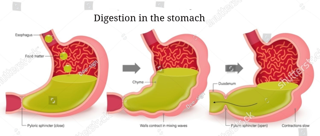
- Food taken through the mouth reaches the stomach through the esophagus. There, mechanical digestion of food begins due to the churning movement of the stomach due to the contraction of the muscular layer of the stomach. Here, all the food is converted from large molecules to small molecules through mechanical digestion.
- After this mechanical digestion has lasted for some time, hydrochloric acid mixes with the food through a gland in the inner wall of the stomach.
- Chemical digestion begins when the chemicals in this stomach juice mix with the food.
- In addition to hydrochloric acid, this gastric juice contains enzymes, mainly pepsin and renin. This pepsin digests large molecules of protein into small molecules. The digestion of protein begins here.
- The rennin present in the gastric juice digests the protein casein present in milk and converts it into paracasein. Thus, the digestion of protein mainly takes place in the stomach.
- The digestion of carbohydrates and fats does not take place in the stomach, only their mechanical digestion takes place here. After the food is partially digested in the stomach, it goes to the small intestine where it undergoes complete digestion and absorption.
- When the food in the stomach mixes with gastric juice, the acidic property of HCL in it destroys the harmful bacteria and viruses present in the food.
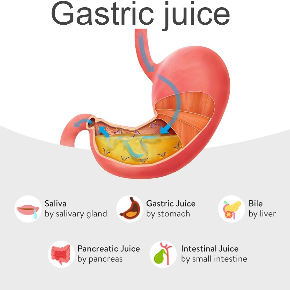
Gastric juice (Gastric juice):

- The stomach is an organ of the digestive system. It forms the largest part of the digestive system and has a capacity of about one and a half to two liters. Its innermost wall is made of a mucous membrane. The glands under this membrane secrete a digestive juice called gastric juice.
- This gastric juice is a clear and colorless liquid. When food reaches the stomach, this juice mixes with it and helps in digestion.
- Salivary amylase, which comes with saliva, mixes with gastric juice in the stomach and its effect is reduced and there is a decrease in the digestion of carbohydrates.
- Gastric glands secrete approximately two liters of gastric juice during the day.
- The composition of gastric juice includes water, mineral salts, mucus, hydrochloric acid, intrinsic factor, pepsinogen, enzymes, etc. is.
Function of the gastric juice:
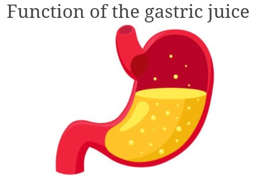
- Water acts to liquefy the food in the stomach.
- Hydrochloric acid makes food acidic It is and works to destroy microorganisms present in the stomach, thus having antiseptic and disinfectant properties and performing a protective function.
- It also provides a medium for the digestion of proteins which occurs due to the conversion of pepsinogen to pepsin.
- Gastric lipase present in gastric juice helps in the digestion of fats to some extent.
- Intrinsic factor is very important for the absorption of vitamin B12.
- The mucus in gastric juice acts as a lubricant and prevents friction between the stomach wall and food.
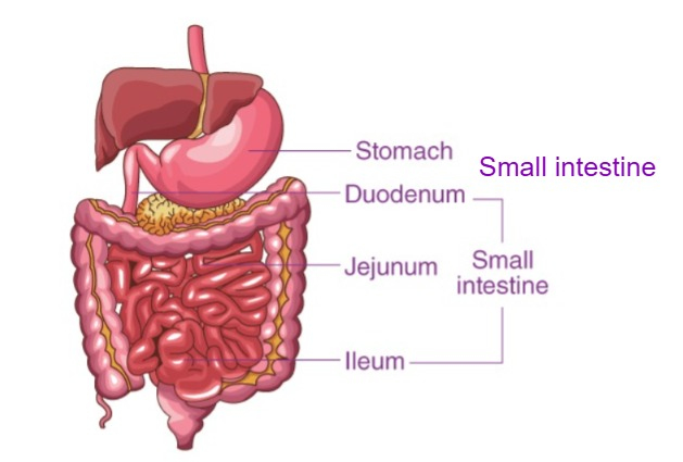
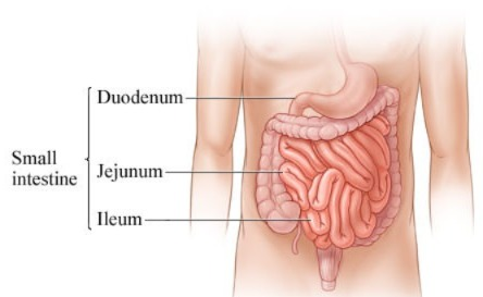
Small intestine:
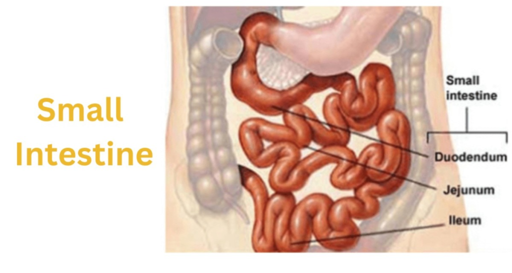
- The small intestine is a tube-like structure in the gastrointestinal tract that starts from the stomach and ends at the ileocecal valve, where it joins the large intestine.
- The small intestine is a part of the abdominal cavity around the umbilical region, surrounded by the large intestine. It is about one inch in diameter and about 20 feet long. It is coiled in the abdominal cavity. The small intestine is divided into three parts.
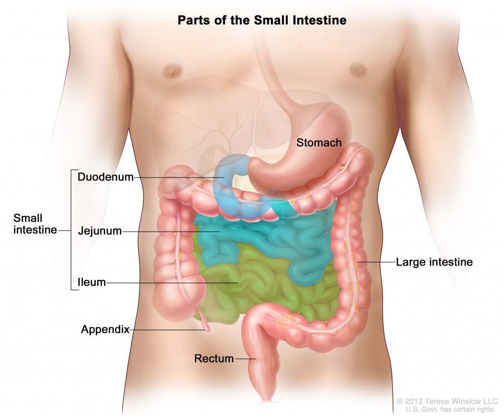
1. Duodenum:
The duodenum is the first part of the small intestine. It starts at the end of the stomach, i.e. the pylorus, and is approximately 25 cm long and is shaped like a C.
2. Jejunum:
It is the part of the small intestine after the duodenum, the upper part of which is connected to the end of the duodenum. From here, this tube-like structure is arranged in a coiled shape downwards. The length of the jejunum is approximately 8 feet. The jejunum connects to the ileum at the bottom.
3. Ileum:
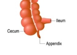
The ileum is the part of the small intestine after the jejunum which connects to the jejunum at the top and is approximately 12 feet long. It is connected to the large intestine by the ileocecal sphincter.
Structure of the Small Intestine Intestine):
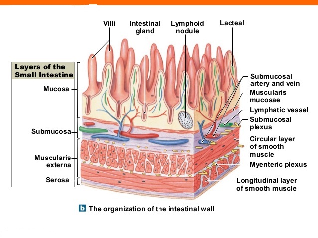
- The small intestine is also made up of four tissue layers, similar to the basic structure of the digestive tract.
- The outermost layer is the peritoneum layer. It is made up of serous membranes and is arranged in a double layer.
- Underneath the peritoneum layer is the muscular layer. The lining of this tract is made up of smooth muscles, with the outer layer made up of longitudinal muscle fibers and the inner layer made up of circular muscle fibers. Due to the contraction of the muscles, peristalsis movement is observed and the content moves forward in the track.
- Below the muscular layer is the submucosal layer, which contains blood vessels, nerves, etc. Some glands are also present in this layer.
- The inner lining of the small intestine is made up of a mucus layer. Between this mucus layer and the submucosal layer is a layer of smooth muscles, which is called the muscularis mucosa. The contraction of these muscle fibers causes the lactic acid to be emptied.
- The lining of the mucosal layer contains permanent fold-like structures called valvulae conniventes. Due to this fold, the contents remain there for a long time and maximum absorption can take place and the digestive juices can act on the food present there for a long time so that proper digestion takes place.
- The mucosal layer has a structure of villi which absorbs the digested food.
- This layer contains some nodules of lymphatic tissue which are called Peyer’s patches which perform a protective function in the intestine.
- The superior part of the small intestine Blood supply is via the mesenteric artery and venous return is via the mesenteric vein.
- Nerve supply is via the sympathetic and parasympathetic nerves.
intestinal Juice:
- Intestinal juice is a clear yellow color. Which is secreted in the amount of one to two liters per day. Its properties are alkaline. Its pH is 7.6 to 8.0.
- Its composition includes water, mineral salts, mucus and enzymes such as maltase, sucrase etc.
- This intestinal juice also includes pancreatic juice and bile. All these juices are called chyme. Which is very important in the digestion of proteins, fats and carbohydrates.
Functions of the Small Intestine:
- The small intestine has a pendulum movement that allows the intestinal contents to mix.
- The fluid in the intestine, called chyme, moves throughout the intestine through peristalsis and helps in digestion.
- The small intestine secretes digestive juices.
- The chemical digestion of proteins, fats, and carbohydrates takes place in it.
- 90% of digested nutrients are absorbed through the wall of the small intestine.
- The segments in the inner layer of the small intestine allow digestive juices and food to mix, digest, and absorb.
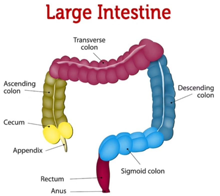
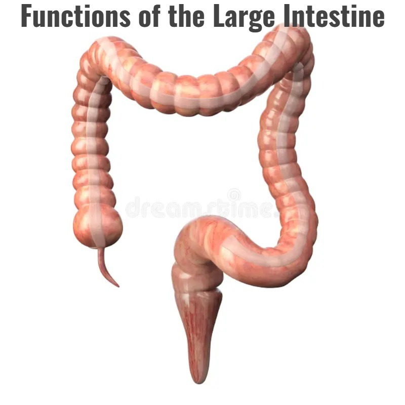
Large intestine:
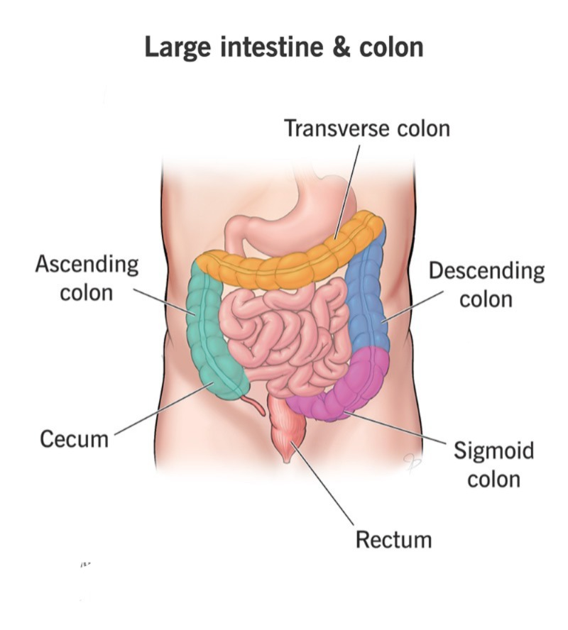
The large interstyle begins at the ileocecal junction and extends to the anus. Its total length is 1.5 meters and is divided into the following parts.

1.Caecum (Cecum)
2.Colon
a. Ascending Colon (Ascending Colon)
b. Transverse Colon (Transverse Colon)
c. Descending Colon (Descending Colon)
d. Sigmoid Colon (Sigmoid Colon)
3. Rectum (Rectum)
4. Anal Canal
A narrow diverticulum is located between the cecum and ileum, known as the vermiform appendix.
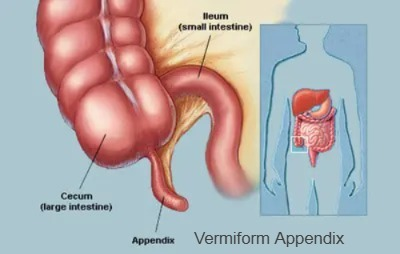
1.Caecum
The caecum is the beginning of a large interstyle It is five to eight centimeters long. It is a blind pouch and is located in the right lower abdominal quadrant.
2.Colon (Colon)
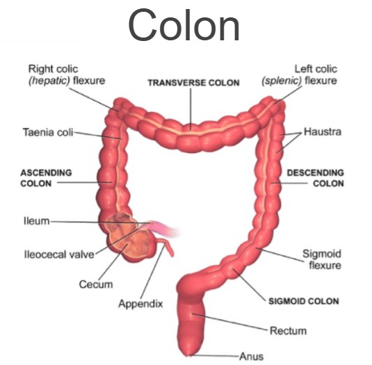
The colon begins with the Ascending Colon. It lies in a vertical position on the right side. From there, it is seen upwards to the border of the liver. The ileum joins the ascending colon near the cecum, where the ileocecal valve is located. Which allows contents to enter the large intestine but does not allow contents to pass in the reverse direction.
Then the Transverse Colon is arranged horizontally in the abdominal cavity. It passes below the liver, stomach, and spleen. It extends from the hepatic flexure to the splenic flexure.
The Descending Colon is in a vertical position and is located in the left side of the abdomen. It is located below the stomach and spleen.
The Sigmoid Colon is located below the iliac crest. It is the last part of the large intestine and is S-shaped.
3. Rectum (Rectum)
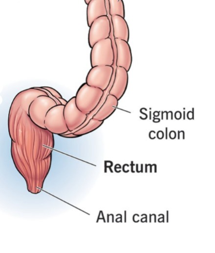
It is approximately 20 cm long piece. Which starts from the sigmoid colon and extends to the anal canal.
4. Anal Canal
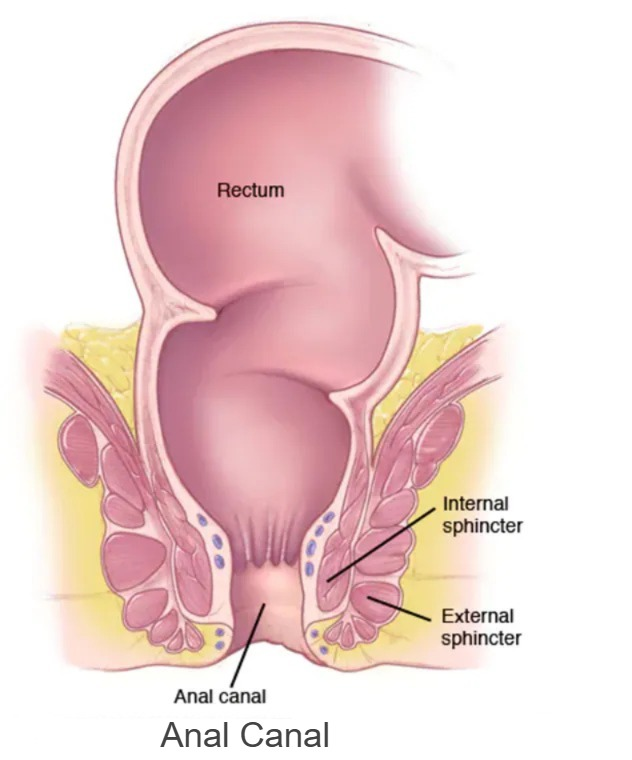
The two to three centimeters of the rectum at the end of the large intestine is called the anal canal. The mucous membrane lining the anal canal is a longitudinal It is arranged in a fold. It is called the anal column. This part contains arteries and veins and there are two sphincter muscles arranged around the anus.
Internal sphincter muscles are smooth muscles. Which are under the control of the autonomic nervous system, i.e. have an involuntary function. External sphincter muscles are skeletal muscles which are under our voluntary control.
Structure of the Large Intestine:
- The structure of the large intestine also has 4 tissue layers, just like the basic structure of the digestive tract.
- In which the mucosal layer is found in the innermost part. This mucosal layer does not have any fusion like the small intestine. This layer contains epithelium tissue and goblet epithelium cells, which function in the absorption of water and secretion of mucus. This layer also contains some lymphatic nodules.
- The submucosal layer of the large intestine has the same basic structural characteristics as the mucosa. Over it lies the muscular layer, which is found in a double layer, the outer layer being made up of longitudinal muscle fibers and the inner layer being made up of circular muscle fibers.
- The outermost layer is the serosa layer. Which is part of the visceral peritoneum.
- The large intestine is supplied with blood by the inferior and superior mesenteric arteries and receives venous return by the superior and inferior mesenteric veins.
Functions of the Large Intestine:
Water And Electrolytes Absorption:
- Most water absorption occurs in the small intestine, but some water and some electrolytes are also absorbed in the large intestine.
- The large intestine absorbs water from the stool, which plays an important role in making the stool semi-solid.
Microbial Activity:
- Some bacteria are present in the large intestine. These bacteria play an important role in the synthesis of vitamin K and folic acid.
- These bacteria can be harmful if they move to other parts of the body and can also cause infection.
- The activity and fermentation of these bacteria produces gas from undigested food, which is passed out of the bowel through the anus in the form of flatus.
Mass Movement (Mass Movement) Movement):
- Peristalsis movement is seen in other parts of the intestine but this type of peristalsis movement is not seen in the large intestine.
- After a long interval, a large strong peristalsis movement is seen in the large intestine due to which the content moves towards the descending colon and sigmoid colon in the intestinal track which is called mass movement.
Defication:
- Due to mass movement in the large intestine, fecal material moves towards the sigmoid colon and rectum in the interstitium, due to which the stretch receptors in the wall of the rectum are stretched and the defecation reflex starts and the process of defecation begins.
- In the defecation reflex, due to the stretching of the wall of the rectum, its receptors are stimulated due to the stretching of the sensory nerve. It transmits impulses to the sacrum in the spinal cord. In addition, the pressure of the diaphragm and the abdominal cavity for the process of defecation helps in the defecation process. Sitting position also helps in the process of defecation. Finally, due to the stimulation of the parasympathetic nervous , the internal sphincter relaxes and stool comes out through it. The external sphincter has voluntary control so that the process of defecation can be stopped for some time through voluntary control, while the internal sphincter has involuntary control. Relaxation of the internal sphincter and external sphincter causes the passage of stool.
Faeces (Faeces):
Faeces is brown in color. This brown color is due to the stercobilin present in it. It has semi-solid and solid properties. The composition of faeces is approximately 60 to 70% water. The excess water is absorbed through the wall of the large intestine. The following things are found in the composition of feces.
- Dead and live microorganisms.
- Dietary fibers.
- Epithelial tissue of the intestinal wall.
- Mucus and undigested food. etc.
Because mucus is secreted through the intestinal wall, stool can pass easily without causing any abrasion to the intestinal wall.