—ENGLISH-ANATOMY UNIT 6 RESPIRATORY SYSTEM
RESPIRATORY SYSTEM (Respiratory System):
INTRODUCTION (Introduction):

For the human body to survive, every cell in the body needs nutrient materials and oxygen to perform its normal functions. The human body cannot survive without it.
Respiration and its types:
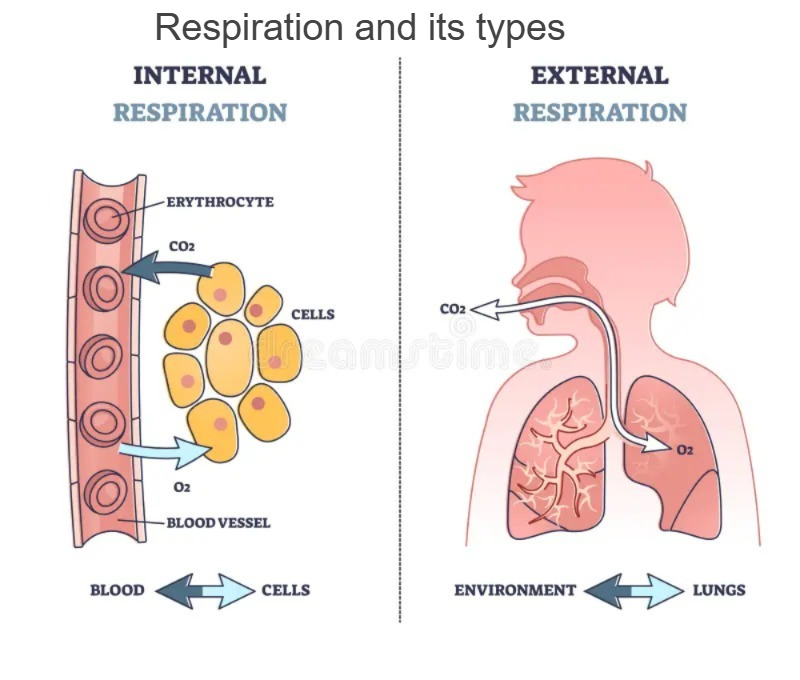
Respiration is the gas exchange that occurs between any two surfaces. In respiration, oxygen enters the body from the atmosphere through inspiration and carbon dioxide, which is accumulated in the body as a waste product, is released through expiration. Thus the action of respiration is seen.
Two main types of respiration are found in the body:
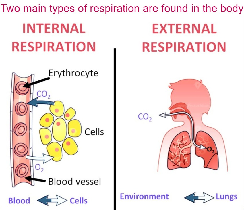
1.Internal Respiration
2.External Respiration
1.Internal Respiration:
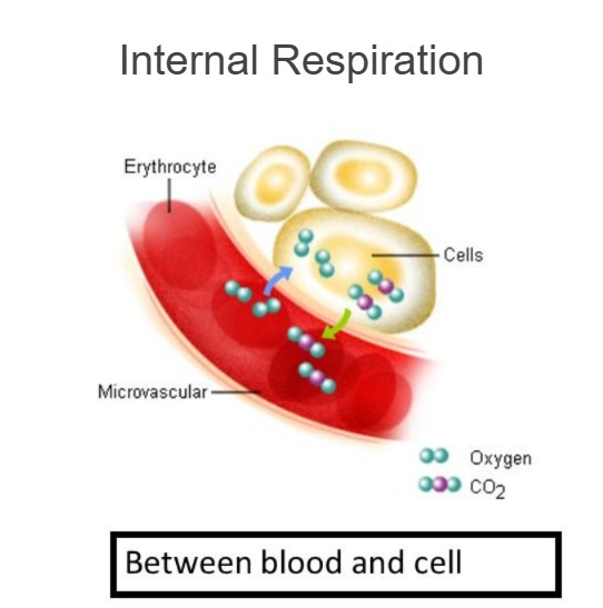
The exchange of oxygen and carbon dioxide between each cell, tissue and the surrounding blood capillaries of the body is called Internal Respiration. This respiration is found all over the body.
2. External Respiration:
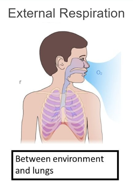
The gas exchange of oxygen and carbon dioxide between the lung tissue, i.e. the alveoli and the surrounding blood capillaries is called external respiration. This respiration occurs in the lungs.
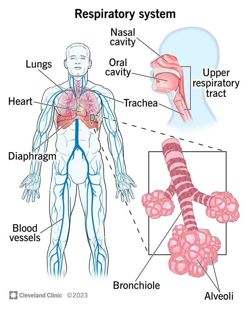
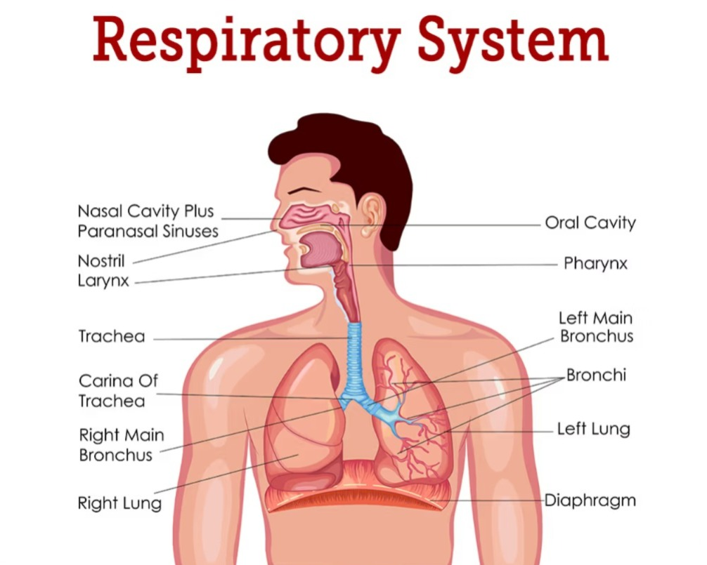
List out the organs of the respiratory system:
The organs located above the respiratory tract outside the thoracic cavity are called the organs of the Upper Respiratory Track. which includes the following organs.
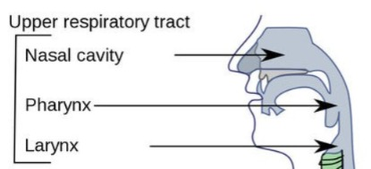
- Nose
- Pharynx
- Larynx
The organs of the respiratory tract within the thoracic cavity are called the lower respiratory tract . Which organs are as follows.
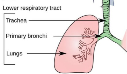
- Trachea
- Bronchi (Right and left)
- Bronchioles
- Terminal Bronchioles
- Terminal Bronchioles
- Alveoli (Alveoli)
- Lungs (Right and left).
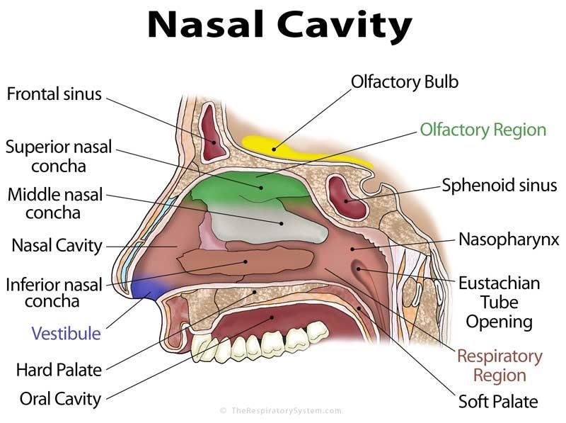
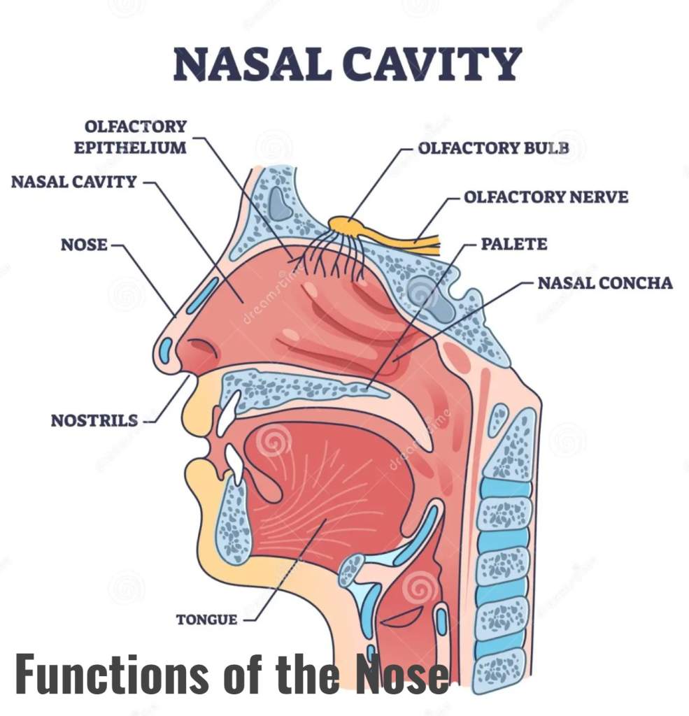
Structure of Nose and its function (Structure of Nose and its function):
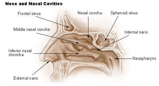
- The nose is an organ at the beginning of the respiratory system. It is located at the front of the face.
- When viewed from the outside, the nose has two openings. These are called external nostrils or nasal striations. There is a partition between these two openings, which has a cartilage-like septum on the front side and a vomer bone on the back side, which is the structure separating the two openings.
- The external nose is covered with skin on the outside and the inner lining is made of mucous membrane.
- It contains pseudo stratified ciliated epithelium cells. Here, goblet cells are present, which secrete mucus. Due to which the inner lining remains moist.
- The nasal cavity is a cavity located in the face, which is arranged above the oral cavity and the cribiform plate of the ethmoid bone of the cranial cavity is present on the upper side of the nasal cavity. In addition, the frontal bone and the sphenoid bone are also found in this part.
- The medial wall of the nasal cavity is formed by cartilage and the vomer bone. This wall is known as the nasal septum.
- The floor of the nose is made up of the soft and hard palates.
- The lateral wall of the nose contains the nasal bones. In addition, a bony structure called the inferior conchae also forms the lateral wall.
- Openings at the back of the nasal cavity, called the posterior nares, are connected to the pharyngeal fossa.
- Hairs are located on the inside and anterior side of the nasal cavity, which warm the air, filter it, and prevent small foreign particles from entering.
Openings into the Nasal cavity (Opening of the nasal cavity):
- There are 2 openings in the front of the nasal cavity, called the anterior nares is called. 2 such openings are located on the posterior side of the nasal cavity, which open into the part of the phalanges. They are called posterior nares.
- In addition, the openings of the sinuses of the bones surrounding it open into the part of the nasal cavity. It is called paranasal sinuses.
Functions of the Nose:
- The nose is an external organ of the respiratory system. Its functions are as follows.
- It performs the act of respiration. In which oxygen-rich air from the external environment enters the lungs through the nose and the air containing carbon dioxide waste from the body exits through the nose.
- The hairs in the inner lining of the nose clean the air. So that no foreign particles enter the respiratory track.
- Due to the hair and vascular mucus membrane in the inner lining of the nasal cavity, the air entering inside becomes warm and reaches the lungs as warm as the body temperature, so that the lung tissue does not get irritated or damaged.
- Due to the presence of goblet epithelium cells in the inner membrane of the nose, it is a moist membrane. Due to the passage of air through it, it becomes moist. So that the lining of the inner mucus membrane does not get damaged or irritated. Thus, it also performs the function of humidification.
- The vestibule structure at the beginning of the nose, which is covered with cells and hair processes, is also found in it. It functions to filter the air entering inside. It prevents dust particles and foreign materials from entering the respiratory tract.
- The nose senses smell, that is, it also performs the function of distinguishing odors. The mucous membrane of the nose contains receptors of the olfactory nerve. When that air enters the nasal cavity, it comes into contact with the odorous or foul-smelling chemicals present in it, which stimulate those receptors, and their impulses travel to the brain via the olfactory nerve, resulting in the sensation of smell. Thus, it also performs the function of smell detection.
Paranasal sinuses and it’s functions:
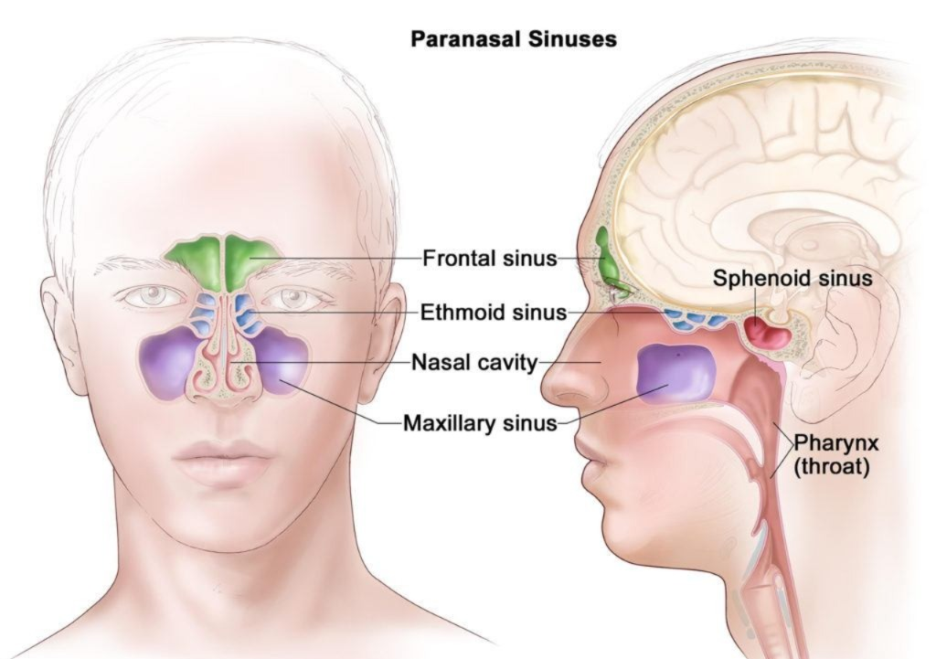
- Sinuses are small foramina in the bones. There are some holes in the bones surrounding the nasal cavity, which open into the nasal cavity. All these sinuses are called paranasal sinuses.
- These sinuses are air-filled cavities and are covered with a mucous membrane. The sinuses associated with the nasal cavity are as follows.
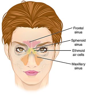
- Maxillary sinuses (maxillary sinuses)
- Frontal sinuses (frontal sinuses)
- Ethmoidal sinuses (ethmoidal sinuses).
- Sphenoid sinuses
All the above bones have two sinuses. Each of these sinuses opens into a major cavity.
Functions of Sinuses:
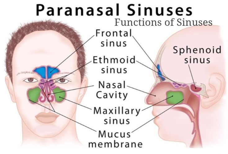
- These sinuses create air field cavities in the bones due to which the bones become lighter in weight.
Makes the skull and face parts lighter. - Moistens and humidifies the air entering during inspiration.
Maintains the air pressure of the nasal cavity. - Identifies surrounding sounds.
Also useful for the sensation of smell.
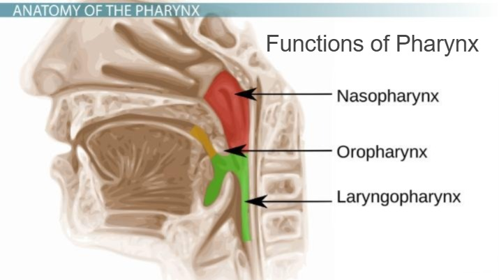
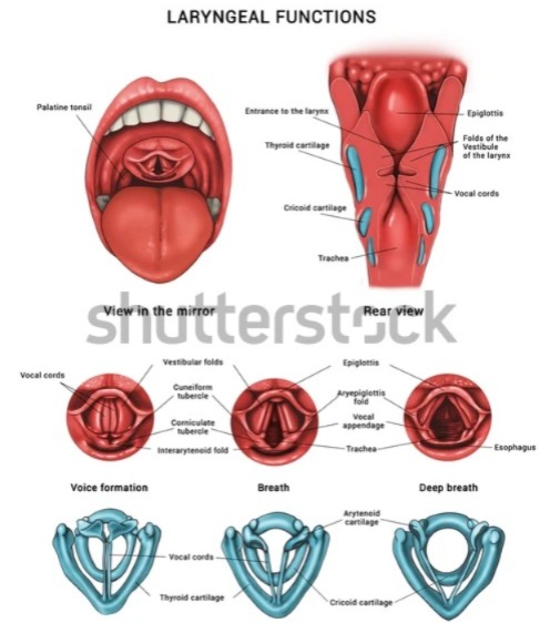
Structure and function of Pharynx:
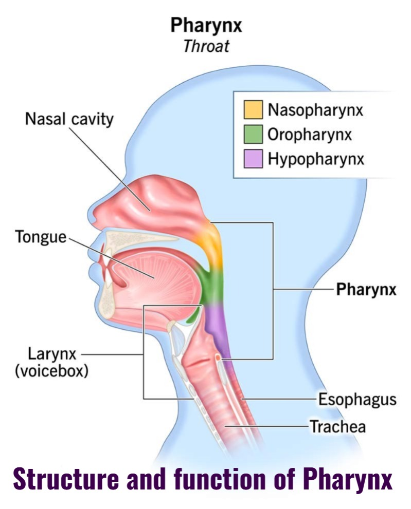
- The larynx is a tube-like structure located in the upper respiratory tract. It is 12 cm long and 3.5 cm wide.
- It is located behind the nose, oral cavity, and larynx. It is located from the base of the occipital bone to the level of the 6th cervical vertebra.
- The pharynx is divided into the following parts based on the structures surrounding it.
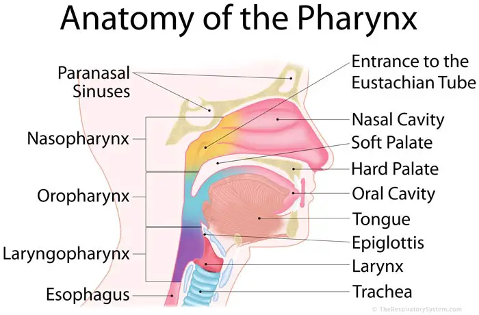
1.Nasopharynx (nose fairings)
2.Oropharynx
3.Laryngopharynx
1.Nasopharynx (nose pharyngs):
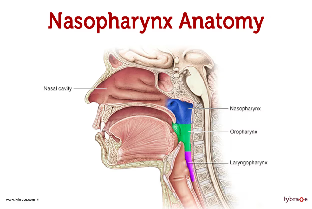
- The nasopharynx is the part of the nasopharynx behind the nasal cavity. The 2 openings of the posterior nares behind the nasal cavity open into the nasopharynx. Other openings also open here, including the 2 openings of the Eustachian tube coming from both sides of the middle ear. In addition, 1 opening of the oropharynx is also found in this area. So, a total of 5 openings are seen in the nasal passages.
- Due to the connection of this part with the middle ear, air pressure is maintained between the passages and the middle ear.
- The wall of the nasal passages is rigid and thick. Therefore, it does not shrink. So it helps in maintaining a patent airway.
- There is a mass of lymphoid tissue on the upper wall of the nasal passages. Which is called Pharyngeal tonsil.
- Ciliated columnar epithelium tissue is found in the lining of this part.
2.Oropharynx (Oropharynx):
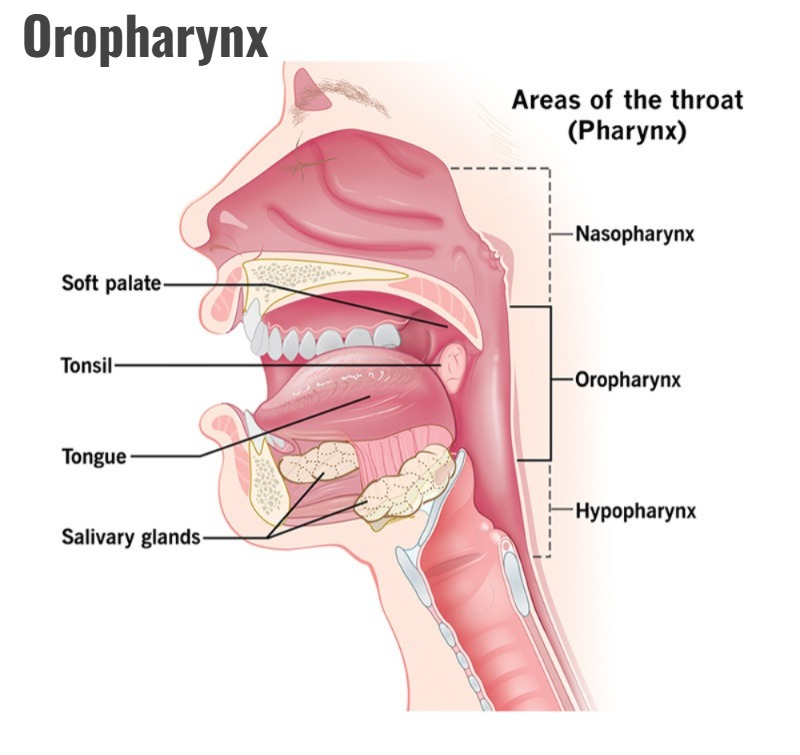
- The pharynx at the back of the oral cavity is called the oropharynx. It is connected to the nasopharynx at the top and forms the back of the mouth. The laryngeal pharynx begins below it.
- The oropharynx is found from the level of the soft palate to the level of the 3rd cervical vertebra, where the hyoid bone is located.
- The oropharynx has masses of lymphoid tissue on both sides of its wall. They are called palatine tonsils. This helps in providing local protection.
- The oropharynx is made up of non-keratinized stratified squamous epithelium tissue.
3. Laryngopharynx (laryngopharyngopharyngitis):
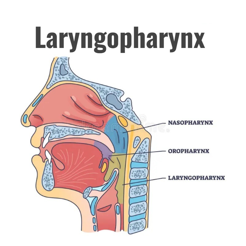
- The part of the larynx next to the larynx is known as the laryngopharings. It is the part starting from the level of the hyoid bone, that is, this part is found from the 3rd to the 6th cervical vertebra.
- On the upper side, it is connected to the oropharynx and on its lower side, the opening of the esophagus is seen.
- The oropharynx and larynx serve as pathways for both the respiratory and digestive tracts.
Structural Layers of Pharynx:
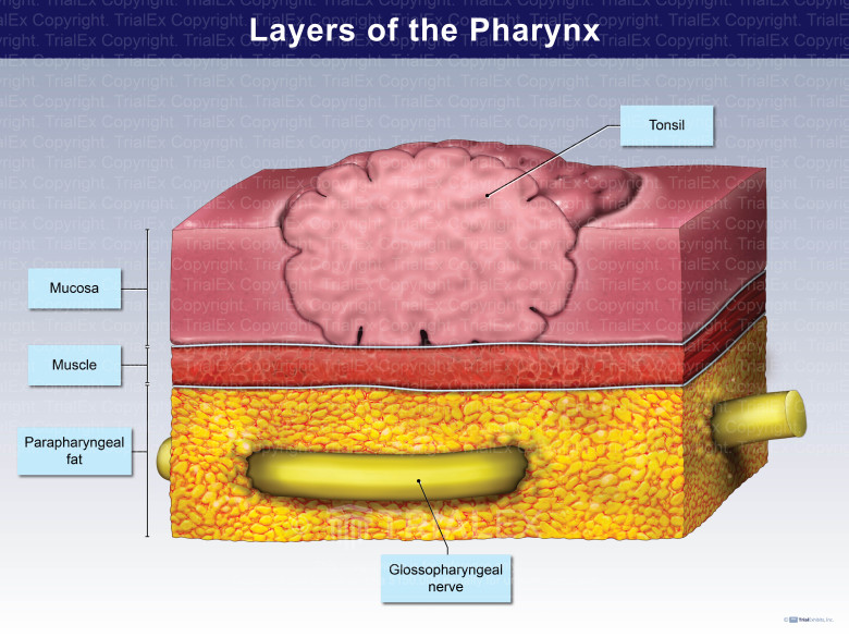
The fairings are a structure made up of muscles and tissue. Which is connected to the function of both the respiratory and digestive systems.
The structure of the fairings consists of three tissue layers arranged from the outside to the inside. Which are as follows.
Muscular tissue Layer.
It is the tissue layer made up of muscles on the outermost side of the fairings. It is a layer made up of involuntary and some voluntary muscles. Most of the muscles are involuntary. These muscles help in the swallowing process.
Fibrous tissue layer.
This is the middle layer in the pharynx. It is a layer made up of many collagen fibers and connective tissue. This layer is thickest in the nasopharynx and then its thickness decreases as it moves towards the oropharynx and larynx. Therefore, the airway of the arch of the nasopharynx remains patent and does not collapse.
Mucous membrane layer.
The innermost lining of the pharynx is made up of a layer of mucous membrane. Ciliated columnar epithelium tissue is found in the nasopharynx and stratified epithelium tissue is found in the oropharynx and laryngopharynx.
This layer forms the continuous inner lining of the mouth, pharynx, larynx, and esophagus.
Functions of Pharynx:
- It helps in the hearing process by balancing the air pressure between the nasopharynx and the middle ear, as it is connected to the eustachian tube.
- It helps in warming the air taken in inspiration. It also humidifies it. So that the air entering the lungs becomes moist and warm as much as body temperature. So that it does not damage the tissue of the inner lining.
- Since the tonsils are located in the part of the pharynx, they help in providing specific defense against microorganisms. Thus, it also functions as a protection.
- Since the olfactory nerve endings are located in the tympanic membrane, it is also important in the perception of taste.
- Due to the elasticity of the tympanic membrane, it also helps in producing the quality of sound. It also functions for speech.
- The tympanic membrane acts as a pathway for both the respiratory and digestive tracts. So it is important in both air passage and food passage.
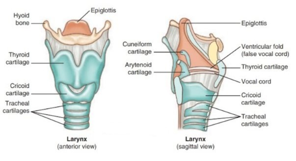
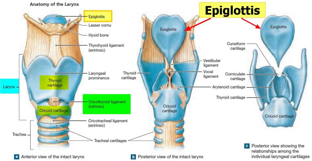
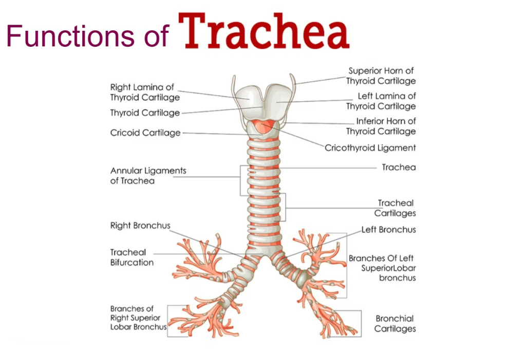
Structure and function of Larynx (Structure and functions of larynx):
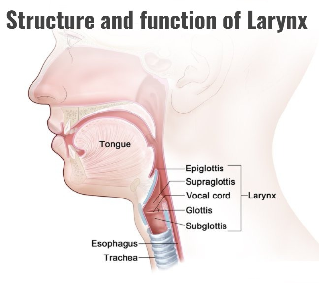
Linguistics is a structure made of cartilage. Which is arranged in the lower part of the fairings and is connected to the trachea below it.
The larynx is also known as the second voice box, because it is the organ that produces sound.
It is located from the hyoid bone, i.e. from the level of the 3rd cervical vertebra to the 6th cervical vertebra.
The structure of the larynx mainly consists of cartilage. This cartilage is made up of chondrocyte cells.
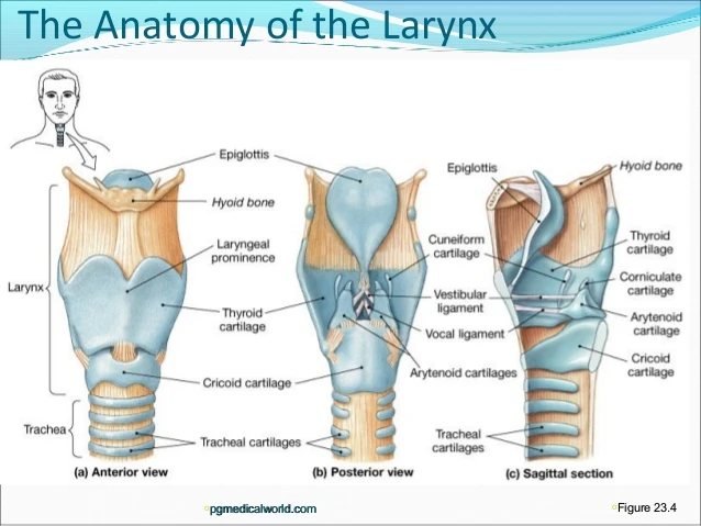
The following cartilages are found in the structure of the larynx:
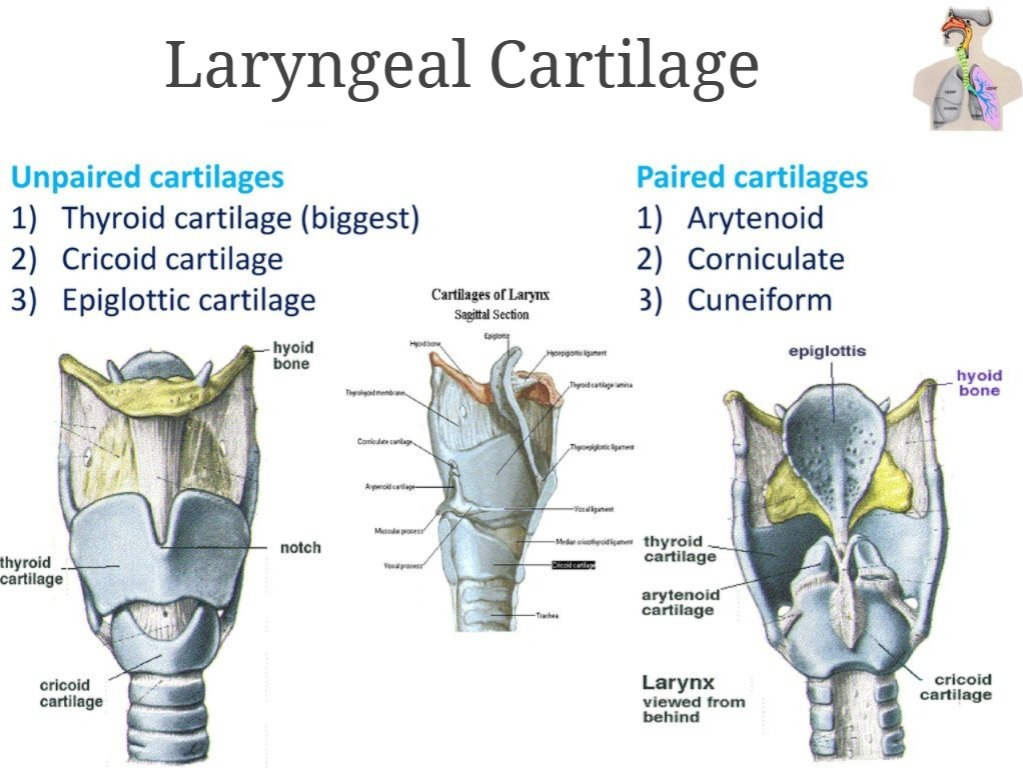
Unpaired cartilages:
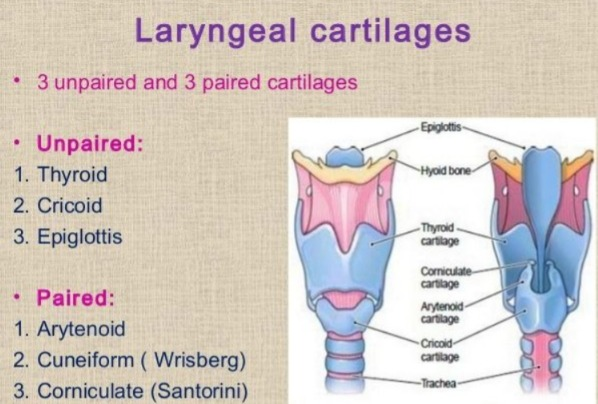
In the structure of the laryngs, there are some cartilages with a single structure, which are as follows.
Thyroid Cartilage:
- The largest cartilage in the structure of the larynx is the thyroid cartilage, which forms the V-shaped structure of the larynx. This part is made of hyaline cartilage.
- The part of the thyroid cartilage on both sides of this cartilage is known as the thyroid lamina and the grooved part in the middle is known as the thyroid notch.
- The processes extending from the thyroid cartilage lamina above and below are known as the superior and inferior cornu of thyroid cartilage.
- This thyroid cartilage The raised part is also known as the Adam’s apple.
Cricoid cartilage:
- This cartilage is arranged like a signet ring on the underside of the thyroid cartilage. Its shape is seen as a truncated ring shape. This cartilage is also made up of hyaline cartilage.
- The cricoid cartilage is connected to the trachea and thyroid cartilage by a ligament.
- When a tracheostomy procedure is performed, the cricoid cartilage is considered as a landmark.
Epiglottis cartilage:
- This is a leaf-shaped structure made of cartilage. It is located on the upper side of the thyroid cartilage. Its upper part is wide and its lower part is narrow.
- During the swallowing process, it moves over the larynx and esophagus. Due to this, food and liquid are stopped from entering the larynx and trachea. Its structure consists of elastic cartilage.
Paired cartilages:

Some cartilages in the structure of the ligaments are paired. These types of cartilage are as follows.
Arytenoid cartilage:
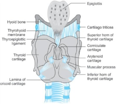
This is made up of hyaline cartilage. It forms part of a triangular shape. Which is attached to the vocal cords and its base is attached to the cricoid cartilage.
Corniculate cartilage:
It is a horn-shaped cartilage made of elastic tissue. It is attached to the anterior part of the arytenoid cartilage. This is the smallest cartilage compared to all cartilages.
Cuneiform cartilage (Cuneiform Cartilage):
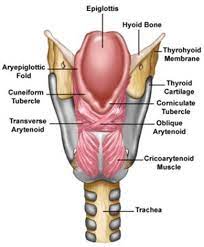
- It is a rod-shaped cartilage made of elastic tissue located near the base of the epiglottis. It is a supporting structure for the epiglottis.
- It is located anterior to the corniculate cartilage and supports the vocal cords.
- The larynx is supplied with blood by the laryngeal artery and has venous return by the laryngeal vessels. supplied with nerve by the laryngeal nerve.
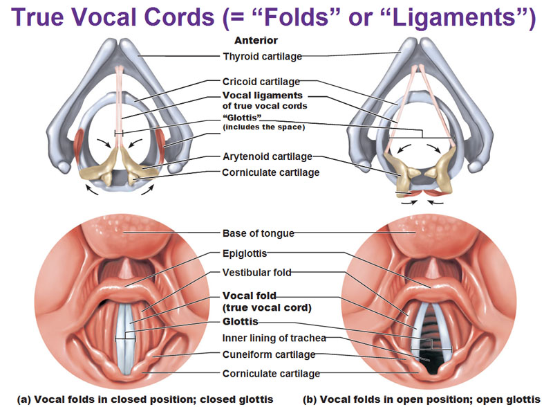
Production of voice:

- The layers of mucus membrane within the laryngs form a pair of two folds. Where the superior pair is called the false vocal cords or ventricular folds and the inferior pair is called the true vocal cords or vocal folds.
- The space between the two ventricular folds is called the rima vestibuli and the space between the two vocal folds is called the rima glottidis.
- When the air pressure is released from the thoracic cavity, both vocal folds straighten and It vibrates. Due to which sound is produced. Due to giving more pressure and tension, high pitch sound is produced. While due to less tension and pressure, low pitch sound can be produced.
- The quality of sound depends on the structure of the oral cavity, teeth, nasal cavity, pharynx, sinuses, vibrating capacity of the vocal cords, etc.
Functions of Larynx (Main function of the larynx):
- The main function of the eardrum is to produce sound.
- The characteristics of sound include pitch, resonance, volume, etc. All these things depend on the structure of the oral cavity, pharynx, larynx, nasal cavity, sinuses, tongue and teeth as well as lips etc.
- The larynx functions as an air passage.
- Since the inner lining of the larynx contains mucus membranes and cilia, it functions to filter the air and warms the air to body temperature and also functions to humidify the air. This prevents damage to the inner mucus membrane.
- All the above organs are considered as organs of the upper respiratory tract, because they are structures outside the thoracic cavity.
The organs inside the thoracic cavity are considered as organs of the lower respiratory tract. The organs involved are as follows.
Structure and Function of Trachea ( Structure and Function of Trachea ):
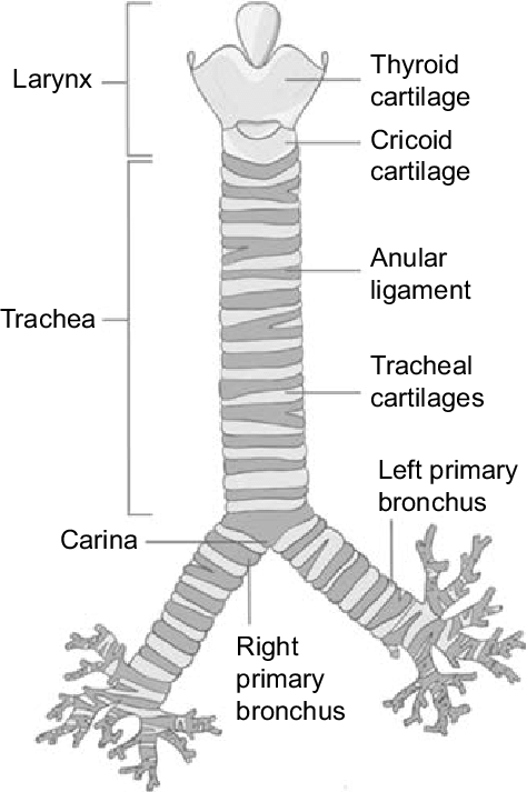
- The trachea is a pipe-like structure that starts at the back of the larynx and ends at the bronchi.
- It is also known as the wind pipe. It functions as the principal air passage. Its length is 12 cm and its diameter is about 2.5 cm.
- The lining of the trachea is made up of C-shaped rings of cartilage. This cartilage is located in the anterior wall of the trachea. The posterior wall does not have such cartilages.
- The anterior wall of the truncus is made up of 16 to 20 incomplete C-shaped cartilages of this type. Due to this cartilage, the strength of the anterior wall increases. So that the truncus does not collapse due to pressure from the front and this cartilage works to provide protection from the front. This cartilage is of the highline type.
There are 4 tissue layers arranged around the truncus which are as follows.
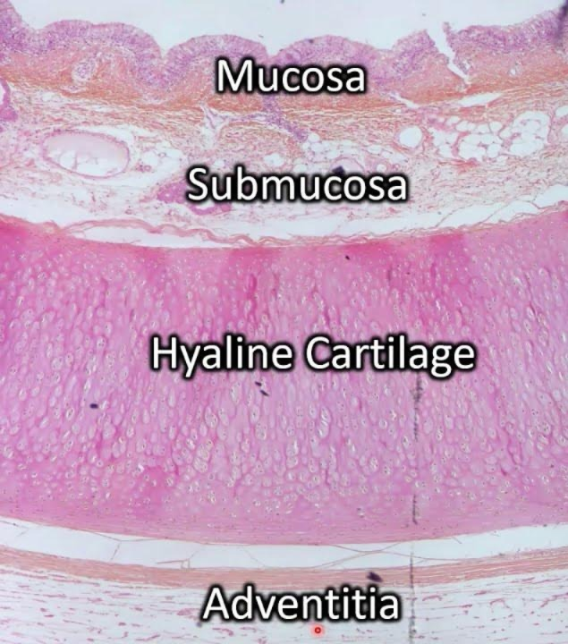
Adventitia:
This tissue layer is located on the outermost side of the trachea. It is made up of areolar connective tissue.
Hyaline cartilage:
This hyaline cartilage is located on the underside of the adventitia layer. This layer contains fibroelastic tissue and C-shaped hyaline cartilages are arranged on the anterior wall of the trachea.
Submucosa:
This layer is located below the hyaline cartilage. It is the layer arranged above the submucosa layer. Mucus glands and their ducts are located in this layer. In addition, blood vessels, lymph vessels and nerves are arranged in this layer.
Mucosa:
This is the layer of the innermost wall of the trachea. It is made up of ciliated columnar epithelium tissue. This layer also contains goblet cells, which secrete mucus. This layer is moist.
This layer helps in humidifying, filtering and warming the air entering the lungs.
Functions of Trachea:
- It acts as the principal air passage.
- It helps in warming, filtering and humidifying the air entering the lungs.
- With the help of cartilage in the front wall of the trachea, the opening inside the trachea remains open, it does not collapse and the patency of the airway is maintained.
- Because the cilia in the lining inside the trachea are arranged in reverse direction It helps in moving the contents, mucus or dust particles etc. accumulated inside the trachea outwards. So that the airway can be kept clear.
- The inner wall of the trachea has nerve endings. Which are sensitive to any irritation. Due to any irritation in the inner lining of the trachea, it stimulates the respiratory center. So that the cough reflex is produced and the mucus and foreign material accumulated inside can be removed by cuffing.
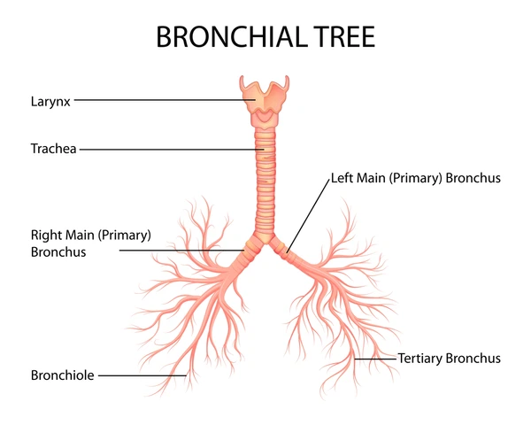
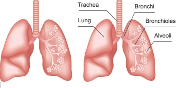
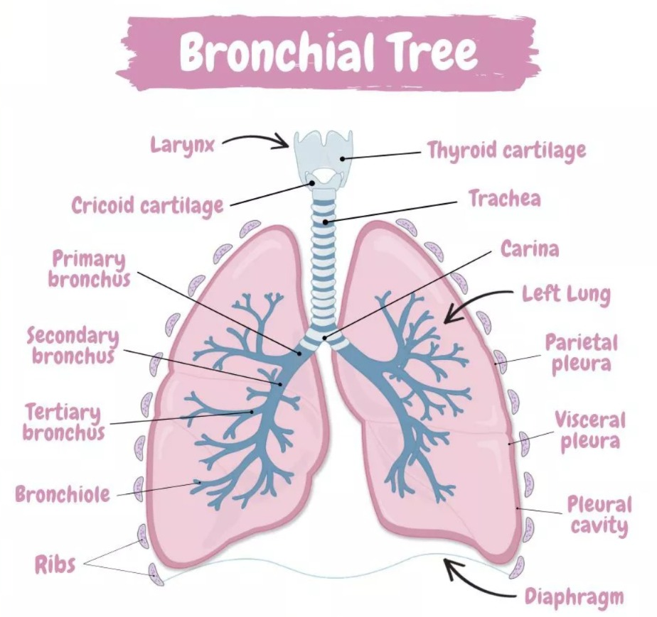
Structure of Bronchi and Bronchioles:
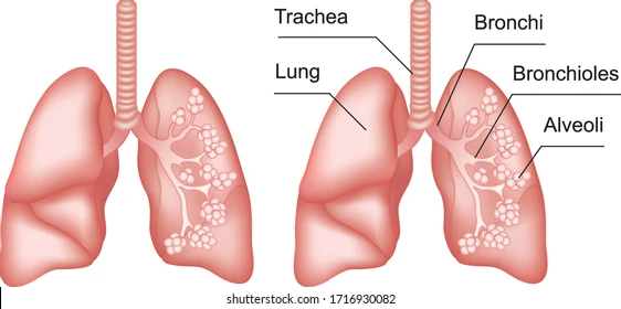
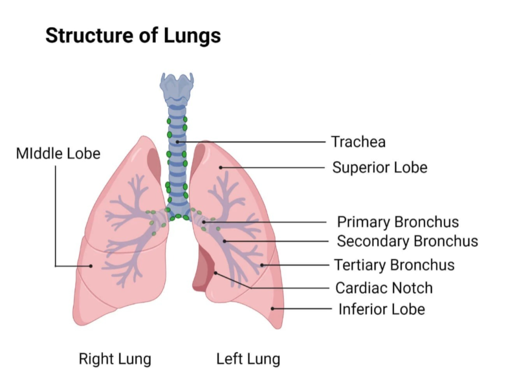
At the level of the 5th thoracic vertebra, the trachea divides into two branches. These branches are called bronchi. The right and left bronchi are called primary bronchi. This is where the branches of the bronchial tree begin.

Right Bronchi :
- The right bronchus is shorter and wider than the left bronchus and is arranged in a more vertical shape. So there is a higher chance of the foreign body getting plugged in the right bronchus.
- It is found to be approximately one inch long. Its wall is lined with ciliated columnar epithelium tissue. Its wall is made of incomplete cartilage.
- The right bronchus continues and divides into three separate branches in the three lobes of the lung, and one branch enters each lobe of the lung. Then it enters the lobe of the lung and divides into small separate branches. It divides into secondary and tertiary bronchi and bronchioles.
- The tertiary bronchi are further divided into grape-shaped alveoli. The lung structure consists of millions of alveoli. As we move towards the alveoli in the bronchial tree, the cartilage disappears.
Left Bronchi:

- The left bronchus is narrower and longer than the right bronchus. Its length is approximately 2 inches. Its structure is also similar to the right bronchus. It branches into two lobes of the left lung and then divides into many branches in each lobe of the lung.
- Oxygenated blood is supplied through the bronchial arteries and deoxygenated blood returns through the bronchial veins. It is supplied by the autonomic nervous system.
Functions of Bronchi and Bronchioles (Structure of Bronchi and Bronchioles):
- It works by warming and humidifying the air.
- It stimulates the cough reflex so that the respiratory tract is protected.
- It keeps the tract patent so that respiration can be taken with ease.
- It works by removing the mucus accumulated in the bronchi through the cough reflex.
- Controls the entry of air taken in during breathing and distributes it equally everywhere.
Structure of Alveoli (Alveoli Structure):
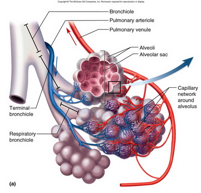
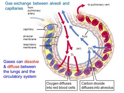
- At the ends of the terminal bronchioles are tiny air sacs, shaped like clusters of grapes. These are called alveoli. This is the primary structure for gas exchange to occur in the lung.
- The wall of the alveoli is very thin and made up of single squamous epithelium tissue. So that the gas exchange of oxygen and carbon dioxide can be done easily by the method of diffusion.
- Around these alveoli is a network of pulmonary blood capillaries. This gas exchange between blood and alveoli is also called external respiration.
- There is a fluid on the surface of the alveoli. Which is called surfactant. This surfactant reduces the surface tension. It prevents the alveoli from collapsing. Prevents the membrane of the alveoli from drying out and also helps in gas exchange.
Functions of Alveoli:
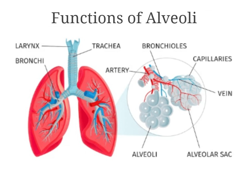
- The main function of the alveoli is gas exchange. It exchanges gases of oxygen and carbon dioxide.
- It humidifies and warms the air because it is surrounded by pulmonary capillaries. Due to this, the air becomes warm and moist.
- Since the membrane of the alveoli is wet, dust particles and foreign materials get plugged there and they also work to remove them from there through coughing and sneezing.
- Connective tissue is present in the walls of the alveoli. So it also contains lymphocytes and plasma cells and it also synthesizes some antibodies, which provide resistance to local foreign bodies and act as a protection.
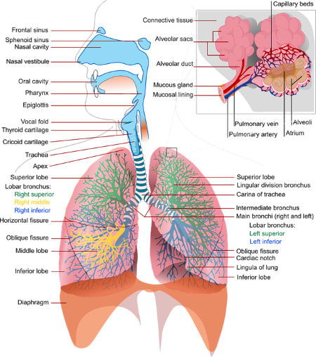
Structure of the Lung:
- The lungs are an important organ of the respiratory system. They are located in the thoracic cavity, one on each side of the mediastinum space, in a total of 2.
- The lungs take in oxygen from the atmosphere and It functions to remove carbon dioxide from the body.
- The lungs are located in the thoracic cavity in number of 2. They are conical shaped.
- The lungs are separated from the thoracic cavity by the heart and the mediastinum space.
- The lungs are made of spongy tissue with many air-filled cavities inside. Its color is found to be brown or gray.
- The right lung weighs approximately 625 grams and the left lung weighs approximately 575 grams. The right lung is heavier in weight and larger in structure than the left lung.
- The lungs are divided into lobes. The right lung has 3 lobes, in which the superior lobe, middle lobe and inferior lobe are seen while the left lung has 2 lobes, the superior lobe and the inferior lobe. These lobes are separated by a fissure. There are two fissures in the right lung. A fissure is seen in the left lung.
- The lungs are classified into the following parts.
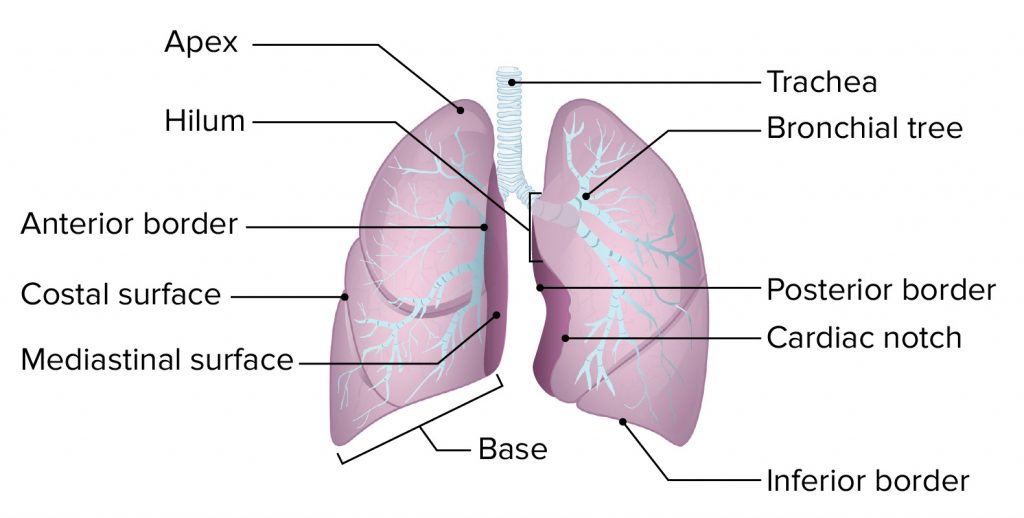
1. Apex:
The triangular and round part of the upper lung is called the apex. It is seen up to the level of the clavicle bone.
2. Base:
The lower broad part of the lung is called the base. This part of the base is attached to the diaphragm at the bottom. This part is concave in shape.
3. Anterior Border:
It is thin. It is shorter than the posterior border. It has a cardiac notch in it. In which the heart part is arranged.
4. Posterior Border:
It is thick. It is found from the 7th cervical vertebra to the 10th thoracic vertebra.
5. Inferior Border:
It is located at the bottom of the lung. It separates the costal surface and the medial surface. The costal surface is large and convex. It is in contact with the costal pleura. It is attached to the ribs and intercostal muscles by costal cartilage.
6. Medial Surface:
It is concave. In its middle part there is a groove called the hilum. This hilum is located at the level of the fifth, sixth and seventh thoracic vertebrae. From this hilum, bronchi, pulmonary blood vessels, lymphatic vessels, and nerves enter and exit each lobe of the lung.
In the middle of the medial surface is the mediastinum space, which separates the two lungs. This space contains structures such as the heart, great vessels, trachea, bronchi, esophagus, etc., which separate the two lungs.
Structure of the Lob of the Lung:
- The lobes of the lung are made up of many lobules. One lobe is separated from the other lobe by a fissure. There is a groove in the middle of the lung called the hilum. The following structures are found inside each lobe from this hilum.
- The bronchi enter each lobe of the lung. After entering, they divide and become secondary bronchi, tertiary bronchi, terminal bronchioles, alveolar sacs and small grape-like alveoli. Thus, this structure is found in a tree-like structure in the lobes of the lungs, which is called the bronchial tree or respiratory tree.
- A network of capillaries of the pulmonary artery and pulmonary vein is spread around these alveoli. Gas exchange takes place here between oxygen in the alveoli and carbon dioxide in the blood capillaries during inspiration. Which is known as external respiration.
- Thus, each lobe of the lung contains a network of bronchial trees, pulmonary vessels capillaries, lymph capillaries, nerves and lung parenchymal tissue.
Pleura:
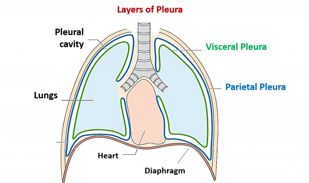
The pleura is a serous membrane surrounding both lungs. It is found in a double layer. The outer layer is called the parietal pleura and the inner layer is called the visceral pleura.
There is a cavity between the parietal pleura and the visceral pleura, which is called the pleural cavity. Here is cirrhotic fluid which is also called pleural fluid.
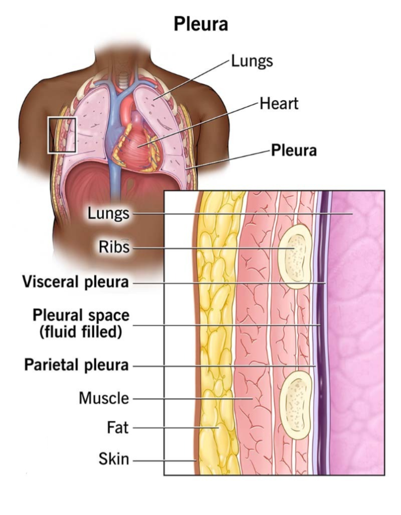
The pleural fluid in the pleural cavity causes friction between the two layers. There is no space for the lungs to expand. The pleural fluid in this cavity is viscous and acts as a lubricant.
The visceral pleura is the layer that is attached to and very close to the lungs. While the parietal pleura is the layer attached to the ribs and muscles.
Difference between right lung and left lung:
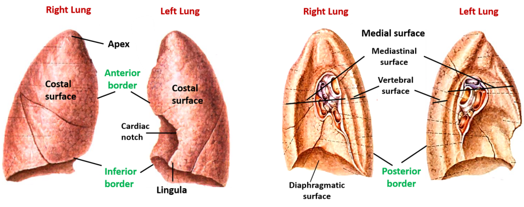
- The right lung has two fissures to separate the lobes while the left lung has only one fissure to separate the lobes.
- The right lung has three lobes, superior, middle and inferior, while the left lung has two lobes, superior and inferior.
- The anterior border of the right lung is straight while the anterior border of the left lung is interrupted because the cardiac notch is present on the left lung side where the heart resides.
- The right lung is heavier and larger in weight. It weighs approximately 625 grams. While the left lung is lighter and smaller in weight. It weighs approximately 575 grams.
- The right lung is short and wide while the left lung is long and narrow.
Function of lung:
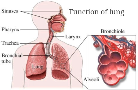
- It is an important organ for respiration.
- Through inspiration, oxygen reaches the lungs and mixes with the blood, providing oxygen to the entire body.
- The body’s waste product carbon dioxide is removed from the body through the act of expiration. is.
- Delivers oxygenated blood to the heart through the pulmonary circulation.
- Excretes excess water from the body through the act of respiration.
- Excretes other gaseous wastes produced in the body from the body through the act of expiration.
Respiratory muscles (respiration muscles) Muscles):
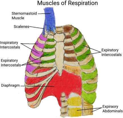
- Respiration means bringing the outside air to the lungs through the act of inspiration and the air accumulated inside the lungs into the outside environment through the act of expiration. Throwing.
- During this action, there is an increase and decrease in the size of the thoracic cavity, which is mainly done by the muscles associated with the act of respiration.
- The muscles of respiration mainly perform involuntary functions. The muscles involved in this action are as follows.
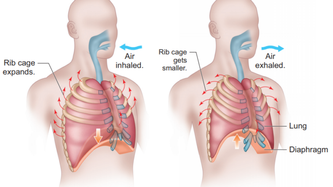
Diaphragm:
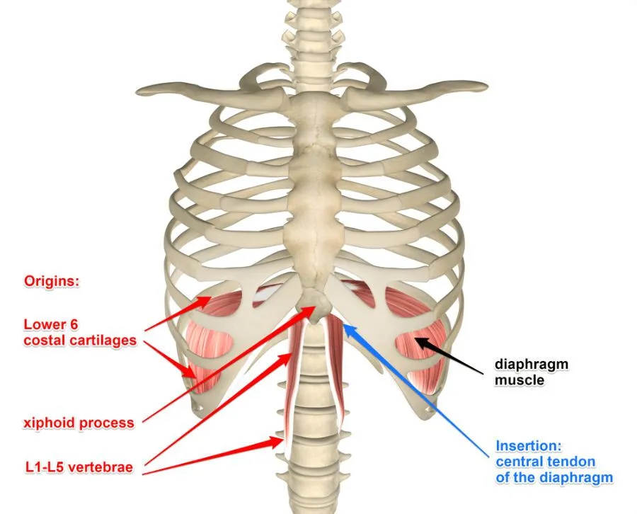
- This muscle located between the thoracic cavity and the abdominal cavity has a dome-shaped structure.
- The diaphragm receives contraction impulses from the phrenic nerve. Due to which its movement up and down is seen.
- The diaphragm is connected to the thoracic vertebrae by a central tendon. It is also connected to the xiphoid process, pleura and ribs on the sides on the upper side. Therefore, due to the movement of the diaphragm, the movement of the chest cavity increases and decreases.
- During the act of inspiration, the diaphragm moves downwards and the size of the thoracic cavity increases, causing the act of inspiration. When the diaphragm returns to its original position and relaxes, the diameter of the thoracic cavity decreases and the act of expiration is observed.
Intercostal muscles:
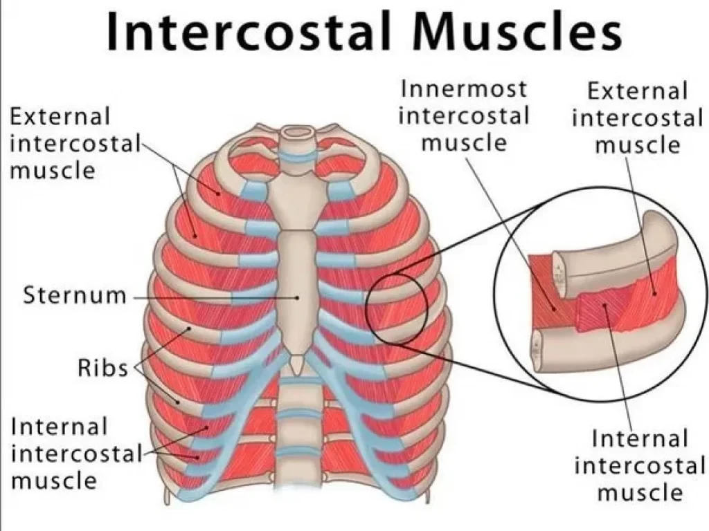
- The space between two ribs is called the intercostal space. The muscles attached to this part are called intercostal muscles. There are total 11 pairs of intercostal muscles. They are arranged in two layers. On the outside are the external intercostal muscles and on the inside are the internal intercostal muscles.
- These intercostal muscles are supplied with impulses by the intercostal nerve. The first rib is fixed and the remaining ribs move towards the first rib due to the movement of the intercostal muscles.
- When the intercostal muscles contract, they increase the diameter of the cavity and when they relax, the size of the thoracic cavity decreases.
- The contraction and relaxation of the above two muscles, the diaphragm and the intercostal muscles, cause the action of inspiration and expiration.
Cycle of respiration (Mechanism of respiration):
- Respiration is the gas exchange between two surfaces. In which air from the atmosphere enters the lungs. Gas exchange between the lung tissue and the blood is called external respiration. The gas exchange between every cell, tissue and blood of the body is called internal respiration.
- During the act of respiration, oxygen enters the lungs through inhalation and carbon dioxide leaves the body through exhalation.
- Usually, the act of respiration occurs 16 to 18 times in a minute.
The following actions are seen in the cycle of respiration.
Inspiration (Inspiration)
Expiration
Pause
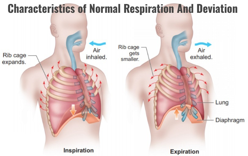
Inspiration (Inspiration):
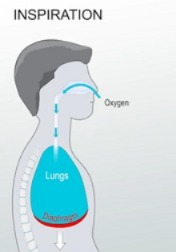
- The act of taking in air from the atmosphere into the lungs through breathing is called inspiration.
- When the brain receives nerve impulses to contract the diaphragm and intercostal muscles, the size of the thoracic cavity increases due to the contraction of the diaphragm and intercostal muscles. The air pressure inside the thoracic cavity decreases so that air from the outside environment can enter the lungs through the act of inspiration. This action is called inspiration.
- When the diaphragm receives contraction impulses, the diaphragm becomes flat and moves downwards and the intercostal muscles contract, causing the ribs and intercostal muscles to move upwards and outwards. This increases the size of the thoracic cavity and creates negative pressure inside the cavity. The action of inspiration can occur when the air pressure in the outside environment is high and the air pressure in the thoracic cavity is low. The act of inspiration is an active process.
Expiration:
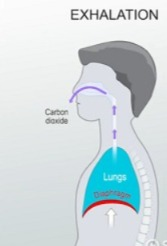
- The process of expelling air from the lungs into the atmosphere is called expiration. The act of expiration is a passive process that begins after the act of inspiration is completed.
- During the act of expiration, the contracted diaphragm and intercostal muscles relax. So that the diaphragm returns to its original position and the ribs come down and inward, the size of the thoracic cavity decreases and the act of expiration occurs. In which air is expelled from the lungs into the atmosphere.
- During the act of expiration, the air pressure in the lungs is greater than the pressure of the atmosphere, so the act of expiration occurs.
Pause (pause):
- This is the relaxed stage of the lungs. In which no inspiration or expiration takes place. This period is called the pause period.
Terminologies related lung volume and vital capacities of the lung:
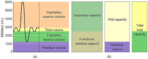
The act of inspiration and expiration is called respiration. Normally 12 to 18 respirations are seen in 1 minute. Spirometer or respirometer is a useful device to measure the air volume during respiration. The terminologies related to this normal respiration and lung capacity are as follows.
Tidal volume:
- The amount of air that enters the lungs during normal inspiration and leaves the lungs during normal expiration during one respiration is called tidal volume. Normally it is 500 ml .
Inspiratory reserve volume (IRV):
- The amount of air that can be inhaled into the lungs in addition to the air taken in during normal inspiration is called inspiratory reserve volume. Its normal amount is 3100ml.
Total inspiratory capacity :
- The total inspiratory capacity in the lungs, i.e. the total of the titral volume and the inspiratory reserve volume, is called the total inspiratory capacity. 500 + 3100 = 3600 This is the total inspiratory capacity of the lung.
Expiratory reserve volume (ERV):
- The maximum amount of air that can be exhaled during a forceful expiration, in addition to the air that can be exhaled during normal expiration, is called the expiratory reserve volume. It is approximately 1200ml .
Residual volume:
- The amount of excess air that still remains in the alveoli during expiratory reserve volume, which is not expelled even during forceful expiration, is called residual volume.
- That amount of air cannot be measured even by a spirometer and this residual volume does not cause alveoli to collapse. Its normal volume is 1200ml .
Vital capacity of lung:
- The sum total of tidal volume, inspiratory reserve volume and expiratory reserve volume is called the vital capacity of the lung. It is approximately 4800 ml.
Anatomical dead space:
- The amount of air in the lower respiratory tract that is not involved in gas exchange. All that space is called anatomical dead space. The air in this space only functions as a conductor (sometimes called a ventilator). Does not perform gas exchange.
Total lung capacity:
- This includes the vital capacity of the lungs plus the residual volume.
- Total lung capacity is estimated As much as 6000ml is found.
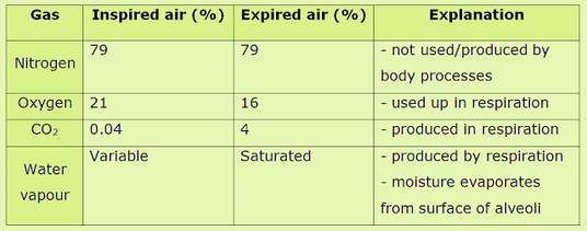
Composition of inspired air and expired air.
Control of Respiration (Control of Respiration):
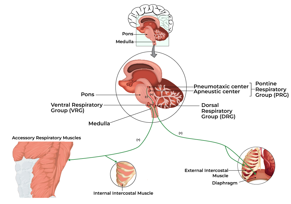
- Respiration is an involuntary process. It is not under the control of the person.
- The process of rhythmic respiration is under the control of the brain. This control structure is as follows.
- The respiratory center located in the medulla oblongata of the brain controls the rate and depth of respiration.
- The pneumotoxic area and the apneustic area are located in the pons veli. The coordination of respiration is also maintained through it.
- The chemoreceptors are located in the medulla oblongata of the brain. These chemoreceptors monitor the amount of carbon dioxide and oxygen in the blood present there.
When there is an increase in the amount of carbon dioxide in the blood, these chemoreceptors are stimulated, i.e. in the case of hypercapnia, these chemoreceptors are stimulated and increase the rate of respiration. - Thus, all the above structures control respiration. So that the amount of oxygen and carbon dioxide in the body is maintained.
Characteristics of Normal Respiration And Deviation:
Characteristics of Normal Respiration:
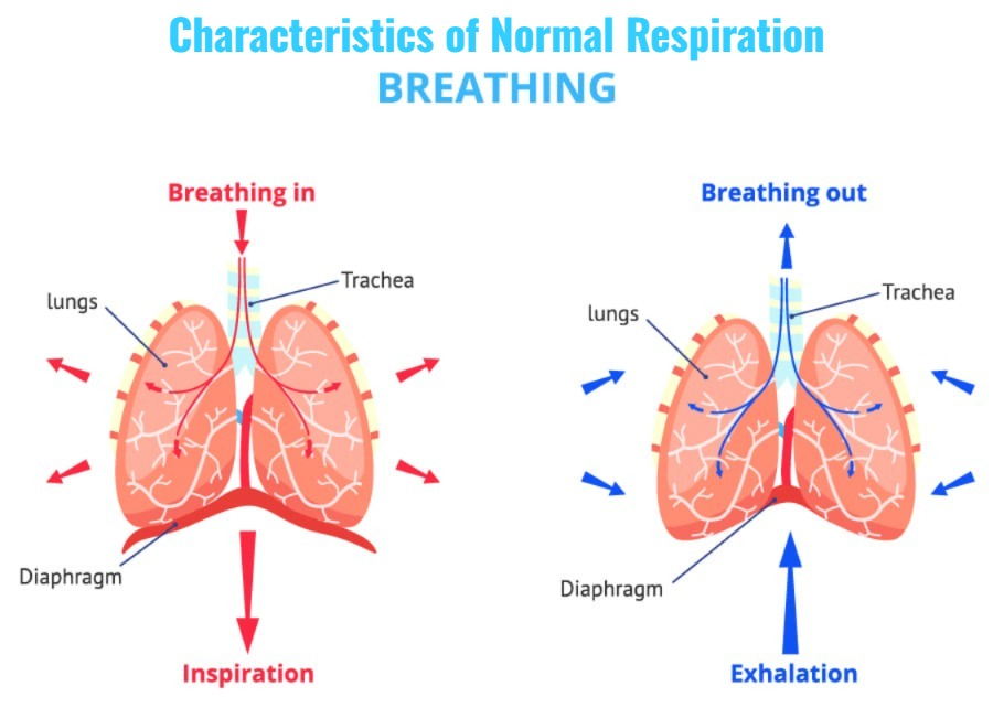
Normal respiration is the natural process of breathing in and out, which is called Eupnea. Normally, this process provides sufficient oxygen to the body and removes carbon dioxide.
Main Symptoms:
1.Respiratory Rate:
Normal breathing rate in adults: 12 to 20 breaths per minute Minutes.
Children have a higher rate (30-60 breaths/minute).
2.Rhythm:
The act of breathing in and out is regular and balanced.
3.Depth:
The breath is neither too deep nor too shallow, balanced.
4.Effortless:
There is no difficulty or difficulty while breathing.
5.Soundless:
Breathing is noiseless, clean and quiet.
Deviations in Respiration:
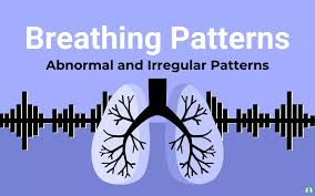
When there is a problem with breathing, the following medical conditions are observed:
1.Tachypnea:
The rate of breathing increases (>20/min in adults).
Cause:Fever, Pain, Anxiety, Exercise.
2.Bradypnea:
The breathing rate is very slow (<12/min in adults).
Cause: Brain injuries, opioid overdose, during sleep.
3.Apnea (Apnea):
Breathing stops completely (10 seconds or more).
Cause:Sleep apnea, cardiac arrest.
4.Dyspnea:
You may experience difficulty breathing.
Cause: Asthma, Heart failure, Pulmonary disease.
5.Orthopnea:
There is difficulty in breathing while sleeping, and it is relieved in a sitting position.
Usually associated with heart failure.
6.Cheyne-Stokes Respiration :
The rate of breathing gradually increases and then decreases, after which breathing stops for a few moments.
Cause:Neurological disorders, heart failure, terminal stages.
7.Kussmaul Breathing:
Deep and rapid breathing.
Cause: Metabolic acidosis, especially diabetic ketoacidosis.
8.Hyperventilation:
Breathing very quickly and deeply.
As a result, the level of carbon dioxide decreases.
Cause:Anxiety, panic attack.
9.Hypoventilation:
Both the rate and depth of breathing decrease.
As a result, oxygen decreases and carbon dioxide increases Is.
Cause: Drug overdose, neurological injury.
Normal respiration requires proper rate, rhythm, depth, and ease. Any change in the breathing pattern can indicate a serious health condition. It is very important to seek medical advice for proper diagnosis and treatment of each deviation.