—ENGLISH-ANATOMY UNIT 4. CVS (PART : 2) HEART
Cardiovascular system:
The Cardiovascular System is a part of the Circulatory System, which includes the Heart, Blood, and the Blood Vessels that transport the Blood. The heart is an important organ of the cardiovascular system that performs a continuous pumping action.
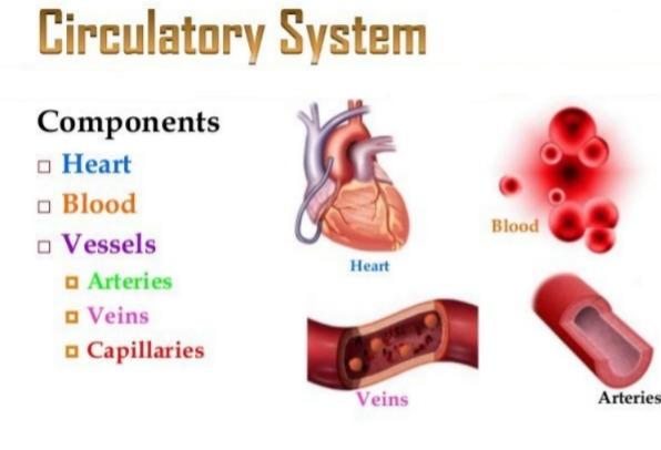
The circulatory system is a transport system of the body, which performs an important function in the body by delivering nutrition and oxygen to every cell and tissue of the body through blood.
To meet all these needs of the body, blood is transported throughout the body and circulates in blood vessels. The heart continuously pumps blood to circulate through the blood vessels.
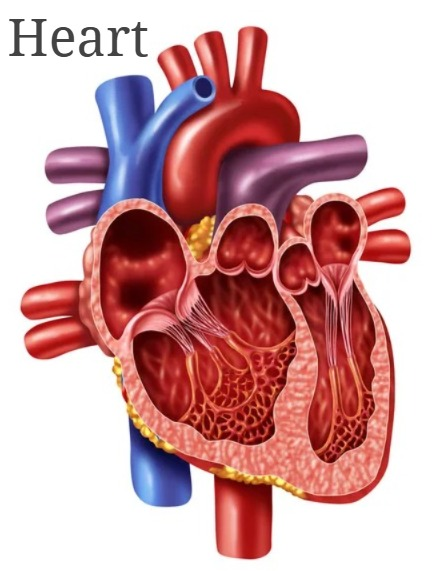
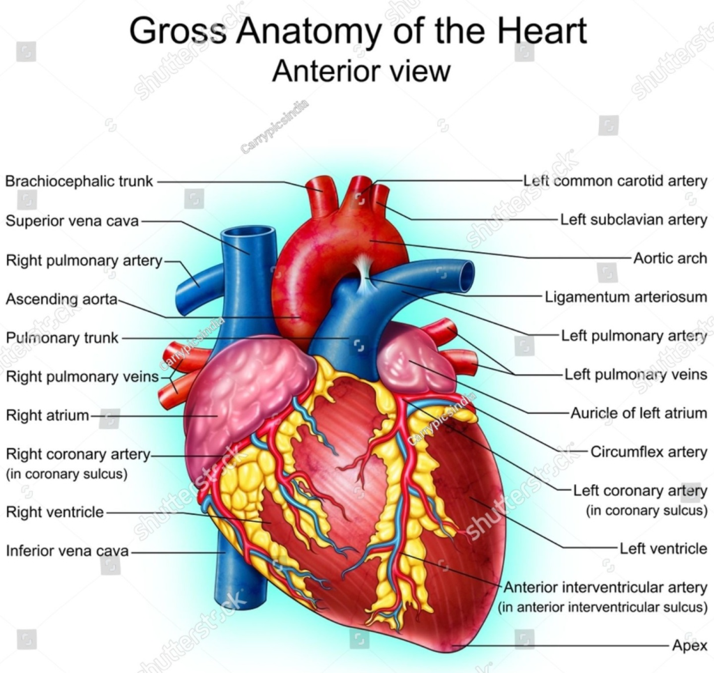
HEART (GROSS STRUCTURE OF THE HEART : Gross Structure of the Heart ):
- The heart is an important organ of the circulatory system. The heart beats continuously throughout human life. Due to its beating, blood circulates continuously in the blood vessels.
- The heart is an organ made up of heart and muscles. Its weight is approximately 310 grams in men and approximately 250 grams in women. The heart beats approximately one lakh times during the day.
Location of the heart:
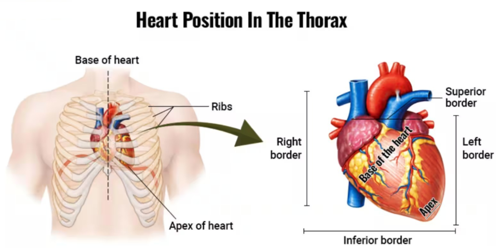
- The heart is located in the thoracic cavity between the two lungs, in the mediastinum space, above the diaphragm.
- The heart is a rough cone-shaped organ. Its upper part is known as the BASE and the lower angled part is known as the APEX.
- The heart is about the size of a human fist. It is 12 cm long, 9 cm wide and 6 cm thick.
- The heart is located in the thoracic cavity, slightly tilted to the left between the two lungs.
- The heart is found in the middle of the thoracic cavity, between the second and fifth intercostal ribs.
Organs surrounding the heart elements):
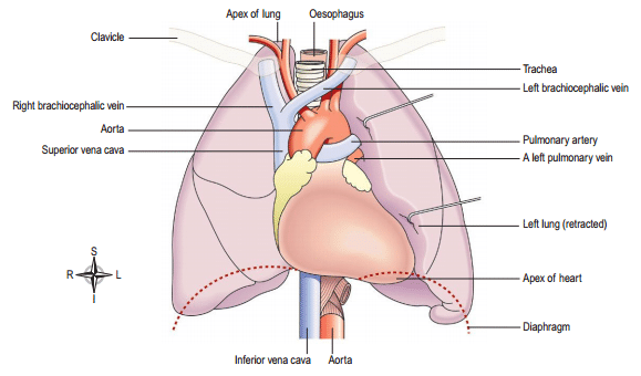
- The heart is located in the thoracic cavity. It has one lung on each of its left and right sides.
- The diaphragm and central tendon are present on the lower side.
- The vaena caeva and aorta and pulmonary artery and pulmonary vein are present on the upper side of the heart.
- The esophagus, trachea, bronchi and bronchioles and the descending aorta and thoracic vertebrae are present on the posterior side of the heart.
- The sternum bone and ribs and intercostal muscles are located on the front side of the heart.
Structure of the heart:
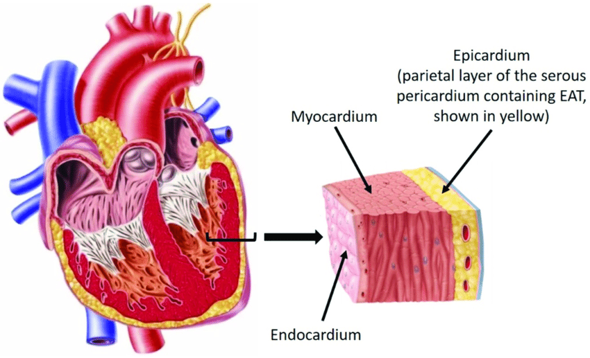
The heart is an organ made up of semi-muscles. Its wall is made up of three types of tissue layers.
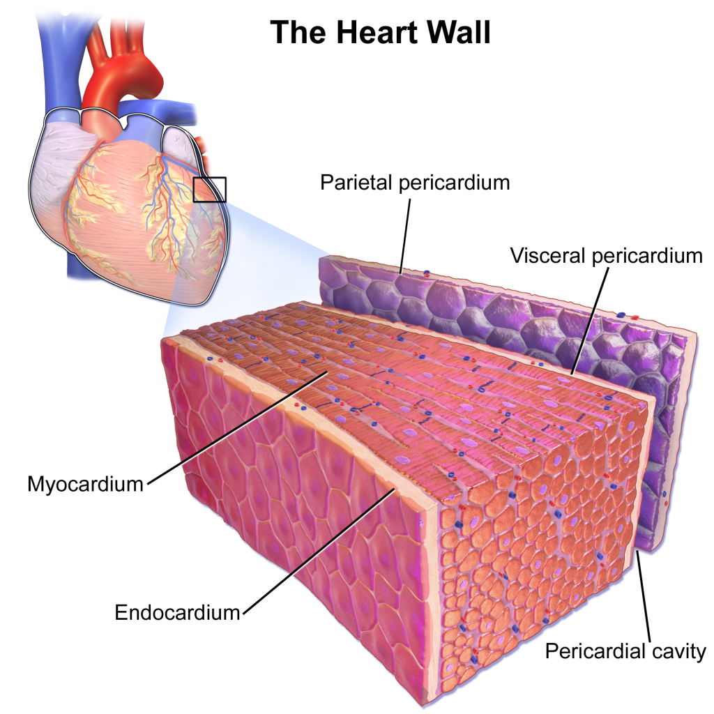
1. Epicardium or pericardium pericardium)
2.Myocardium
3.Endocardium
The outermost layer of the heart wall is called the epicardium or pericardium.
1.Epicardium or pericardium (epicardium or Pericardium):
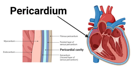
- It is thin and transparent and covers the heart from the outside. It is made of fibrous connective tissue. In which the outermost layer of fibrous tissue is present and the serous membrane is found in a double layer on the inner side of the fibrous tissue. The outer layer of the serous membrane is known as the parietal and the inner layer is known as the visceral pericardium layer.
- The space between the parietal and visceral pericardial layers is called the pericardial space. This space contains a fluid called serous fluid or pericardial fluid. Which prevents friction between the two layers.
- This outer pericardium layer works to protect the heart from the outside and this layer is also found around the vessels coming out of the heart.
2.Myocardium (Myocardium):
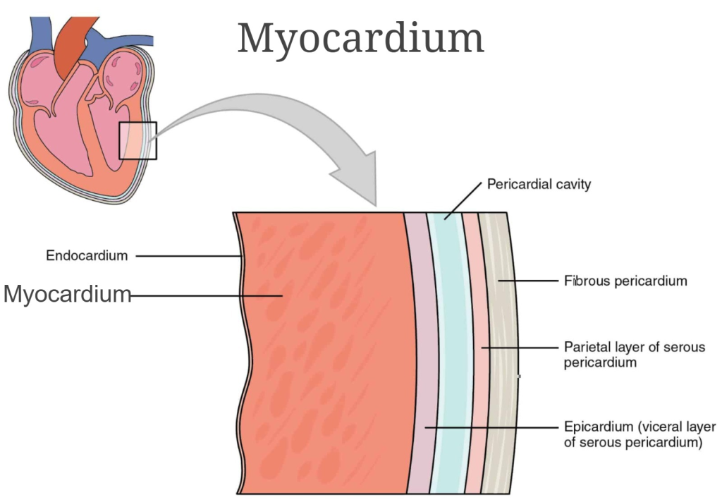
- The myocardium is the middle layer of the heart. It lies below the pericardium. It is made up of special types of cardiac muscle tissue. The pumping action of the heart is seen due to the contraction of these muscles.
- This layer of myocardium is thin at the base and thick at the apex. The wall layer of the left ventricle is also thicker than the wall of the right ventricle.
- The contraction of these muscles is an involuntary action, which causes the pumping action of the heart. Its control is done by the autonomic nervous system and the conducting system in the heart.
3.Endocardium (endocardium):
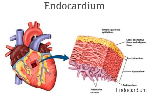
- It is the innermost layer of the heart. It is the layer in contact with the blood. This layer is made up of epithelial tissue and connective tissue. This layer is smooth and shiny, which is important for blood to flow easily within the heart. This layer also covers the valves inside the heart and is also found continuously in the inner walls of the blood vessels leaving the heart.
Chambers of the Heart:

- The heart is mainly divided into two parts. The right side and the left side.
- The septum of the heart is located between these right and left sides. This is again divided into four chambers by a valve between the right and left sides of the heart, above the valve and below the valve.
- The two chambers above the valve are called atrium or auricle. The two chambers below the valve are called ventricles. Thus, the heart is divided into four chambers.
- The right side of the heart contains deoxygenated blood and the left side of the heart contains oxygenated blood. After birth, there is no connection between the right and left sides. Before birth, there is an opening between the two atria called the foramen ovale, but it closes after birth.
Valve of the heart:
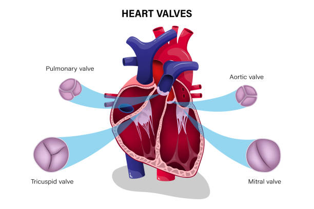
The tissue flaps inside the heart are called valves. There are two types of valves in the heart.
1.Atrioventricular valve
2.Semilunar valve
1.Atrioventricular valve (Atrioventricular valve) Valve):
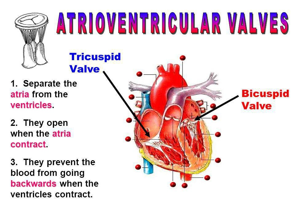
- These valves are located in the heart between the atrium and the ventricle. The valve between the right atrium and the ventricle is called the tricuspid valve. This valve is made up of three tissue flaps.
- The valve between the left atrium and the ventricle is called the bicuspid valve or mitral valve. This valve is made up of two tissue flaps.
- The hanging ends of both the bicuspid and tricuspid valves are attached to the inner wall of the ventricle of the heart by the chordi tendini and papillary muscles. This strengthens the valve and prevents it from opening in the opposite direction.
2.Semilunar valve:
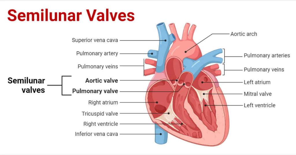
- Since this valve is C-shaped or crescent-shaped, it is called semilunar valve. These valves are present in the aorta and pulmonary artery. The valve present in the aorta is called aortic valve and the valve present in the pulmonary artery is called pulmonary valve. Which are located at the opening of their vessels. This valve also opens in only one direction.
- Both the above atrioventricular valves open when the pressure in the atrium increases, that is, during the contraction of the atrium, and blood flows from the atrium to the ventricle. And during the contraction of the ventricle, the atrioventricular valve closes and the semilunar valve opens and due to this, blood flows into the aorta and pulmonary artery.
- Thus, due to the opening and closing of these valves, blood circulation in the heart can occur. This valve opens in one direction so that blood cannot flow in the opposite direction.
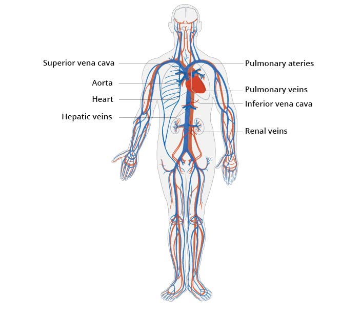
Openings of the Heart:
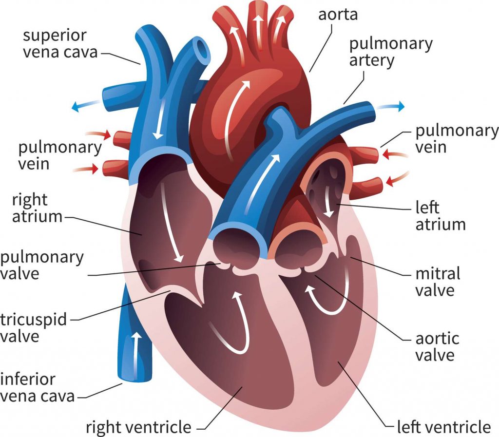
Mainly large blood vessels are connected to the heart. Through which blood comes from the body to the heart and is pumped from the heart and the blood goes out to the body. The vessels and their openings are as follows.
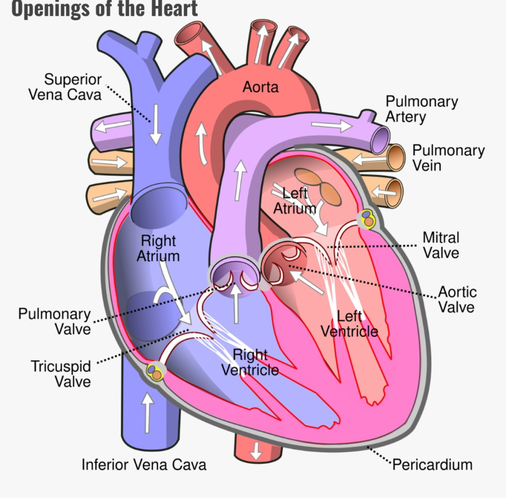
Superior Venacava..(Superior Venacava.).
These blood vessels bring deoxygenated blood from the upper part of the thoracic cavity, head and neck to the right atrium of the heart. They are one in number.
Inferior Venacava..(Inferior Venacava.).
These blood vessels bring deoxygenated blood from the inferior part of the body and the lower part of the thoracic cavity and open into the right atrium of the heart. It is one in number.
Pulmonary artery..
It carries deoxygenated blood from the right ventricle of the heart out of the heart to the lungs. Its number is one.
Pulmonary vein..
Two pulmonary veins from both lungs bring oxygenated blood to the left atrium of the heart. Its number is four.
Aorta..(Aorta.).
It takes oxygenated blood from the left ventricle of the heart and circulates it throughout the body. Its number is one.
Thus, a total of eight blood vessels and their openings are directly connected to the heart. Which is connected to blood circulation in the heart.
Blood circulation in the heart:
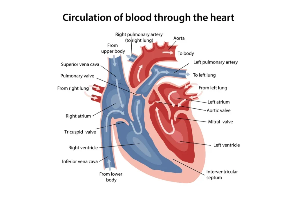
- Deoxygenated blood from different areas of the body enters the heart through the superior and inferior vena cava into the right atrium of the heart. At the same time, oxygenated blood from the lungs enters the left atrium of the heart through four pulmonary veins. Thus, both the atria of the heart are filled with blood at the same time.
- Then, due to the contraction of both the atria of the heart, both the atrioventricular valves, i.e. the bicuspid valve and the tricuspid valve, open, causing both the atria to empty and both the ventricles to fill with blood.
- The right ventricle contains deoxygenated blood while the left ventricle contains oxygenated blood. Then, due to the contraction of both ventricles, the blood from the right ventricle goes to the lungs through the pulmonary artery through the opening of the pulmonary valve, and the blood from the left ventricle goes to the lungs through the opening of the aortic valve, and oxygenated blood circulates throughout the body. Thus both ventricles are emptied.
- Then after both the atria in the heart are filled with blood again, this cycle continues continuously and blood circulation occurs.
Blood supply of the heart:
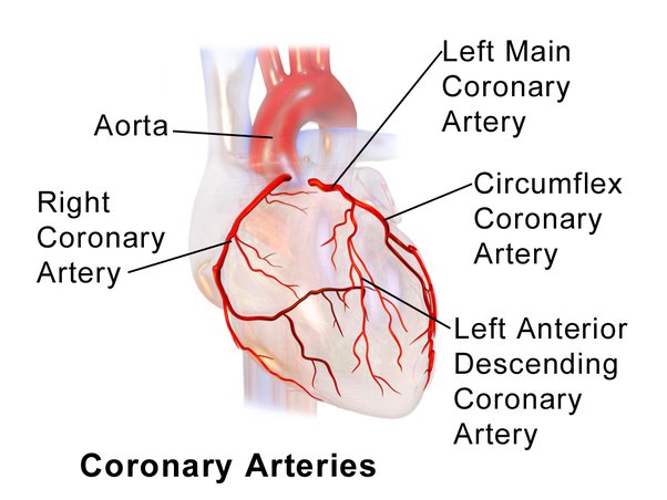
- The aorta, which comes out of the left ventricle of the heart, immediately branches off from it on both sides, the right and left coronary arteries, which provide the heart with oxygenated blood supply. These coronary arteries are branches of the ascending aorta.
- The amount of blood the heart pumps out through the aorta during a single contraction Of this, five percent of the blood is supplied to the heart through the coronary arteries.
- These coronary arteries divide into many branches and supply oxygenated blood to all parts of the heart.
- The deoxygenated blood in the heart is collected by small coronary veins that drain the deoxygenated blood into the coronary sinuses, and all this deoxygenated blood drains directly into the right atrium of the heart through the coronary sinuses.
Functions of the Heart:
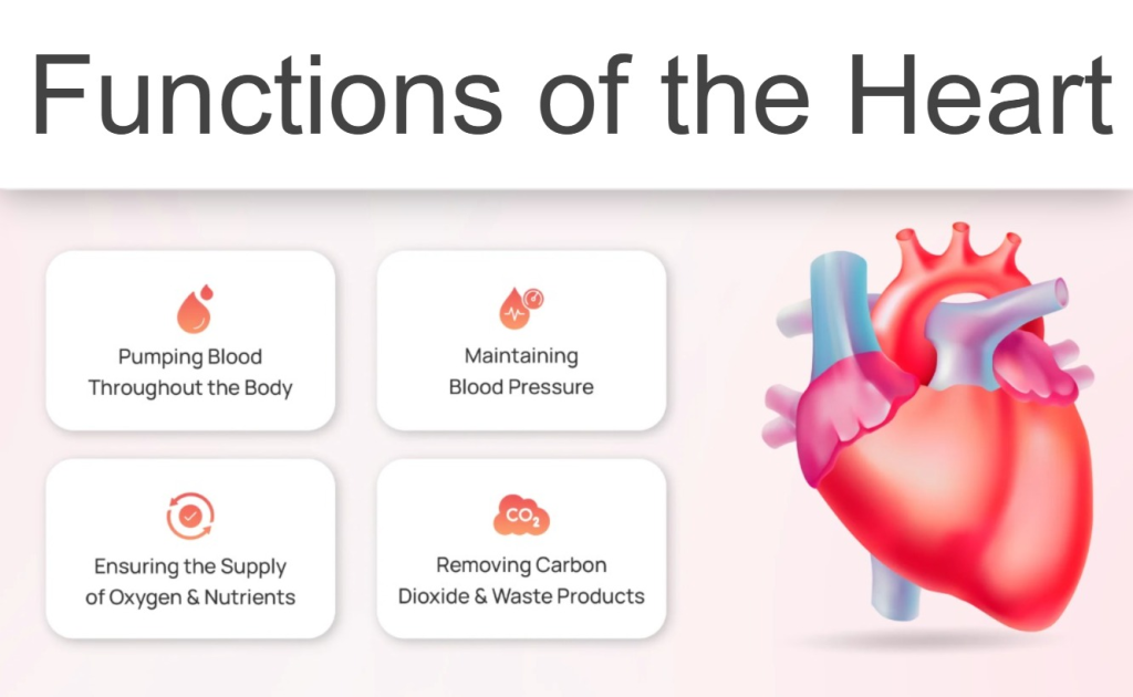
- The heart supplies oxygenated blood to all the organs and tissues of the body.
- The heart is an important organ of the cardiovascular system. Without which the human body cannot survive, it functions as a vital organ.
- The heart circulates deoxygenated blood to the lungs so that the blood can be oxygenated and purified.
- Circulations like pulmonary circulation and systemic circulation are regulated by the heart.
- The heart also regulates the heart rate according to the needs of the body and body temperature.
- The heart circulates blood to every part of the body It also regulates the body’s temperature by delivering it to the body’s excretory organs.
- Since the heart delivers blood to the body’s excretory organs, the blood can be filtered and waste products can be removed from the blood.
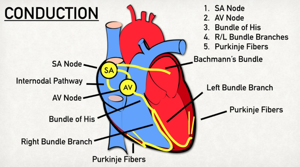
Conducting System of the Heart:
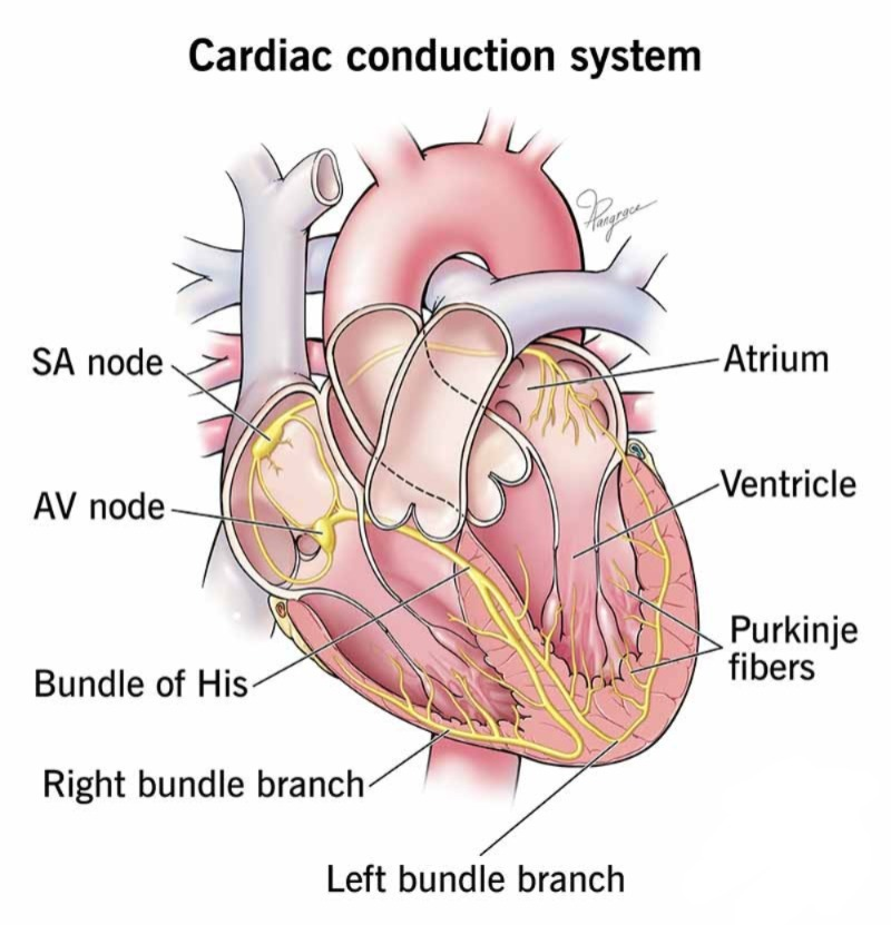
The heart has a system for impulse transmission within its own myocardium layer; it does not directly require an external nerve supply to function.
The myocardium layer in the inner wall of the heart contains special types of neuromuscular cells, which have the characteristic of generating impulses and transmitting the impulses further to the myocardium layer. Which causes the contraction and relaxation of the heart muscles. The structure of this conducting system is as follows.
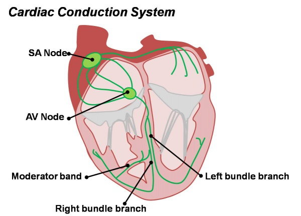
1.S.A. NODE S. A. node (sino atrial node)..
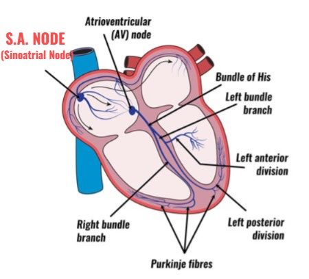
- S.A. The node is a specialized neuromuscular cell mass located in the myocardium layer near the opening of the superior vena cava in the wall of the right atrium. It is also known as the pacemaker because it is the unit that generates the impulses for the contraction of the heart.
- The impulses generated by the S. A. node are transmitted further to the myocardium and the contraction of the myocardium muscles of the heart occurs.
- S. A. The node regulates the heart rate.
2.A.V. NODE A. V. node (atrio ventricular node)..
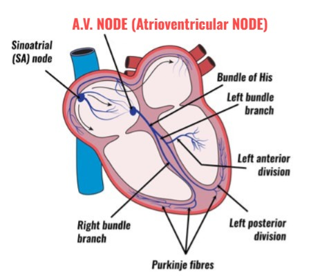
- A. V. The node is a mass of specialized neuromuscular cells located near the septum of the heart near the atrioventricular valve.
- The node generates impulses to the myocardium to contract, which are received by the AV node and transmitted to the septum of the ventricle.
3.Atrioventricular Bundle (A.V. Bundal) or Bundle of His:
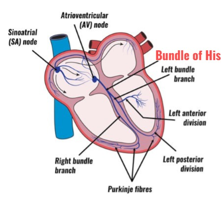
- The atrioventricular bundle (bundle of His) is a network of fibers in the layer of myocardial muscles that begins at the A. V. node and from there descends to the septum of the ventricle. This network of fibers divides into two branches and is arranged as Parkin’s fibers throughout the wall of the ventricle.
- This network of fibers in the wall of the right ventricle is known as the right bundle branch. The network of these fibers in the wall of the left ventricle is known as the left bundle branch.
- Thus, the network of these fibers is arranged all the way to the apex of the heart and the impulses coming from the AV node pass through these fibers and reach the apex of the heart and the contraction of the heart muscles occurs.
Cardiac Cycle of the Heart:
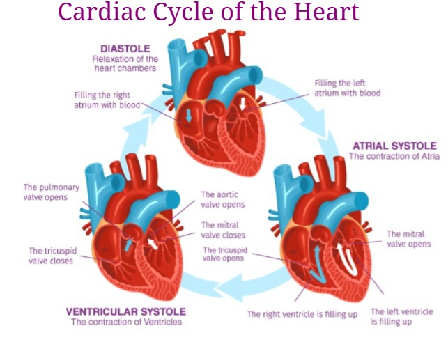
The heart is a continuously pumping organ. The pumping action of the heart is called its cardiac cycle. In a healthy person, cardiac cycles, i.e. pumping action, occur 68 to 72 times in a minute. It takes 0.8 seconds to complete a cardiac cycle. This function is continuously performed by the heart of a living human being.
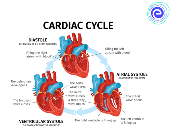
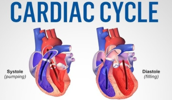
Contraction and relaxation of the heart muscles occur through impulses generated from the S. A. node of the heart. Contraction is known as Systole and relaxation is known as Diastole.
The following events occur in the cardiac cycle.
1.Atrial systole:
Atrial systole means the simultaneous contraction of both atria, which takes 0.1 seconds.
In this atrial systole, when both atria are filled with blood, an impulse is generated by the S. A. node and this impulse reaches the A. V. node. During this time, it takes 0.1 seconds and both atria contract simultaneously, both atrioventricular valves open and both atria are emptied of blood and both ventricles are filled with blood. This phase is called atrial systole.
2.Ventricular systole ( ventricular systole ):
Ventricular systole is the simultaneous contraction of both ventricles. It takes 0.3 seconds for it.
During ventricular systole, when both ventricles are filled with blood, impulses from the A. V. node reach the bundle of His and Purkinje fibers, i.e., the heart’s atrium.
During this time, it takes 0.3 seconds and both ventricles contract simultaneously. The blood from both ventricles respectively goes to the lungs through the pulmonary artery and the blood from the left ventricle circulates throughout the body through the aorta. This phase is called ventricular systole.
3.Complete cardiac diastole:
Complete cardiac diastole means the simultaneous relaxation of both atria and both ventricles i.e. all four chambers of the heart. This process takes 0.4 seconds.
During the period of complete cardiac diastole, there is no electrical activity in the heart. The muscles of the myocardium are relaxed. During this period, both the atria and both the ventricles dilate, that is, relax. Both the atria fill with blood again during this period. This relaxation period is 0.4 seconds. This is called complete cardiac diastole.
Thus, it takes 0.8 seconds to complete a complete cardiac cycle and blood circulation occurs due to the contraction and relaxation of the heart.
Factors affecting heart rate:
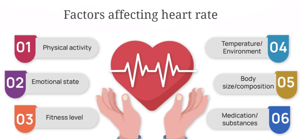
Heart rate, i.e. the action of the heart beating, in a normal healthy person the heart beats 68 to 72 times in a minute. The beating of the heart is mainly controlled by the cardiac center located in the medulla of the brain.
In addition, the heart rate is also affected by the automatic nervous system and stimulation of the vagus nerve.
The factors that affect the heart rate are as follows.
Position:
Changing the position of a person can cause a general change in the heart rate. For example, the heart rate may be different in sleeping position, sitting position and standing position.
Circulating Chemicals:
Circulating chemicals are different hormones, chemicals, electrolytes, etc. secreted in the body. The increase or decrease in the amount of all these can cause an increase or decrease in the heart rate.
Age:
Age is an important factor affecting the heart rate. Changes in heart rate are seen according to different ages. For example, the heart rate of newborns is highest while adults have different heart rates. Changes in heart rate are also seen in old age people.
Emotional Status:
Emotional changes affect heart rate. Changes in heart rate are seen in situations of happiness and sadness.
Fear and Anxiety:
A sudden increase in heart rate is seen during fear or anxiety.
Exercise:
During exercise, the heart rate also increases due to increased blood circulation and oxygen demand in the body.
Gender:
Generally, there is a slight difference in the heart rate of men and women. Which may be due to hormones and metabolic activity.
Temperature:
Due to the increase in body temperature, the heart rate also increases.
Heart sound:
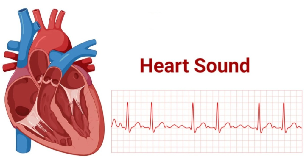
- The sound produced due to the closure of the valves in the heart is known as a heart sound. This heart sound is seen due to the closure of the atrioventricular valve and semilunar valve in the heart.
- The sound heard in the heart when the atrioventricular valve closes is called the Lub sound. It is also known as the first sound. It is a dull sound and is a long sound.
- When the semilunar valve closes in the heart, the second heart sound is heard which is called the dub sound. It has Short and Sharp (Short And Sharp) type characteristics.
- No sound is heard in the heart except lub and dub. If any additional sound is heard apart from this sound, it is called Murmur Sound. Which indicates any abnormality related to the heart valves.
Blood pressure:
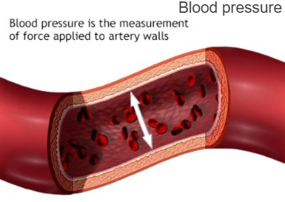
The pressure created by the flow of blood on the walls of blood vessels is called blood pressure.
Normal blood pressure in a healthy person is found to be 120/80 mm/hg.
Blood pressure is measured by applying force to the wall of the artery. The equipment used to measure this is a sphygmomanometer. is.
Blood pressure is measured in two parts.
1. Systolic Blood Pressure
2. Diastolic blood pressure (Diastolic Pressure
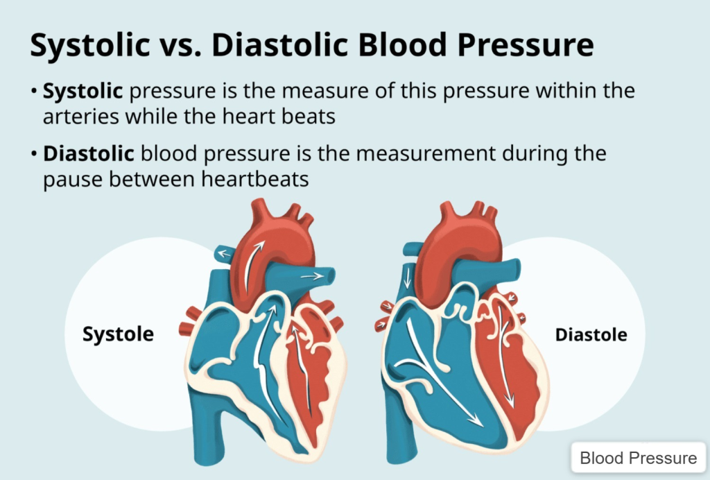
1. Systolic blood pressure (Systolic Pressure):
This is the highest level of blood pressure. It is obtained by measuring the blood flow passing through the artery wall during the contraction of the heart. Which is normally found to be around 120 mmhg.
2. Diastolic Blood Pressure:
It is the lowest blood pressure. When the heart is in the relaxed phase, the pressure of the blood passing through the artery is measured and is called diastolic blood pressure. It is usually found to be 80 mmhg.
The difference between systolic blood pressure and diastolic blood pressure is called Pulse Pressure. Normally, normal pulse pressure is 40 mmhg .
Electrocardiogram (ECG):
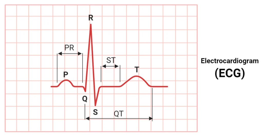
- The procedure of printing the electrical activity of the heart on a graph of paper is called Electrocardiogram.
- The normal activity of the heart and any abnormalities related to it can be known from the waves of Electrocardiogram.
- A normal electrocardiogram has five waves: P, QRS, T.
- In which P wave shows the contraction of the atrium.
- QRS complex shows the dilation of the atrium and contraction of the ventricle.
- T wave shows the dilation of the ventricle.
- We can also calculate the heart rate from a normal ECG.
- If more than 100 hard beats are recorded in a minute, the condition is called Tachycardia. If the pulse is less than 60, the condition is called Bradycardia.
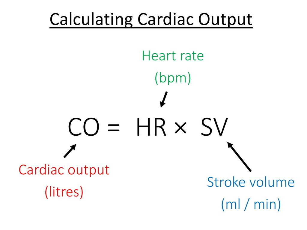
Cardiac output:
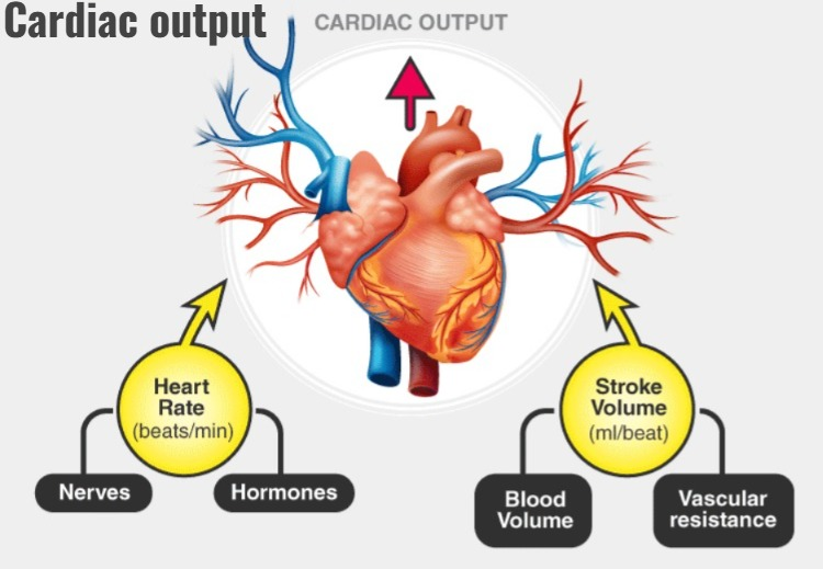
- The amount of blood that flows from the left ventricle of the heart to the aorta in one minute is called cardiac output.
- The amount of blood that flows into the aorta during one contraction of the left ventricle is called the stroke volume.
- The stroke volume of a healthy adult is 70 ml
- If If a healthy person has a heart rate of 72/minute and a stroke volume of 70ml, then their work output is approximately five litres per minute.
Cardiac Output = Stroke Volume * Heart Rate