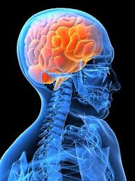
MHN-2 UNIT-12 Nursing management of organic brain disorders
Nursing management of organic brain disorders
Prevalence and Incidence of Organic Brain Disorders
🔍 Definition
- Organic Brain Disorders (OBDs): These are brain-related conditions caused by physical damage or dysfunction of the brain tissue, not due to psychiatric causes.
Examples: Dementia, Delirium, Alzheimer’s disease, Traumatic Brain Injury (TBI), etc. - Incidence: The number of new cases of a disease in a given population during a specified time.
- Prevalence: The total number of existing cases (both new and old) at a specific point in time or over a period.
📊 Prevalence and Incidence of Major Organic Brain Disorders:
1. Dementia (including Alzheimer’s Disease)
- Prevalence:
- Global: Over 55 million people (WHO, 2023)
- India: Around 4 million cases
- Incidence:
- Worldwide: About 10 million new cases/year
- Increases significantly after age 65
🔸 Alzheimer’s disease accounts for 60–70% of dementia cases.
2. Delirium
- Prevalence:
- Hospitalized elderly: 14% to 24%
- ICU patients: Up to 80%
- Incidence:
- Post-surgery (elderly): 15–53%
- Among terminally ill: 80–90% before death
🔸 Often underdiagnosed but highly prevalent in hospital and postoperative settings.
3. Traumatic Brain Injury (TBI)
- Prevalence:
- Global: Over 69 million people/year
- India: Approx. 1.5 to 2 million people/year
- Incidence:
- Road accidents major cause in India
- Males (15–45 years) most affected
4. Wernicke-Korsakoff Syndrome (Alcohol-related brain disorder)
- Prevalence: 1-2% in the general population
- High risk: Chronic alcoholics
- Often undiagnosed and overlaps with other disorders.
🎯 Summary Table
| Disorder | Global Prevalence | Incidence |
|---|---|---|
| Dementia | 55+ million | 10 million new/year |
| Delirium | 14–80% (varies by setting) | High in hospitalized elderly |
| TBI | 69 million/year | 1.5–2 million/year in India |
| Wernicke-Korsakoff | ~1–2% (general pop.) | Common in alcoholics |
🧠 Classification of Organic Brain Disorders
Organic Brain Disorders (OBDs), also called organic mental disorders, are conditions that cause disturbances in brain function due to structural damage, disease, or dysfunction of the brain tissue.
They are classified based on etiology (cause), duration (acute vs chronic), and clinical features.
✅ Main Classification
I. Acute Organic Brain Disorders
These develop suddenly and may be reversible if treated early.
- Delirium
- Sudden onset of confusion, disorientation
- Fluctuating consciousness
- Causes: infections, metabolic imbalances, medications, alcohol withdrawal
- Amnestic Disorders (Short-term memory loss)
- Sudden memory disturbance with intact attention
- Causes: trauma, alcohol (Korsakoff syndrome), hypoxia
II. Chronic Organic Brain Disorders
These develop gradually and are often irreversible.
- Dementia
- Progressive decline in memory, judgment, language, and other cognitive functions
- Types:
- Alzheimer’s disease
- Vascular dementia
- Lewy body dementia
- Frontotemporal dementia
- Degenerative Neurological Diseases (with cognitive or behavioral effects)
- Parkinson’s disease (with dementia)
- Huntington’s disease
- Multiple sclerosis (with cognitive decline in some cases)
III. Substance-Induced Organic Mental Disorders
Caused by chronic or acute exposure to substances that damage brain function.
- Alcohol-induced: Wernicke’s encephalopathy, Korsakoff syndrome
- Drug-induced: Delirium, psychosis due to sedatives, stimulants, or hallucinogens
- Heavy metals: Lead or mercury poisoning
IV. Trauma-Related Brain Disorders
Caused by physical injury to the brain.
- Traumatic Brain Injury (TBI): Can cause memory loss, cognitive deficits, personality changes
- Post-concussion syndrome
- Chronic Traumatic Encephalopathy (CTE) – often in athletes
V. Infection-Related Organic Disorders
Due to infections that damage the brain or CNS.
- Encephalitis – viral inflammation of brain tissue
- Meningitis – infection of meninges (brain coverings)
- Neurosyphilis
- HIV-associated neurocognitive disorder (HAND)
VI. Metabolic & Nutritional Disorders Affecting the Brain
These affect brain function due to deficiency or systemic dysfunction.
- Hypoxia (low oxygen)
- Hypoglycemia or hyperglycemia
- Vitamin B1 (thiamine) deficiency – Wernicke’s encephalopathy
- Liver failure, renal failure – Hepatic or uremic encephalopathy
VII. Tumors and Space-Occupying Lesions
- Brain tumors (benign or malignant)
- Increase intracranial pressure → cause cognitive or neurological symptoms
VIII. Epilepsy-Related Disorders
- Temporal lobe epilepsy – causes behavioral and memory changes
- Post-ictal confusion (after seizures)
📌 Summary Table
| Category | Examples |
|---|---|
| Acute OBDs | Delirium, Amnestic Syndrome |
| Chronic OBDs | Alzheimer’s, Parkinson’s with dementia |
| Substance-Induced | Alcoholic dementia, Drug-induced psychosis |
| Trauma-Related | TBI, CTE |
| Infection-Related | Encephalitis, Meningitis, HIV dementia |
| Metabolic/Nutritional | Hypoxia, Hypoglycemia, Thiamine deficiency |
| Tumors & Lesions | Brain tumor, Increased ICP |
| Epilepsy-Related | Post-ictal states, Temporal lobe epilepsy |
🩺 NURSING ASSESSMENT HISTORY TAKING IN ORGANIC BRAIN DISORDERS
🔶 Purpose:
- Identify cause, duration, and progression of the brain disorder.
- Understand how the condition is affecting cognitive, emotional, and physical functions.
- Plan appropriate nursing care, safety precautions, and caregiver education.
✅ 1. GENERAL INFORMATION
Obtain from the patient and/or caregiver.
- Name, age, sex, education
- Occupation
- Marital and family status
- Residence (urban/rural)
- Socioeconomic background
✅ 2. PRESENTING COMPLAINTS
Ask questions like:
- When did the problem start?
- How has it progressed (sudden or gradual)?
- Has there been any confusion, memory loss, disorientation, behavioral changes, language difficulty?
🧠 Example Complaints:
- Forgetting names or places
- Getting lost in familiar areas
- Hallucinations or delusions
- Unusual aggression or withdrawal
- Difficulty performing daily tasks (ADLs)
✅ 3. HISTORY OF PRESENT ILLNESS
- Onset: Sudden (e.g., delirium) or gradual (e.g., dementia)
- Duration: Acute (hours/days) or chronic (months/years)
- Progression: Static, worsening, fluctuating
- Any associated symptoms:
- Fever
- Headache
- Seizures
- Loss of consciousness
- Behavioral disturbances
✅ 4. PAST MEDICAL HISTORY
- History of:
- Hypertension, diabetes, stroke
- Head injury or trauma
- Seizure disorders
- Infections (e.g., meningitis, encephalitis)
- HIV, syphilis, or other systemic illnesses
- Hospitalizations and past treatments
- History of psychiatric illness
✅ 5. FAMILY HISTORY
- Any family members with:
- Dementia
- Parkinsonism
- Epilepsy
- Mental illness
- Genetic or hereditary conditions
🧬 Helpful in Alzheimer’s, Huntington’s, etc.
✅ 6. PERSONAL HISTORY
- Sleep: Disturbed? Night wandering?
- Appetite: Increased, decreased, or unchanged
- Bowel and bladder habits
- Substance use:
- Alcohol, smoking, drug abuse (very important!)
- Sexual behavior
- Daily functioning: Need help with dressing, eating, toileting?
✅ 7. COGNITIVE FUNCTION ASSESSMENT
- Orientation: Time, place, person
- Memory: Immediate, recent, and remote
- Attention span and concentration
- Judgment: Give simple scenarios
- Language ability
- Visuospatial ability
- Use tools like:
- Mini-Mental State Examination (MMSE)
- Montreal Cognitive Assessment (MoCA)
✅ 8. MENTAL STATUS EXAMINATION (MSE)
- Appearance & behavior
- Mood and affect
- Thought content: Hallucinations, delusions?
- Perception
- Insight and judgment
- Speech: Coherence, rate, volume
✅ 9. PHYSICAL AND NEUROLOGICAL ASSESSMENT
- Vital signs (fever? BP?)
- Cranial nerve examination
- Motor function: Strength, gait, balance
- Reflexes: Brisk, absent, abnormal?
- Coordination tests: Finger-nose, heel-shin
- Sensory testing
✅ 10. INVESTIGATIONS (As ordered by physician)
- Blood tests: CBC, electrolytes, liver/renal function
- Vitamin B12, thyroid profile
- Imaging: CT scan, MRI brain
- EEG (for seizure or delirium)
- CSF analysis (if infection suspected)
- Serological tests for HIV/syphilis
💡 NURSE’S ROLE:
- Observe carefully and document changes in behavior or cognition
- Collect data from family or caregivers when patient is confused
- Prioritize safety – fall prevention, avoid wandering
- Encourage routine, memory aids, emotional support
- Educate family on disease process and caregiving tips
🧠 Physical Assessment of Organic Brain Disorders
(For conditions like dementia, delirium, traumatic brain injury, Wernicke’s encephalopathy, etc.)
🔶 Objectives of Physical Assessment:
- Identify neurological deficits
- Detect underlying medical causes
- Monitor progression of cognitive and functional decline
- Ensure patient safety and holistic care planning
✅ 1. General Appearance and Behavior
- Level of consciousness (LOC):
- Alert, drowsy, stuporous, or comatose
- Facial expressions & body posture
- Grooming and hygiene
- Signs of restlessness or agitation
- Cooperation and response to commands
✅ 2. Vital Signs
- Temperature – Fever may suggest infection (e.g., encephalitis, meningitis)
- Pulse – Irregularities may be seen in autonomic dysfunction
- Blood pressure – Elevated in stroke or head trauma
- Respirations – Abnormal patterns (Cheyne-Stokes, etc.)
- SpO₂ – Hypoxia can worsen cognitive status
✅ 3. Level of Consciousness and Orientation
- Use the Glasgow Coma Scale (GCS):
- Eye Opening (1–4)
- Verbal Response (1–5)
- Motor Response (1–6)
- Total: 3–15
- Assess Orientation to:
- Time
- Place
- Person
- Disorientation is common in delirium and dementia
✅ 4. Cranial Nerve Examination
(For brainstem and central nervous system function)
| Cranial Nerve | Function | What to assess |
|---|---|---|
| CN I – Olfactory | Smell | Often skipped unless head trauma |
| CN II – Optic | Vision | Visual acuity, fields |
| CN III, IV, VI – Oculomotor, Trochlear, Abducens | Eye movement | Pupil size, reactivity, nystagmus |
| CN V – Trigeminal | Facial sensation | Corneal reflex, jaw strength |
| CN VII – Facial | Facial expression | Asymmetry, droop |
| CN VIII – Vestibulocochlear | Hearing & balance | Whisper test, Romberg |
| CN IX, X – Glossopharyngeal & Vagus | Gag, swallowing | Hoarseness, palate movement |
| CN XI – Spinal Accessory | Neck & shoulder movement | Shrug test |
| CN XII – Hypoglossal | Tongue movement | Deviation, atrophy |
✅ 5. Motor Function
- Muscle strength: 0–5 scale (paralysis to normal)
- Muscle tone: Flaccid, spastic, rigid?
- Gait and balance: Observe walking, Romberg test
- Involuntary movements: Tremors, chorea (e.g., Parkinson’s, Huntington’s)
- Coordination tests:
- Finger-to-nose
- Heel-to-shin
- Rapid alternating movements
✅ 6. Sensory Function
- Light touch
- Pain (pinprick)
- Temperature
- Vibration
- Position sense (proprioception)
🧠 Abnormalities may suggest stroke, trauma, or degenerative disease.
✅ 7. Reflexes
- Deep tendon reflexes (DTRs):
- Biceps, triceps, patellar, Achilles
- Graded 0 (absent) to 4+ (hyperactive)
- Babinski’s sign:
- Present in upper motor neuron lesion (e.g., in dementia with frontal lobe damage)
✅ 8. Cognitive and Mental Status Screening
(May overlap with mental status exam but part of physical neuro assessment)
- Mini-Mental State Examination (MMSE) or MoCA
- Memory
- Attention
- Language
- Calculation
- Visual-spatial skills
- Abstract thinking
✅ 9. Other Observations
- Skin condition: Pressure sores (especially in immobile or unaware patients)
- Signs of trauma: Bruises, swelling, CSF leak (head injury)
- Nutrition and hydration status
- Bladder & bowel function
- Mobility and fall risk assessment
📝 Summary Checklist:
| System/Function | Key Assessment Points |
|---|---|
| Appearance | Alertness, grooming, behavior |
| Vitals | Fever, BP, Pulse, SpO₂ |
| LOC & GCS | Eye, motor, verbal responses |
| Cranial nerves | Pupils, facial movements, reflexes |
| Motor | Strength, tone, gait |
| Sensory | Light touch, pain, proprioception |
| Reflexes | DTRs, Babinski |
| Cognition | Orientation, memory, MMSE/MoCA |
| Others | Skin, nutrition, trauma, bladder/bowel |
👩⚕️ Nurse’s Role:
- Monitor changes over time
- Report early signs of deterioration
- Maintain safety, prevent falls or injury
- Involve family, provide support & education
🧠 Mental Assessment of Organic Brain Disorders (OBDs)
Focuses on evaluating cognitive, emotional, behavioral, and perceptual functions of the patient — affected due to brain damage, disease, or dysfunction.
🔶 Purpose of Mental Assessment:
- To evaluate cognitive decline, thought disorders, and emotional state
- To differentiate organic from functional (psychiatric) conditions
- To plan appropriate nursing care and medical interventions
✅ 1. Mental Status Examination (MSE)
The MSE is a structured method to assess a patient’s mental functioning at a point in time.
🟠 A. Appearance and Behavior
- Grooming: Clean/unkempt, appropriate/inappropriate dress
- Facial expression: Blank, anxious, smiling, fearful
- Psychomotor activity: Agitation, retardation, restlessness
- Level of consciousness (LOC): Alert, drowsy, stuporous
- Eye contact: Maintained, poor, avoidant
- Cooperation: Cooperative, withdrawn, hostile
🧠 In delirium, behavior may be hyperactive or hypoactive.
🟠 B. Speech
- Rate: Normal, fast, slow
- Volume: Loud, soft
- Tone: Monotonous, normal
- Fluency: Slurred, broken, pressured
- Relevance: Coherent or disorganized
🗣 Slurred or incoherent speech may suggest neurological damage
🟠 C. Mood and Affect
- Mood (subjective): Ask the patient – “How are you feeling?”
- Anxious, sad, irritable, elated?
- Affect (objective):
- Appropriate/inappropriate
- Flat, blunted, labile (rapidly shifting)
🧠 Dementia may show shallow or labile affect.
🟠 D. Thought Process and Content
- Stream: Logical, tangential, circumstantial
- Content:
- Delusions (false beliefs): Paranoia, grandiosity
- Obsessions
- Suicidal or homicidal thoughts
🧠 Paranoid delusions are common in Lewy body dementia.
🟠 E. Perception
- Hallucinations:
- Visual (common in delirium, Lewy body dementia)
- Auditory (less common in organic disorders)
- Illusions (misinterpretation of real stimuli)
- Depersonalization or derealization
🟠 F. Cognitive Functions
| Function | What to Assess | Example |
|---|---|---|
| Orientation | Time, Place, Person | Ask: What is today’s date? Where are you now? |
| Attention | Concentration span | Ask to spell ‘WORLD’ backward |
| Memory | Immediate, Recent, Remote | Recall 3 objects, last meal, childhood |
| Language | Naming, comprehension, repetition | Ask to name an object, follow commands |
| Abstract Thinking | Interpret proverbs | “What does ‘Don’t cry over spilt milk’ mean?” |
| Visuospatial Skills | Drawing task | Ask to copy a figure or clock |
🧠 Tools: Mini-Mental State Examination (MMSE), MoCA, Addenbrooke’s Cognitive Examination (ACE)
🟠 G. Insight and Judgment
- Insight:
- Does the patient recognize their condition?
- Absent, partial, or full insight
- Judgment:
- Ability to make decisions
- Ask: “What would you do if you found a sealed envelope on the road?”
🧠 Often impaired in dementia, partially intact in delirium.
✅ 2. Behavioral Observations Specific to Organic Disorders
| Disorder | Common Behavioral Signs |
|---|---|
| Delirium | Acute confusion, fluctuating LOC, agitation, hallucinations |
| Dementia | Memory loss, poor judgment, word-finding difficulty |
| TBI (Traumatic Brain Injury) | Aggression, impulsivity, emotional lability |
| Wernicke-Korsakoff | Confabulation (fabricated memories), ataxia |
📋 Mental Status Assessment Documentation Format
| Domain | Observations |
|---|---|
| Appearance & Behavior | Disoriented, unkempt |
| Speech | Slurred, slow |
| Mood | Flat |
| Affect | Blunted |
| Thought Process | Coherent but slowed |
| Thought Content | No delusions |
| Perception | Visual hallucinations present |
| Orientation | Disoriented to time |
| Memory | Recent memory impaired |
| Judgment | Poor |
| Insight | Lacking awareness |
👩⚕️ Nurse’s Role:
- Use simple language and repetition
- Provide emotional support
- Monitor risk of injury, wandering, or aggression
- Maintain consistent routine
- Involve family in care and educate about the illness
🧠 Neurological Assessment in Organic Brain Disorders
A systematic evaluation of the central and peripheral nervous systems to assess the structure and function of the brain, especially when it is affected by diseases like dementia, delirium, brain injury, stroke, Wernicke’s encephalopathy, etc.
🎯 Objectives of Neurological Assessment:
- Detect neurological deficits (motor, sensory, cognitive, or autonomic)
- Identify the site and extent of brain dysfunction
- Provide baseline data for ongoing evaluation
- Assist in diagnosis and planning of nursing/medical care
✅ Components of a Complete Neurological Assessment
🔶 1. Level of Consciousness (LOC)
- Most important indicator of cerebral function.
- Use the Glasgow Coma Scale (GCS):
| Response | Score |
|---|---|
| Eye Opening (E) | 1–4 |
| Verbal Response (V) | 1–5 |
| Motor Response (M) | 1–6 |
| Total Score | 3 (deep coma) to 15 (fully alert) |
- Check for alertness, drowsiness, stupor, coma.
🔶 2. Orientation
- Ask the patient:
- Time (day, date, year)
- Place (current location, city)
- Person (name, relatives, caregiver)
- Disorientation is common in delirium, dementia, brain injury.
🔶 3. Pupillary Assessment (PERRLA)
- Pupils Equal, Round, Reactive to Light and Accommodation
- Abnormalities:
- Unequal pupils (anisocoria) – increased ICP
- Fixed and dilated – brain herniation
- Sluggish response – cranial nerve III (oculomotor) involvement
🔶 4. Cranial Nerve Examination
| Cranial Nerve | Function | Signs of Damage in OBDs |
|---|---|---|
| CN I (Olfactory) | Smell | Loss of smell (trauma) |
| CN II (Optic) | Vision | Visual field defects |
| CN III, IV, VI | Eye movements | Diplopia, nystagmus |
| CN V | Facial sensation, chewing | Weakness, loss of reflex |
| CN VII | Facial muscles | Facial asymmetry |
| CN VIII | Hearing, balance | Vertigo, hearing loss |
| CN IX, X | Swallowing, gag | Absent gag reflex |
| CN XI | Shoulder movement | Weakness in shrugging |
| CN XII | Tongue movement | Deviation, fasciculation |
🔶 5. Motor Function
- Muscle Strength (graded 0–5)
- Muscle Tone:
- Flaccidity → lower motor neuron lesion
- Spasticity/rigidity → upper motor neuron lesion
- Involuntary Movements:
- Tremors, chorea, myoclonus seen in Parkinsonism, Huntington’s, etc.
- Coordination Tests:
- Finger-to-nose test
- Heel-to-shin test
- Rapid alternating movements
🔶 6. Sensory Function
- Test bilaterally and symmetrically:
- Light touch
- Pain (pinprick)
- Temperature
- Vibration sense (using tuning fork)
- Position sense (proprioception)
🧠 Dysfunction in these areas may indicate damage to sensory cortex, thalamus, or peripheral nerves.
🔶 7. Reflexes
- Deep tendon reflexes (DTRs): Biceps, triceps, patellar, Achilles
- Graded from 0 (absent) to 4+ (hyperreflexia)
- Plantar Reflex (Babinski sign):
- Positive in upper motor neuron lesion (abnormal in adults)
- Superficial reflexes: Corneal, abdominal
🔶 8. Gait and Balance
- Ask patient to:
- Walk in a straight line
- Perform heel-to-toe walking
- Stand with eyes closed (Romberg test)
- Gait abnormalities:
- Shuffling (Parkinson’s)
- Ataxic (cerebellar damage)
- Unsteady (vestibular dysfunction)
🔶 9. Cerebellar Function Tests
- Rapid Alternating Movements
- Point-to-point movements
- Romberg test: Tests proprioception and cerebellum
- Look for:
- Intention tremor
- Dysdiadochokinesia (inability to perform alternating movements)
🔶 10. Autonomic Function (if needed)
- Heart rate variability
- Blood pressure fluctuations (orthostatic hypotension)
- Bladder and bowel control
- Sweating abnormalities
🧠 Affected in advanced dementia, Parkinson’s disease.
📝 Summary Chart of Key Findings in OBDs:
| Disorder | Common Neurological Signs |
|---|---|
| Delirium | Fluctuating LOC, disorientation, tremors |
| Dementia | Memory loss, slow gait, poor coordination |
| TBI | Altered LOC, unequal pupils, seizures |
| Wernicke’s Encephalopathy | Ataxia, nystagmus, ophthalmoplegia |
| Stroke (organic cause) | Hemiplegia, speech defects, facial droop |
👩⚕️ Nurse’s Role in Neurological Assessment
- Perform hourly or shift-wise neuro checks if needed
- Recognize early warning signs of deterioration
- Maintain safety: prevent falls, aspiration, seizures
- Assist with diagnostic tests: CT scan, MRI, EEG
- Document findings in detail and report promptly
🧠 Treatment Modalities of Organic Brain Disorders (OBDs)
Organic Brain Disorders are caused by physical or structural abnormalities of the brain due to trauma, infection, stroke, toxins, or neurodegeneration. Treatment is multimodal, focusing on:
- Managing the underlying cause
- Relieving symptoms
- Improving function and quality of life
🔶 Classification of Treatment Modalities
✅ 1. Pharmacological Treatment
A. Cognitive Enhancers
Used in dementia and Alzheimer’s disease:
- Cholinesterase inhibitors:
- Donepezil, Rivastigmine, Galantamine
🔸 Improve memory, attention, behavior
- Donepezil, Rivastigmine, Galantamine
- NMDA receptor antagonist:
- Memantine
🔸 Slows down cognitive decline
- Memantine
B. Antipsychotics
Used to manage agitation, hallucinations, or delusions (with caution):
- Risperidone, Olanzapine, Quetiapine
🧠 Use in low doses to avoid side effects like sedation and falls
C. Antidepressants
For depression, anxiety, common in chronic OBDs:
- SSRIs: Sertraline, Escitalopram
🔸 Safer in elderly than tricyclics
D. Sedatives/Hypnotics
- Benzodiazepines: Used very cautiously in delirium (short term only)
- Melatonin: For sleep disorders in dementia
E. Vitamins & Supplements
- Thiamine (Vitamin B1) – for Wernicke’s encephalopathy
- Vitamin B12, Folic Acid – if deficiency-related
- Omega-3 – neuroprotective in some studies
✅ 2. Non-Pharmacological Therapies
A. Cognitive Rehabilitation/Therapy
- Memory training, problem-solving tasks
- Use of reminder cues, alarms, labeled environments
- Reality orientation therapy (use of calendars, clocks, familiar photos)
B. Behavioral Therapy
- Managing aggression, wandering, disinhibition
- Positive reinforcement, structured routines
C. Occupational Therapy
- Helps in ADL (Activities of Daily Living) training
- Use of assistive devices for dressing, grooming, feeding
D. Speech and Language Therapy
- For aphasia, dysarthria, word-finding difficulty
- Especially important after stroke or in Alzheimer’s
E. Physical Therapy (Physiotherapy)
- Maintain mobility, prevent contractures
- Balance training, fall prevention
- Improve coordination in cerebellar or Parkinson’s-related OBDs
✅ 3. Environmental & Supportive Management
- Safe and familiar environment – prevent confusion and agitation
- Adequate lighting, minimize noise and clutter
- Bed rails, anti-slip mats to prevent falls
- Structured daily routine to reduce anxiety
✅ 4. Family and Caregiver Support
- Educating caregivers about the illness
- Counseling and emotional support
- Training in behavioral management techniques
- Encouraging participation in support groups
✅ 5. Psychotherapy
- Useful in early-stage dementia or mild OBDs
- Focused on adjustment, grief, coping strategies
✅ 6. Social and Community Interventions
- Day-care centers, memory clinics
- Legal guidance on advance directives, guardianship
- Disability certification and government support schemes
✅ 7. Hospitalization (When Needed)
- For acute delirium, aggressive or suicidal behavior
- Detoxification (e.g., alcohol withdrawal)
- Severe depression or psychosis
✅ 8. Surgical and Interventional Treatments (Rare Cases)
- Shunt surgery for Normal Pressure Hydrocephalus (a type of reversible dementia)
- Tumor removal (if space-occupying lesion causing cognitive symptoms)
- Deep Brain Stimulation (DBS) – used in Parkinson’s with cognitive decline
📋 Summary Table
| Modality | Examples |
|---|---|
| Drugs | Donepezil, Memantine, SSRIs, Antipsychotics, Vitamins |
| Therapies | Cognitive, Behavioral, Physical, Occupational, Speech |
| Supportive | Safe environment, Routine, Family education |
| Community-based | Support groups, Legal aid, Social support |
| Surgical (selected cases) | Shunts, Tumor surgery, DBS |
👩⚕️ Nursing Role in Management
- Monitor drug side effects
- Reinforce memory strategies
- Educate caregivers and reduce caregiver burden
- Prevent injuries, falls, infections
- Maintain nutritional status, hydration
- Document behavioral changes and report promptly
🧠 Nursing Management of Organic Brain Disorders (OBDs)
Organic brain disorders (e.g., dementia, delirium, traumatic brain injury, Wernicke’s encephalopathy) affect cognition, behavior, orientation, memory, and physical abilities due to structural/functional brain changes. Nurses play a central role in care, safety, education, and rehabilitation.
🔶 Goals of Nursing Management:
- Maintain safety and prevent complications
- Promote cognitive and functional abilities
- Support emotional and behavioral stability
- Educate and involve caregivers/family
- Prevent further brain damage or deterioration
✅ 1. Nursing Assessment
Start with a thorough assessment:
🩺 A. History Taking
- Onset, duration, and progression of symptoms
- Past medical/neurological illness
- Substance abuse history
- Medication use
- Family history of similar disorders
🧠 B. Physical and Neurological Assessment
- Vital signs, GCS score
- Motor/sensory function, reflexes
- Pupillary reaction, muscle strength, coordination
🧠 C. Mental Status Examination (MSE)
- Orientation to time, place, person
- Memory (recent and remote)
- Mood, behavior, speech, thought content
- Use of MMSE or MoCA scoring
✅ 2. Nursing Diagnoses (Examples)
| Problem | Related to | Evidenced by |
|---|---|---|
| Risk for injury | Cognitive impairment, disorientation | Wandering, poor judgment |
| Impaired memory | Organic brain dysfunction | Forgets recent events |
| Disturbed thought process | Neurological damage | Disorganized thinking, hallucinations |
| Self-care deficit | Cognitive and motor impairment | Inability to dress/feed self |
| Impaired verbal communication | Brain damage | Word-finding difficulty |
| Caregiver role strain | Chronic care demands | Expressed stress, fatigue |
✅ 3. Nursing Interventions and Rationale
🟡 A. Ensure Patient Safety
- Keep bed in low position; use side rails
- Remove sharp/dangerous objects
- Supervise during ambulation or toileting
- Use ID bracelet for identification
- Provide calm, structured environment
Rationale: Patients may be disoriented, impulsive, or wander off.
🟡 B. Enhance Cognitive Function
- Use clocks, calendars, photos, and familiar objects
- Speak clearly, use simple words
- Encourage reality orientation (repeating date/time/place)
- Use memory aids (notes, labels)
Rationale: Supports orientation and memory recall.
🟡 C. Support ADLs (Activities of Daily Living)
- Assist in feeding, dressing, grooming as needed
- Encourage independence with supervision
- Use adaptive tools or occupational therapy referrals
Rationale: Promotes self-worth and maintains function.
🟡 D. Manage Behavior and Mood
- Stay calm and reassuring during agitation
- Distract, do not argue if hallucinating or delusional
- Maintain consistent caregivers and routines
- Use behavioral therapy techniques for aggression or restlessness
Rationale: Reduces confusion and behavioral outbursts.
🟡 E. Provide Nutrition and Hydration Support
- Monitor fluid and food intake
- Offer frequent small meals
- Assist in feeding if needed
- Check for swallowing difficulty (risk of aspiration)
Rationale: Prevents dehydration, malnutrition, aspiration.
🟡 F. Promote Rest and Sleep
- Maintain regular sleep-wake cycle
- Reduce environmental noise at night
- Avoid caffeine or sedatives (unless prescribed)
- Use soothing techniques: music, soft lighting
Rationale: Sleep disturbances are common and worsen confusion.
🟡 G. Family and Caregiver Education
- Teach disease progression and behavior management
- Support emotional and physical care planning
- Encourage use of community services or support groups
Rationale: Reduces caregiver stress and improves care continuity.
🟡 H. Monitor Medication Effects
- Observe for side effects (e.g., sedation, dizziness)
- Educate patient/caregiver on proper drug use
- Report adverse reactions
Rationale: Older adults are prone to medication toxicity.
✅ 4. Evaluation
- Patient remains free from injury
- Shows improvement/stability in cognitive function
- Performs ADLs with or without assistance
- Demonstrates emotional stability
- Family/caregiver expresses better understanding and reduced stress
👩⚕️ Nurse’s Role in Long-Term Care
| Area | Nurse’s Role |
|---|---|
| Acute settings | Monitor, treat delirium, prevent complications |
| Rehabilitation | Help regain function, memory training |
| Long-term care | Provide support, prevent decline |
| Hospice/palliative | Comfort measures, caregiver support |
📋 Example Nursing Care Plan Formation
| Nursing Diagnosis | Goal | Interventions | Rationale | Evaluation |
|---|---|---|---|---|
| Risk for injury related to disorientation | Patient will remain safe during hospital stay | – Supervise ambulation – Use call bell – Keep environment uncluttered | To prevent falls and accidents | No injury reported during stay |
🧠 Follow-Up, Home Care, and Rehabilitation of Organic Brain Disorders
Organic Brain Disorders (like dementia, delirium, traumatic brain injury, Wernicke’s encephalopathy) require long-term, individualized care. After hospital discharge, patients need regular follow-up, structured home care, and multidisciplinary rehabilitation to promote function and prevent complications.
✅ 1. Follow-Up Care
🎯 Goals:
- Monitor progress or decline
- Evaluate medication effectiveness and side effects
- Manage comorbidities
- Provide ongoing caregiver support
🔄 Regular Follow-Up Should Include:
- Neurological assessments
- Cognitive function testing (MMSE, MoCA)
- Review of medications and side effects
- Nutritional and hydration status
- Sleep pattern and behavior evaluation
- Address any new symptoms: aggression, falls, incontinence, etc.
🔔 Frequency:
- Initially every 2–4 weeks, then monthly or quarterly, based on condition stability
✅ 2. Home Care Management
🏠 A. Environment Modification
- Safe, clutter-free space
- Install grab bars, anti-slip mats, bed rails
- Label drawers, doors, use clocks/calendars
- Night lights to reduce disorientation
- Remove dangerous items (knives, medications)
🛏️ B. Daily Routine & Supervision
- Fixed schedule for meals, hygiene, sleep
- Supervise medication intake, personal hygiene, and meals
- Monitor for wandering, agitation, sleep disturbances
- Use memory aids: reminder cards, pill boxes
🧴 C. Basic Nursing Care
- Monitor vital signs, intake/output
- Assist with bathing, dressing, toileting
- Prevent bedsores, infections, dehydration
- Observe for behavioral changes and report promptly
✅ 3. Rehabilitation
🎯 Purpose:
- Regain or maintain independent function
- Improve cognitive and physical abilities
- Enhance communication and social interaction
- Reduce caregiver burden
🧠 A. Cognitive Rehabilitation
- Reality orientation therapy
- Reminiscence therapy
- Puzzles, games, brain exercises
- Use memory notebooks, visual cues
🧍 B. Physical Rehabilitation
- Physiotherapy: Maintain strength, balance, prevent contractures
- Gait training, fall prevention techniques
- Occupational therapy for self-care skills
🗣️ C. Speech and Language Therapy
- Relearn language or speech skills
- Practice word recall, articulation
👪 D. Psychosocial Rehabilitation
- Supportive psychotherapy for early-stage dementia
- Social engagement (day-care centers, community groups)
- Address mood changes (depression, anxiety)
✅ 4. Family & Caregiver Education
🧑🤝🧑 Teach Caregivers:
- Disease nature and progression
- Behavior management techniques
- How to give medications safely
- Red flags: falls, sudden confusion, hallucinations
- Emergency response steps
💬 Provide Emotional Support:
- Offer counseling or connect with support groups
- Encourage respite care to avoid burnout
✅ 5. Use of Assistive Devices and Technology
- Walker, wheelchair, handrails
- Pill organizers, digital alarms
- GPS tracking devices (for wanderers)
- Video monitoring (if needed)
✅ 6. Community Support & Resources
- Day-care centers for the elderly
- Home nursing services
- Legal advice on guardianship or advance directives
- Financial aid or government schemes (disability pension, insurance)
✅ 7. Palliative and End-of-Life Care (Advanced Cases)
- Comfort measures over curative treatment
- Symptom control: pain, agitation, incontinence
- Emotional and spiritual support
- Decision-making support for family
📋 Nursing Responsibilities Across Settings:
| Phase | Nurse’s Role |
|---|---|
| Follow-Up | Monitor condition, report changes, adjust care |
| Home Care | Educate caregiver, maintain hygiene, monitor meds |
| Rehabilitation | Coordinate therapies, encourage participation |
| Terminal Stage | Ensure comfort, support dignity and family |
🌟 Key Points for Nurses:
- Maintain continuity of care across hospital → home → rehab
- Encourage realistic goals
- Document and communicate all changes in behavior or function
- Act as a link between family, physician, and therapist
etiology of delirium

refers to the various causes and contributing factors that lead to this acute neuropsychiatric syndrome. Delirium is characterized by sudden onset, fluctuating course, inattention, and disturbance in cognition. It often results from a complex interaction of multiple factors—especially in vulnerable individuals like the elderly, critically ill, or those with pre-existing brain disease.
1. Predisposing Factors (Baseline vulnerabilities):
These factors increase the risk of developing delirium when a person is exposed to an acute insult:
- Advanced age
- Pre-existing cognitive impairment (e.g., dementia)
- Chronic medical conditions (e.g., heart failure, kidney or liver disease)
- History of stroke or neurological disorders
- Poor nutritional status
- Sensory impairment (e.g., vision or hearing loss)
- Substance use history
- Functional dependence
2. Precipitating Factors (Acute insults or triggers):
These are the immediate causes or events that directly provoke the onset of delirium.
A. Infections
- Urinary tract infections (UTIs) – common in elderly
- Pneumonia
- Sepsis
- Meningitis/encephalitis
B. Metabolic & Electrolyte Imbalances
- Hypoglycemia or hyperglycemia
- Hyponatremia or hypernatremia
- Hypocalcemia
- Hypoxia
- Hepatic or renal failure
- Thyroid dysfunction
C. Medications & Toxins
- Anticholinergic drugs (e.g., atropine, diphenhydramine)
- Benzodiazepines
- Opioids
- Corticosteroids
- Polypharmacy
- Withdrawal states (alcohol, benzodiazepines)
- Drug intoxication or overdose
D. CNS Causes
- Stroke
- Subdural hematoma
- Traumatic brain injury
- Seizures (especially non-convulsive status epilepticus)
- Tumors or metastases
E. Environmental & Iatrogenic Causes
- ICU setting
- Surgery and anesthesia (especially cardiac or orthopedic surgeries)
- Use of physical restraints
- Sleep deprivation
- Pain
- Sensory deprivation or overload
F. Substance Use & Withdrawal
- Alcohol withdrawal (Delirium Tremens)
- Sedative-hypnotic withdrawal
- Illicit drug use (e.g., cocaine, hallucinogens)
3. Pathophysiology (Brief Overview)
Though the exact mechanism is not fully understood, several hypotheses exist:
- Neurotransmitter imbalance: especially reduced acetylcholine and increased dopamine
- Inflammatory cytokines: systemic inflammation can affect the blood-brain barrier and neuronal function
- Oxidative stress
- Disruption in sleep-wake cycle regulation
- Cerebral metabolic insufficiency
Mnemonic: DELIRIUM for Common Causes
D – Drugs
E – Electrolyte disturbances
L – Lack of drugs (withdrawal)
I – Infection
R – Reduced sensory input
I – Intracranial pathology
U – Urinary retention or fecal impaction
M – Myocardial and pulmonary (e.g., MI, hypoxia)
The psychopathology of delirium refers to the underlying psychological and neurobiological processes that explain the symptoms and clinical presentation of delirium. While the exact mechanisms are complex and not fully understood, a combination of neurotransmitter dysregulation, neuroinflammation, stress responses, and brain network dysfunction contributes to the disorder.
🧠 Psychopathology of Delirium – In Detail
1. Neurotransmitter Imbalance
This is considered a core mechanism of delirium.
🔽 Reduced Acetylcholine (ACh)
- Acetylcholine is essential for attention, memory, and arousal.
- Delirium often occurs in settings of cholinergic deficiency:
- Anticholinergic drug use (e.g., atropine, diphenhydramine)
- Hypoxia, hypoglycemia, and metabolic disturbances that impair ACh synthesis
- Also seen in Alzheimer’s disease, which increases delirium risk
🔼 Increased Dopamine
- Excess dopamine may lead to hallucinations, delusions, agitation, and psychosis-like symptoms.
- Antipsychotics (dopamine antagonists) are often used to manage hyperactive delirium.
Other neurotransmitters involved:
- Serotonin: Imbalance can cause sleep disturbances, agitation.
- GABA: Withdrawal from GABAergic drugs (like alcohol or benzodiazepines) can cause delirium.
- Glutamate: Excitotoxicity may contribute in some cases.
2. Neuroinflammation
- Systemic infections or inflammation (e.g., sepsis, pneumonia) lead to release of cytokines (IL-1, IL-6, TNF-alpha).
- These cytokines can cross the blood-brain barrier or affect the vagal nerve, causing:
- Microglial activation
- Disruption of neurotransmitter synthesis
- Impaired synaptic function
- Results in altered cognition and consciousness.
3. Stress and the HPA Axis
- Delirium often occurs during medical stress (e.g., surgery, ICU stay).
- Activation of the hypothalamic-pituitary-adrenal (HPA) axis increases cortisol.
- Hypercortisolemia impairs hippocampal function, leading to:
- Attention deficits
- Memory impairment
- Sleep-wake cycle disruption
4. Brain Network Dysfunction
📉 Default Mode Network (DMN)
- This network is involved in self-awareness, attention, and consciousness.
- In delirium, there is disruption in connectivity between the DMN and other attentional networks.
🧠 Functional disconnection
- Impaired integration between frontal cortex (executive function) and thalamus (arousal relay center).
- Explains the fluctuating levels of attention and consciousness seen in delirium.
5. Sleep-Wake Cycle Disruption
- Common in delirium: patients often have day-night reversal, fragmented sleep, or insomnia.
- Melatonin secretion is disturbed.
- Sleep deprivation worsens cognitive impairment and may trigger delirium.
6. Oxidative Stress & Mitochondrial Dysfunction
- Impaired brain metabolism reduces ATP availability, especially in vulnerable areas (e.g., prefrontal cortex).
- Contributes to:
- Cognitive dysfunction
- Neuronal injury
- Neuroinflammation
Clinical Correlation to Symptoms
| Symptom | Underlying Psychopathology |
|---|---|
| Inattention, poor memory | ↓ Acetylcholine, cortical and hippocampal dysfunction |
| Hallucinations, delusions | ↑ Dopamine, frontal cortex disruption |
| Sleep-wake cycle disturbance | Melatonin dysregulation, HPA axis activation |
| Fluctuating consciousness | Thalamocortical network impairment, inflammation |
| Agitation or lethargy | Imbalance in neurotransmitters, especially dopamine and GABA |
| Disorganized thinking | Cortical disconnection, frontal lobe hypofunction |
🩺 Clinical Manifestations of Delirium (In Detail)
Delirium presents as an acute, fluctuating disturbance of attention and cognition, with a broad spectrum of psychological and physical symptoms.
🔹 Core Features (as per DSM-5 criteria)
- Disturbance in attention and awareness
- Reduced ability to direct, focus, sustain, and shift attention
- Easily distracted, poor concentration
- Disorientation to time, place, person
- Develops over a short period of time
- Usually hours to a few days
- Tends to fluctuate during the day (worse at night – “sundowning”)
- Additional cognitive disturbance
- Memory impairment (especially recent memory)
- Disorganized thinking, incoherent speech
- Language disturbance: word-finding difficulty, slurred or nonsensical speech
- Visuospatial deficits
- Disturbance is not better explained by another neurocognitive disorder (like dementia)
- Evidence of a medical cause or substance-related etiology
🔹 Types of Delirium (Clinical Subtypes)
- Hyperactive Delirium(Excited/Agitated form)
- Restlessness, agitation
- Mood lability
- Hallucinations (often visual)
- Combativeness, pulling out tubes or IVs
- Hypervigilance, insomnia
- Hypoactive Delirium(Quiet/Silent form)
- Lethargy, slowed movements
- Apathy or withdrawal
- Decreased responsiveness
- Easily missed or mistaken for depression
- Mixed Delirium
- Alternates between hyperactive and hypoactive states
- Most common in clinical settings
🔹 Detailed Clinical Signs by Domain
| Domain | Manifestations |
|---|---|
| Consciousness | Fluctuating levels – drowsy, stuporous, or hyper-alert |
| Attention | Inability to focus, easily distracted |
| Orientation | Disoriented to time > place > person |
| Perception | Visual hallucinations, illusions (e.g., seeing shadows as people) |
| Thinking | Disorganized, rambling, incoherent thoughts |
| Memory | Poor short-term memory, confabulation |
| Language | Slurred, incoherent, irrelevant, or pressured speech |
| Sleep-Wake Cycle | Fragmented sleep, day-night reversal, insomnia |
| Psychomotor Behavior | Agitation or retardation; tremors or purposeless movements |
| Affect & Mood | Anxiety, fear, irritability, euphoria, or apathy |
| Insight & Judgment | Impaired; patient may not recognize their altered state |
🔹 “Red Flags” Suggesting Delirium (esp. in Elderly)
- Sudden change in mental status
- Disorganized speech
- New-onset paranoia or hallucinations
- Patient “not acting like themselves”
- Wandering or unusual behavior
- Sudden decline in function or refusal to eat
🔹 Behavioral Clues in Different Settings
- In hospital/ICU: Pulling out catheters, refusing medication, shouting, or being unusually silent
- Post-surgery: Delayed recovery, confusion upon waking
- In elderly at home: Accusations of theft, talking to imaginary people, increased falls
🔹 Fluctuation Pattern (Very Important Diagnostic Clue)
- Symptoms wax and wane throughout the day.
- Patients may appear lucid during part of the day and confused or agitated at other times.
✅ Diagnostic Criteria of Delirium (DSM-5)
A. Disturbance in Attention and Awareness
- Attention: Reduced ability to direct, focus, sustain, or shift attention
- Awareness: Reduced orientation to the environment (e.g., may not know where they are, time of day, etc.)
B. Develops Over a Short Period of Time
- Typically develops over hours to a few days
- Represents a change from baseline mental status
- Tends to fluctuate in severity throughout the day (often worse at night)
C. Additional Disturbance in Cognition
Includes at least one of the following:
- Memory impairment (particularly short-term)
- Disorientation
- Language disturbance (e.g., incoherent or irrelevant speech)
- Visuospatial impairment
- Perceptual disturbance (e.g., hallucinations, illusions)
D. The Disturbances Are Not Better Explained by:
- A pre-existing, established, or evolving major neurocognitive disorder (like dementia)
- Or occurring in the context of severely reduced level of arousal (e.g., coma)
E. There Is Evidence That the Disturbance Is Caused by:
- A medical condition (e.g., infection, metabolic imbalance)
- Substance intoxication or withdrawal
- Toxin exposure
- Or multiple etiologies
🩺 Assessment Tools for Delirium Diagnosis
Though DSM-5 is the formal diagnostic guideline, the following tools are commonly used in clinical practice to detect and assess delirium
🔹 Confusion Assessment Method (CAM) – Most Widely Used Tool
CAM Diagnostic Algorithm:
Delirium is diagnosed if the patient has:
- Acute onset and fluctuating course
AND - Inattention
AND EITHER - Disorganized thinking
OR - Altered level of consciousness
✅ CAM is quick and bedside-friendly, often used by nurses and doctors in general wards or ICUs.
🔹 CAM-ICU
- Adapted version for non-verbal, mechanically ventilated ICU patients
🔹 Delirium Rating Scale-Revised-98 (DRS-R-98)
- Detailed scoring system for severity and subtype differentiation
- More common in research settings
🔹 4AT Tool (for Rapid Assessment)
- Useful for quick screening in emergency or geriatric settings
| Item | Assessment |
|---|---|
| Alertness | Normal, drowsy, or agitated |
| AMT4 | Age, date of birth, place, current year |
| Attention | Ask to say months of year backward |
| Acute change or fluctuating course | History of changes in cognition or behavior |
Score ≥4 suggests delirium.
🧠 Delirium vs. Dementia – Key Diagnostic Distinction
| Feature | Delirium | Dementia |
|---|---|---|
| Onset | Acute (hours to days) | Insidious (months to years) |
| Course | Fluctuating, reversible | Progressive, irreversible |
| Attention | Markedly impaired | Usually preserved in early stages |
| Consciousness | Altered (clouded) | Clear until late stages |
| Hallucinations | Common (often visual) | Rare (except in Lewy body dementia) |
| Sleep-wake cycle | Severely disrupted | Less affected early on |
🩺 TREATMENT MODALITIES OF DELIRIUM (IN DETAIL)
🔶 1. Identification and Treatment of Underlying Cause
The most important principle in managing delirium is to find and reverse the underlying cause(s).
✅ Common Investigations:
- CBC: Infection, anemia
- Electrolytes: Sodium, potassium, calcium
- Liver/renal function tests
- Blood glucose
- Urinalysis and culture
- Chest X-ray: Pneumonia
- ECG & cardiac enzymes: For MI
- CT/MRI brain: If focal neurological signs or head injury
- Drug levels or toxicology screen (if overdose suspected)
🔷 2. Non-Pharmacological Management (First-line)
These are cornerstone interventions to reduce symptom severity, prevent complications, and aid recovery.
🌿 Supportive & Environmental Strategies
- Reorientation: Use clocks, calendars, and familiar objects
- Adequate lighting: Reduce shadows and confusion
- Minimize noise and interruptions
- Assign a familiar caregiver or staff member
- Ensure use of hearing aids and glasses
🛏️ Physical Care
- Hydration & nutrition: Encourage oral intake or IV fluids
- Correct sensory deficits
- Mobilization: Encourage walking and activity
- Pain control: Avoid overuse of opioids
- Sleep hygiene: Nocturnal calm, avoid unnecessary awakenings
⚠️ Avoid:
- Physical restraints (unless absolutely necessary)
- Foley catheters unless needed
- Polypharmacy
🔶 3. Pharmacological Management (Used cautiously)
💊 When to use medications:
- Severe agitation or distress
- Danger to self or others
- Prevent interruption of essential medical care (e.g., pulling out IVs)
- Distressing hallucinations or delusions
Note: Use lowest effective dose for shortest duration.
🔹 Antipsychotics (Preferred in most cases)
| Drug | Dose (starting) | Notes |
|---|---|---|
| Haloperidol | 0.25–1 mg PO/IV/IM | Most studied; watch for QT prolongation |
| Risperidone | 0.25–0.5 mg PO | Atypical; less EPS than haloperidol |
| Olanzapine | 2.5–5 mg PO/IM | Sedating; avoid in liver impairment |
| Quetiapine | 12.5–25 mg PO | Useful in Parkinson’s or Lewy body dementia |
⚠️ Avoid in patients with Parkinson’s or Lewy body dementia (except quetiapine) due to risk of worsening motor symptoms.
🔹 Benzodiazepines
- Not first-line for most delirium
- Indicated only in:
- Alcohol or benzodiazepine withdrawal delirium
- Severe agitation unresponsive to antipsychotics
| Drug | Example Dose | Notes |
|---|---|---|
| Lorazepam | 0.5–1 mg PO/IM/IV | Short-acting, safe in liver dysfunction |
| Diazepam | 5–10 mg PO/IV | Used in withdrawal states |
⚠️ Drugs to Avoid in Delirium (may worsen it):
- Anticholinergics (e.g., diphenhydramine)
- Benzodiazepines (except in withdrawal)
- Opioids (use with caution)
- Corticosteroids (can precipitate delirium)
🔷 4. Prevention of Delirium
Especially important in high-risk patients (elderly, post-operative, ICU).
🌟 Prevention Strategies:
- Avoid unnecessary medications
- Maintain hydration and nutrition
- Early mobilization
- Minimize sleep disruptions
- Regular reorientation
- Avoid sensory deprivation (use glasses/hearing aids)
- Treat pain adequately
📌 Monitoring & Follow-up
- Delirium may last days to weeks — monitor daily for improvement or complications
- Educate caregivers: It’s often reversible, but some patients may have lingering cognitive effects
- Assess for post-delirium depression, PTSD, or cognitive impairment, especially in elderly
🧠 Etiology of Dementia
Dementia is a clinical syndrome characterized by a progressive decline in cognitive functions such as memory, language, executive function, visuospatial skills, and social cognition. It can arise from multiple causes, broadly divided into primary (neurodegenerative) and secondary (reversible or systemic).
🔶 1. Primary Neurodegenerative Causes (Most Common)
These involve progressive neuronal degeneration without a clearly identifiable external cause. They make up the majority of dementia cases.
🧩 A. Alzheimer’s Disease (AD) – 60-70% of all dementia cases
- Etiology: Accumulation of amyloid-beta plaques and tau protein tangles → neuronal death
- Risk factors: Age, APOE4 gene, family history, head injury, Down syndrome
- Features: Insidious onset, memory loss (early), language and visuospatial impairment
🧠 B. Vascular Dementia
- Caused by reduced blood flow to the brain (multiple infarcts, strokes)
- Risk factors: Hypertension, diabetes, smoking, atrial fibrillation, hyperlipidemia
- Features: Stepwise progression, focal neurological signs, executive dysfunction
⚖️ C. Lewy Body Dementia (LBD)
- Caused by accumulation of alpha-synuclein (Lewy bodies) in the cortex
- Features: Visual hallucinations, fluctuating cognition, Parkinsonian features, REM sleep behavior disorder
- Very sensitive to antipsychotics (can worsen)
🔄 D. Frontotemporal Dementia (FTD)
- Degeneration of frontal and temporal lobes
- Onset often <65 years
- Subtypes:
- Behavioral variant (disinhibition, apathy)
- Language variants (semantic or nonfluent aphasia)
- Associated with tau or TDP-43 proteinopathies
⚠️ E. Parkinson’s Disease Dementia
- Dementia developing more than one year after Parkinson’s motor symptoms
- Caused by dopaminergic and cholinergic deficits
- Similar to Lewy body dementia but different in timing and presentation
🔷 2. Secondary Causes of Dementia (Potentially Treatable)
These are non-degenerative causes that lead to dementia-like symptoms. Identifying and treating these early can prevent progression or even reverse symptoms.
🦠 A. Infectious Causes
- HIV-associated dementia
- Neurosyphilis
- Tuberculosis meningitis
- Progressive multifocal leukoencephalopathy (PML)
- Prion diseases: Creutzfeldt–Jakob disease (rapidly progressive)
🧪 B. Metabolic & Nutritional Deficiencies
- Vitamin B12 deficiency – demyelination, subacute combined degeneration
- Thiamine deficiency (Wernicke-Korsakoff) – seen in chronic alcoholism
- Hypothyroidism
- Hypoglycemia
- Hypercalcemia
- Liver or renal failure (hepatic or uremic encephalopathy)
💊 C. Drug-induced Dementia
- Chronic use of:
- Benzodiazepines
- Anticholinergics
- Sedatives or hypnotics
- Alcohol or drug abuse
- Chemotherapy-related cognitive impairment (“chemo brain”)
🧠 D. Structural Brain Lesions
- Normal pressure hydrocephalus (NPH): Triad of dementia, gait disturbance, urinary incontinence
- Subdural hematoma (especially in elderly)
- Brain tumors
- Traumatic brain injury (TBI)
☢️ E. Toxic Causes
- Heavy metal poisoning: Lead, mercury, arsenic
- Chronic exposure to solvents
🔺 3. Genetic Causes of Dementia
- Familial Alzheimer’s disease: Mutations in APP, PSEN1, PSEN2
- Huntington’s disease: CAG repeat expansion in HTT gene
- Frontotemporal dementia: MAPT, GRN, C9ORF72 mutations
- Often have earlier onset and more aggressive progression
🧬 4. Risk Factors for Dementia
These do not directly cause dementia but increase the likelihood of its development:
| Modifiable Risk Factors | Non-modifiable Risk Factors |
|---|---|
| Hypertension | Age (strongest risk factor) |
| Diabetes mellitus | Family history/genetics |
| Smoking | Down syndrome (for early AD) |
| Obesity | Female sex (Alzheimer’s higher) |
| Physical inactivity | |
| Social isolation | |
| Low education | |
| Depression | |
| Hearing loss |
📌 Summary Table
| Etiology Type | Examples |
|---|---|
| Primary (Neurodegenerative) | Alzheimer’s, Vascular, FTD, Lewy Body, Parkinson’s |
| Infectious | HIV, syphilis, prion disease |
| Metabolic/Nutritional | B12 deficiency, hypothyroidism, liver/kidney failure |
| Toxic/Drug-induced | Alcohol, anticholinergics, heavy metals |
| Structural | NPH, TBI, subdural hematoma |
| Genetic | Familial AD, Huntington’s, FTD mutations |
🧠 PSYCHOPATHOLOGY OF DEMENTIA
Dementia is not just memory loss—it’s a progressive syndrome that reflects widespread brain dysfunction due to neuronal degeneration, synaptic loss, neurotransmitter changes, and structural and functional network breakdown. Each type of dementia involves different regions and pathological processes.
🔶 1. Structural and Functional Brain Changes
🧩 A. Cortical and Subcortical Atrophy
- Alzheimer’s Disease (AD): Starts in the hippocampus and medial temporal lobes → spreads to parietal and frontal cortices
- Frontotemporal Dementia (FTD): Affects frontal and anterior temporal lobes
- Lewy Body Dementia (LBD): Involves cortical areas and subcortical nuclei
- Vascular Dementia: Affects multiple regions depending on infarcts
These changes disrupt networks responsible for memory, attention, language, emotion regulation, and executive functions.
🔷 2. Neurotransmitter Dysregulation
Neurotransmitter deficits are central to the psychopathology of dementia, especially in producing behavioral and cognitive symptoms.
| Neurotransmitter | Effect in Dementia |
|---|---|
| Acetylcholine ↓ | Impaired memory, attention (mainly in AD & LBD) |
| Dopamine ↓ or ↑ | Movement issues, psychosis, hallucinations (in LBD and Parkinson’s dementia) |
| Serotonin ↓ | Depression, aggression, anxiety, appetite changes |
| Glutamate ↑ (excitotoxicity) | Neuronal injury and cognitive decline |
| GABA ↓ | Agitation, seizures, disinhibition |
🔶 3. Neuropathological Hallmarks (Disease-Specific)
🧠 A. Alzheimer’s Disease
- Amyloid-beta plaques: extracellular protein deposits
- Neurofibrillary tangles (NFTs): tau protein accumulation inside neurons
- Leads to synaptic failure, neuronal death, and brain atrophy
⚠️ B. Frontotemporal Dementia
- Abnormal tau or TDP-43 protein accumulation
- Leads to frontal lobe dysfunction: behavioral disinhibition, lack of empathy, language decline
🌀 C. Lewy Body Dementia
- Alpha-synuclein aggregates (Lewy bodies) in neurons
- Causes hallucinations, attention fluctuations, Parkinsonism
🧩 D. Vascular Dementia
- Ischemic injury from strokes or microinfarcts
- Patchy, stepwise damage depending on the vascular territory involved
🔷 4. Cognitive and Emotional Dysfunction
The anatomical and neurochemical disruptions cause characteristic cognitive and psychiatric symptoms:
| Brain Region Affected | Psychological Manifestations |
|---|---|
| Hippocampus | Memory loss, disorientation |
| Prefrontal Cortex | Poor judgment, executive dysfunction, personality change |
| Parietal Lobe | Visuospatial deficits, apraxia |
| Temporal Lobe (left) | Language dysfunction (aphasia) |
| Temporal Lobe (right) | Recognition problems (agnosia, prosopagnosia) |
| Frontal Lobe (orbitofrontal) | Disinhibition, impulsivity, emotional blunting |
| Amygdala & limbic system | Emotional dysregulation, fear, agitation, mood changes |
🔶 5. Behavioral and Psychological Symptoms of Dementia (BPSD)
These are secondary to underlying brain changes and network dysfunction:
- Apathy – common in AD and FTD
- Depression and anxiety
- Aggression and agitation
- Hallucinations – particularly in Lewy Body Dementia
- Delusions/paranoia – often persecutory
- Wandering and disinhibition
- Sleep-wake cycle disturbances (circadian rhythm disruption)
BPSD is often the most distressing aspect for caregivers and is linked to caregiver burnout and institutionalization.
🔷 6. Cognitive Reserve & Vulnerability
- Individuals with higher education, cognitive engagement, and social interaction tend to show symptoms later even with similar brain pathology.
- This concept of cognitive reserve shows how lifestyle factors modulate the expression of dementia pathology.
📌 Summary of Psychopathological Mechanisms
| Mechanism | Effect |
|---|---|
| Neuronal loss | Decline in brain function |
| Synaptic dysfunction | Impaired connectivity and processing |
| Neurotransmitter imbalance | Mood, behavior, and memory changes |
| Protein aggregates (e.g., tau) | Disrupt cellular function and cause cell death |
| Vascular damage | Focal deficits, executive dysfunction |
| Frontal lobe dysfunction | Apathy, disinhibition, poor planning |
🧠 CLINICAL MANIFESTATIONS OF DEMENTIA
🔷 1. Core Cognitive Symptoms
These represent the defining features of dementia and typically worsen progressively over time.
| Cognitive Domain | Manifestations |
|---|---|
| Memory Impairment | – Recent memory loss (early sign) – Repeating questions – Misplacing items – Later: long-term memory loss |
| Language Disturbance (Aphasia) | – Difficulty finding words (anomia) – Reduced vocabulary – Comprehension problems – Fluent but meaningless speech |
| Executive Dysfunction | – Poor planning/organization – Impaired problem-solving – Difficulty with abstract thinking – Rigid or impulsive behavior |
| Visuospatial Impairment | – Difficulty recognizing objects or faces (agnosia) – Getting lost in familiar places – Problems with spatial orientation |
| Attention and Concentration Deficits | – Easily distracted – Difficulty following conversations or tasks |
| Disorientation | – Confusion about time, place, and sometimes identity |
🔶 2. Behavioral and Psychological Symptoms of Dementia (BPSD)
These are very common and often the most distressing for caregivers. They may fluctuate and vary with dementia type and stage.
| Symptom | Examples |
|---|---|
| Apathy | Loss of interest, initiative, or emotional engagement |
| Depression | Sadness, crying, guilt, withdrawal |
| Anxiety | Nervousness, restlessness, excessive worry |
| Agitation/Aggression | Yelling, hitting, resistiveness to care |
| Hallucinations | Usually visual, common in Lewy Body Dementia |
| Delusions/Paranoia | Beliefs like theft or infidelity |
| Wandering | Aimless walking, leaving home |
| Sleep disturbances | Insomnia, day-night reversal |
| Disinhibition | Inappropriate language or social behavior |
🔷 3. Functional Impairments
Loss of independence is a hallmark of dementia and progresses as the disease worsens.
🏠 Activities of Daily Living (ADLs)
- Early stage: Trouble with complex tasks (managing finances, shopping, planning)
- Moderate stage: Problems with cooking, dressing, hygiene
- Advanced stage: Fully dependent on others for basic care
🔶 4. Neurological and Motor Signs
These are more prominent in some types of dementia and help differentiate between them.
| Sign | Associated Type |
|---|---|
| Parkinsonism (tremors, rigidity, gait issues) | Parkinson’s dementia, Lewy Body dementia |
| Myoclonus or seizures | Advanced Alzheimer’s, Creutzfeldt–Jakob disease |
| Gait disturbances | Normal pressure hydrocephalus, vascular dementia |
| Apraxia (inability to perform learned movements) | Alzheimer’s, FTD |
| Dysphagia or mutism (late stage) | End-stage dementia |
🔷 5. Staging of Dementia (Generalized View)
| Stage | Features |
|---|---|
| Early | – Mild memory loss – Word-finding difficulty – Still independent – Subtle disorientation |
| Moderate | – Worsening memory – Getting lost – Needing help with daily tasks – Mood and behavior changes |
| Severe | – Inability to communicate – Complete dependence – Incontinence – Bedridden in later stages |
🔍 Clinical Presentations by Dementia Type
| Type | Key Features |
|---|---|
| Alzheimer’s Disease | Memory loss early, gradual decline, language and orientation issues |
| Vascular Dementia | Stepwise decline, focal deficits, emotional lability |
| Frontotemporal Dementia (FTD) | Early personality and behavior changes, disinhibition, language problems |
| Lewy Body Dementia | Visual hallucinations, fluctuating cognition, Parkinsonian signs |
| Parkinson’s Disease Dementia | Motor symptoms first, then cognitive decline, hallucinations |
| Normal Pressure Hydrocephalus (NPH) | Triad: gait disturbance, urinary incontinence, memory loss |
📌 Red Flags in Dementia Diagnosis
- Sudden onset: Consider vascular or secondary causes
- Rapid progression: Think of prion disease or metabolic issues
- Early hallucinations or Parkinsonism: Suggest Lewy Body Dementia
- Early personality change or disinhibition: Suggest FTD
🔹 A. Evidence of Significant Cognitive Decline
From a previous level of performance in one or more cognitive domains:
- Complex attention
- Executive function
- Learning and memory
- Language
- Perceptual-motor
- Social cognition
Based on:
- Concern from the individual, informant, or clinician
- AND
- Substantial impairment in cognitive performance, preferably documented by standardized neuropsychological testing or another qualified clinical assessment
🔹 B. The Cognitive Deficits Interfere with Independence in Everyday Activities
- Require assistance with complex tasks like managing finances, medications, or transportation
- Markedly affects work or social functioning
🔹 C. The Deficits Do Not Occur Exclusively in the Context of Delirium
- The individual should not be actively delirious during assessment
🔹 D. The Deficits Are Not Better Explained by Another Mental Disorder
- Such as major depressive disorder, schizophrenia, etc.
🟠 DSM-5 Criteria for Mild Neurocognitive Disorder
(Considered a pre-dementia stage)
🔍 Diagnostic Subtypes in DSM-5
Once Major NCD is diagnosed, the etiology or underlying cause should be specified, such as:
| Type of Dementia | Specific Features in DSM-5 |
|---|---|
| Alzheimer’s Disease | Gradual onset and progressive decline; genetic testing or imaging may support |
| Vascular Dementia | Stepwise decline; history of cerebrovascular events |
| Frontotemporal Dementia | Prominent behavior/language symptoms early |
| Lewy Body Dementia | Visual hallucinations, Parkinsonism, fluctuating attention |
| Traumatic Brain Injury | Onset post-head injury |
| HIV-associated | Direct CNS impact of HIV |
| Parkinson’s Disease Dementia | Dementia develops after motor symptoms |
| Huntington’s Disease | Genetic testing confirms diagnosis |
| Prion Disease | Rapid progression, myoclonus |
| Substance/Medication-induced | Related to chronic use or withdrawal |
| NPH, tumors, or other medical conditions | Based on imaging or clinical context |
📋 Additional Tools Used in Diagnosis
Though DSM-5 provides formal criteria, clinicians often use the following for screening and evaluation:
🧠 Cognitive Screening Tools
| Tool | Use |
|---|---|
| MMSE (Mini-Mental State Examination) | General cognitive screening (cut-off <24/30) |
| MoCA (Montreal Cognitive Assessment) | More sensitive for early cognitive changes |
| Clock Drawing Test, Mini-Cog | Quick bedside assessments |
| Neuropsychological testing | For in-depth evaluation across domains |
🧪 Investigations to Rule Out Reversible Causes
- CBC, electrolytes, thyroid function, B12, LFTs, RFTs
- Brain imaging (CT/MRI) to assess atrophy, strokes, tumors
- HIV, syphilis testing in certain contexts
📌 Key Points to Remember
- Dementia = progressive, non-reversible cognitive decline (in most cases)
- Must affect independence in daily life
- Rule out delirium, depression, and other mimics
- Classify based on underlying etiology for proper management
🧠 TREATMENT MODALITIES OF DEMENTIA
🔶 1. Non-Pharmacological Interventions (First-line approach)
These are foundational therapies for dementia management and can significantly improve quality of life, reduce behavioral symptoms, and support function.
✅ A. Cognitive and Behavioral Interventions
- Cognitive stimulation therapy (CST): Group activities and exercises to enhance memory and problem-solving.
- Reality orientation: Use of clocks, calendars, and personal items to reduce confusion.
- Reminiscence therapy: Talking about past experiences with prompts like photos/music.
- Validation therapy: Accepting and validating the patient’s feelings rather than correcting them.
🏡 B. Environmental Modifications
- Reduce clutter and noise to avoid confusion.
- Install nightlights, grab bars, and safety locks.
- Use memory aids (labels, checklists, alarms).
👪 C. Caregiver Support and Education
- Training on handling behavioral symptoms and communication.
- Emotional support and respite care to prevent caregiver burnout.
- Support groups and counseling services.
🎯 D. Occupational and Physical Therapy
- Tailored exercise programs to maintain mobility and prevent falls.
- Training for Activities of Daily Living (ADLs) to maintain independence longer.
🔷 2. Pharmacological Treatment
🧩 A. Cognitive Symptom Management
| Drug Class | Examples | Use | Notes |
|---|---|---|---|
| Cholinesterase Inhibitors | Donepezil, Rivastigmine, Galantamine | Mild to moderate Alzheimer’s, Lewy Body, Parkinson’s Dementia | GI side effects; avoid in bradycardia |
| NMDA Antagonist | Memantine | Moderate to severe Alzheimer’s | Can be used alone or with ChE inhibitors |
These drugs may delay cognitive decline, but do not reverse dementia.
😠 B. Managing Behavioral and Psychological Symptoms (BPSD)
Used only when non-drug approaches fail and symptoms are severe or dangerous.
| Symptom | Drug Options | Notes |
|---|---|---|
| Agitation, aggression, hallucinations | Risperidone, Quetiapine, Olanzapine | Use lowest dose; monitor for stroke risk in elderly |
| Depression | SSRIs (e.g., Sertraline, Citalopram) | Avoid TCAs (anticholinergic) |
| Anxiety, insomnia | Short-term use of Trazodone or Melatonin | Avoid benzodiazepines |
| Severe aggression (rare) | Valproate or Carbamazepine (off-label) | Monitor for side effects |
❌ Drugs to Avoid in Dementia
- Anticholinergics (e.g., diphenhydramine)
- Benzodiazepines (increase confusion, fall risk)
- Tricyclic antidepressants
- Typical antipsychotics (e.g., Haloperidol – use cautiously)
🔶 3. Treatment of Underlying and Comorbid Conditions
- Correct reversible causes: B12 deficiency, hypothyroidism, depression
- Manage vascular risk factors:
- Control blood pressure, diabetes, cholesterol
- Encourage smoking cessation and physical activity
🧬 4. Disease-Specific Treatments
| Type of Dementia | Targeted Treatment |
|---|---|
| Alzheimer’s Disease | ChE inhibitors ± Memantine |
| Lewy Body Dementia | Avoid typical antipsychotics; use Quetiapine if needed |
| Frontotemporal Dementia | No approved drugs; manage behaviors with SSRIs, antipsychotics |
| Vascular Dementia | Control vascular risk factors, antiplatelets if history of stroke |
| Parkinson’s Disease Dementia | Rivastigmine is preferred; avoid antipsychotics if possible |
| Normal Pressure Hydrocephalus | Surgical intervention: VP shunt can improve cognition and gait |
🔷 5. Advanced Care Planning & Palliative Care
- Discuss goals of care, advance directives, and legal planning early in the disease.
- Ensure comfort and dignity in advanced stages.
- Involve multidisciplinary palliative care teams as needed.
📌 Summary: Multidimensional Treatment Approach
| Domain | Approach |
|---|---|
| Cognitive decline | ChE inhibitors, Memantine |
| Behavioral symptoms | Non-drug first, then targeted meds |
| Functional loss | Occupational/physical therapy |
| Caregiver burden | Education, respite, counseling |
| Comorbid illness | Screen and treat proactively |
| Safety & planning | Environment, legal planning, palliative care |
🧠 MANAGEMENT OF DEMENTIA
🔶 1. Comprehensive Clinical Assessment
🧪 A. Diagnose and Identify the Type
- Detailed history from patient and caregiver
- Cognitive testing (e.g., MMSE, MoCA)
- Rule out reversible causes: B12 deficiency, hypothyroidism, depression, medication effects
- Neuroimaging (CT/MRI) to assess structural changes
🧬 B. Classify Type of Dementia
- Alzheimer’s Disease
- Vascular Dementia
- Lewy Body Dementia
- Frontotemporal Dementia
- Parkinson’s Disease Dementia
- Others (HIV-associated, NPH, etc.)
🔷 2. Non-Pharmacological Management (First-line and Ongoing)
✅ A. Cognitive and Functional Support
- Cognitive stimulation therapy (CST)
- Reality orientation and memory aids (clocks, labels, calendars)
- Encourage structured routines and daily activities
🏡 B. Environmental Modifications
- Ensure home safety (handrails, adequate lighting, remove hazards)
- Use of assistive devices: hearing aids, visual aids, pill organizers
👪 C. Caregiver Education and Support
- Teach communication strategies and behavior management
- Encourage breaks and respite care
- Refer to support groups and counseling services
🏃 D. Physical Activity and Occupational Therapy
- Encourage regular physical activity to improve mood and mobility
- OT to support independence in ADLs (eating, bathing, dressing)
🌙 E. Sleep Hygiene
- Consistent sleep-wake schedule
- Limit caffeine, evening stimulation, and daytime naps
🔶 3. Pharmacological Management
Used when appropriate, especially for cognitive symptoms and behavioral disturbances.
💊 A. For Cognitive Symptoms
| Drug Class | Medication | Indications | Notes |
|---|---|---|---|
| Cholinesterase Inhibitors | Donepezil, Rivastigmine, Galantamine | Mild-to-moderate AD, Parkinson’s dementia | May cause GI upset, bradycardia |
| NMDA Receptor Antagonist | Memantine | Moderate-to-severe AD | Can be combined with ChEIs |
These drugs may slow cognitive decline but do not reverse it.
💊 B. For Behavioral and Psychological Symptoms (BPSD)
Used only when non-drug methods fail and behavior is dangerous or distressing.
| Symptom | Drug Class | Examples | Notes |
|---|---|---|---|
| Agitation, aggression | Atypical antipsychotics | Risperidone, Quetiapine | Use short-term; risk of stroke and mortality in elderly |
| Depression | SSRIs | Sertraline, Citalopram | Safer than TCAs |
| Anxiety, sleep issues | Trazodone, Melatonin | Avoid benzodiazepines due to fall and confusion risk |
🔷 4. Management of Comorbidities
- Control vascular risk factors: HTN, diabetes, high cholesterol
- Treat pain, infections, constipation, and sensory impairments
- Monitor for polypharmacy and reduce unnecessary medications
🔶 5. Advanced Care Planning
- Discuss prognosis, preferences, and goals of care early
- Plan for:
- Legal decisions (power of attorney, living will)
- Financial planning
- Long-term care arrangements
🔷 6. Palliative and End-of-Life Care
- Focus on comfort, dignity, and quality of life
- Address feeding issues, skin care, pain relief
- Avoid aggressive medical interventions in late stages unless clearly beneficial
📌 Summary Management Plan
| Domain | Strategy |
|---|---|
| Diagnosis | Confirm type, rule out reversible causes |
| Cognitive symptoms | Cholinesterase inhibitors, memantine |
| Behavioral issues | Non-drug first, then targeted meds |
| Function and mobility | OT, PT, daily activity encouragement |
| Caregiver support | Education, respite, support groups |
| Safety | Home modifications, routine monitoring |
| Legal/palliative planning | Early advance care planning |
🧠 Etiology of Amnestic Disorders
Amnestic disorders can result from various neurological, systemic, psychiatric, and toxic causes that disrupt memory-related brain structures, primarily the medial temporal lobes (especially the hippocampus) and diencephalon (mammillary bodies, thalamus).
🔹 1. Neurological Causes (Structural Brain Damage)
A. Stroke
- Infarction in the hippocampus, thalamus, or basal forebrain can impair memory consolidation.
B. Head Trauma
- Traumatic Brain Injury (TBI), especially when it affects temporal lobes, can lead to post-traumatic amnesia.
- Common in closed head injuries.
C. Brain Tumors
- Especially those affecting limbic structures, such as in temporal lobes or third ventricle region.
D. Epilepsy
- Temporal lobe epilepsy is associated with episodic memory dysfunction.
- Transient epileptic amnesia (TEA): brief, recurrent memory loss episodes.
E. Neurodegenerative Diseases
- Early stages of:
- Alzheimer’s disease (especially with hippocampal atrophy)
- Frontotemporal dementia (temporal variant)
- Lewy body dementia (can cause fluctuating memory impairment)
🔹 2. Nutritional and Metabolic Causes
A. Thiamine Deficiency → Wernicke-Korsakoff Syndrome
- Common in chronic alcoholics
- Wernicke’s encephalopathy (acute) → Korsakoff’s syndrome (chronic)
- Causes severe anterograde and retrograde amnesia, confabulation, and lack of insight
B. Hypoxia or Anoxia
- Cardiac arrest or severe respiratory failure may damage hippocampi
- Results in global amnesia, especially anterograde
C. Hypoglycemia
- Prolonged low blood sugar can affect medial temporal lobes
🔹 3. Infections
- Herpes Simplex Encephalitis: Tropism for temporal lobes causes profound memory loss
- HIV-associated neurocognitive disorder (HAND)
- Neurosyphilis, tuberculosis, or Cryptococcus (in immunocompromised individuals)
🔹 4. Toxic and Substance-Related Causes
- Chronic Alcohol Abuse → Korsakoff syndrome
- Benzodiazepines, barbiturates, anesthetics, and anticholinergics can impair memory (especially short-term)
- Heavy metals: Lead, mercury exposure
🔹 5. Psychiatric Conditions (Functional/Non-organic Causes)
Though these don’t involve structural brain damage, memory disturbances can occur in:
A. Dissociative Amnesia
- Typically retrograde and related to psychological trauma
- Patient is unaware of specific autobiographical information
B. Depression (Pseudodementia)
- Memory complaints common, but testing often shows inconsistent effort
- Improved with treatment of depression
🔹 6. Autoimmune and Paraneoplastic Limbic Encephalitis
- Often involves autoantibodies against neuronal proteins
- Causes subacute onset of memory loss, confusion, seizures
- Seen in cancers like small cell lung carcinoma
🔹 7. Transient Global Amnesia (TGA)
- Sudden, temporary memory loss (usually <24 hours)
- Preserved identity and alertness
- Triggered by stress, exertion, or temperature change
- No clear cause, possibly vascular or migraine-related
📌 Summary Table: Etiologies of Amnestic Disorders
| Category | Examples |
|---|---|
| Neurological | Stroke, TBI, tumors, epilepsy |
| Nutritional/Metabolic | Thiamine deficiency, hypoxia, hypoglycemia |
| Infectious | Herpes encephalitis, HIV, neurosyphilis |
| Substance-induced | Alcohol, benzodiazepines, heavy metals |
| Psychiatric | Dissociative amnesia, depression |
| Autoimmune | Paraneoplastic limbic encephalitis |
| Others | Transient global amnesia |
🧠 PSYCHOPATHOLOGY OF AMNESTIC DISORDERS
Amnestic disorders are primarily characterized by persistent memory impairment, especially anterograde amnesia (inability to form new memories), and to a lesser extent, retrograde amnesia (loss of previously formed memories). Other cognitive functions (attention, language, executive function) are relatively preserved.
🔷 1. Core Brain Structures Involved
The key psychopathology of amnestic disorders arises from damage or dysfunction in specific brain regions involved in encoding, consolidation, and retrieval of memory.
🧩 A. Medial Temporal Lobe
- Hippocampus: Central to the formation of new episodic memories
- Entorhinal cortex and parahippocampal gyrus: Gateways to memory consolidation
🧠 B. Diencephalon
- Mammillary bodies (hypothalamus)
- Mediodorsal thalamus
- Part of the Papez circuit → critical for declarative memory
🧠 C. Basal Forebrain
- Nucleus basalis of Meynert and cholinergic pathways:
- Essential for attention and memory encoding
🔶 2. Neurotransmitter Dysfunction
Memory impairment in amnestic disorders is influenced by imbalances in key neurotransmitter systems:
| Neurotransmitter | Role in Memory | Pathological Change |
|---|---|---|
| Acetylcholine (ACh) | Facilitates memory encoding and attention | ↓ in Korsakoff syndrome, Alzheimer’s |
| Glutamate | Synaptic plasticity, long-term potentiation (LTP) | Excitotoxicity can damage hippocampus |
| GABA | Inhibitory regulation; important in TLE | Benzodiazepines enhance GABA → transient amnesia |
| Dopamine | Motivation and emotional memory | Disrupted in limbic system dysfunction |
🔷 3. Types of Memory Affected
Amnestic disorders primarily impair explicit (declarative) memory, which includes:
❌ Affected:
- Episodic memory: Personal experiences (e.g., events, names, places)
- Semantic memory (sometimes): General world knowledge
✅ Spared:
- Procedural memory: Motor skills and habits (e.g., riding a bike)
- Short-term/working memory (partially spared in mild cases)
🔶 4. Pathological Mechanisms by Cause
| Cause | Pathophysiology |
|---|---|
| Korsakoff Syndrome | Thiamine deficiency → mammillary body and thalamic degeneration → severe anterograde amnesia and confabulation |
| Herpes Simplex Encephalitis | HSV-1 targets temporal lobes → necrosis of hippocampus → profound episodic memory loss |
| Hypoxia/Ischemia | Hippocampus highly vulnerable to oxygen deprivation → acute anterograde amnesia |
| Head Trauma (TBI) | Damage to temporal lobes or diencephalic structures → both anterograde and retrograde amnesia |
| Transient Global Amnesia (TGA) | Temporary disruption in hippocampal function, possibly due to venous congestion or migraine-like mechanisms |
| Epilepsy (Temporal Lobe) | Repeated seizures disrupt hippocampal networks → memory blackouts or chronic deficits |
🔷 5. Psychological and Behavioral Features
Amnestic disorders often present with unique psychological manifestations:
| Feature | Description |
|---|---|
| Confabulation | Fabricated or distorted memories to fill gaps (common in Korsakoff syndrome) |
| Lack of insight (anosognosia) | Unawareness of memory deficits |
| Emotional flattening | Reduced emotional response in chronic cases |
| Preserved social skills | Despite memory loss, basic social behaviors often remain intact |
| Disorientation | Temporal or spatial confusion due to episodic memory loss |
🧠 Neural Circuit: The Papez Circuit
Involved in memory consolidation and emotional processing:
- Hippocampus → Fornix → Mammillary bodies → Thalamus → Cingulate gyrus → Entorhinal cortex → Hippocampus
Damage at any point in this loop → memory encoding or retrieval impairment
📌 Summary of Psychopathological Mechanisms
| Mechanism | Outcome |
|---|---|
| Hippocampal damage | Inability to form new long-term memories (anterograde amnesia) |
| Diencephalic injury | Combined anterograde and retrograde amnesia |
| Cholinergic dysfunction | Poor encoding, attention deficits |
| Glutamate excitotoxicity | Neuronal injury and memory impairment |
| GABAergic enhancement | Temporary memory suppression (drug-induced) |
🧠 CLINICAL MANIFESTATIONS OF AMNESTIC DISORDERS
🔶 1. Core Symptom: Memory Impairment
✅ A. Anterograde Amnesia (hallmark feature)
- Inability to form new memories after the onset of the disorder
- Difficulty learning or retaining new information
- Example: Forgetting conversations or events minutes after they happen
⚠️ B. Retrograde Amnesia (often present)
- Loss of previously acquired memories, especially recent ones
- May spare older, deeply consolidated memories
- Severity varies by cause (e.g., more profound in Korsakoff syndrome)
🔷 2. Preserved Cognitive Functions (Key Diagnostic Clue)
Unlike dementia, many cognitive abilities are relatively intact, especially early on or in isolated amnestic syndromes:
| Function | Status |
|---|---|
| Attention | Usually preserved |
| Language | Generally intact |
| Executive function | Mostly intact (unless frontal involvement) |
| Visuospatial skills | Typically unaffected |
| Intelligence (IQ) | Preserved |
This pattern helps differentiate amnestic disorder from dementia, where multiple cognitive domains are impaired.
🔶 3. Confabulation (especially in Korsakoff syndrome)
- Patient fills in memory gaps with fabricated or distorted information without intent to deceive
- More common in diencephalic amnesia (e.g., thalamus, mammillary body damage)
- May vary from plausible to bizarre stories
🔷 4. Lack of Insight (Anosognosia)
- The patient is often unaware of their memory problem
- Denial or minimization of deficits is common, especially in Korsakoff syndrome
- Can contribute to poor compliance and safety risks
🔶 5. Disorientation
- Time disorientation is most prominent (e.g., doesn’t know the date, time of day)
- May extend to place and situation
- Personal identity is typically preserved
🔷 6. Repetition & Forgetfulness
- Repeats the same question or story multiple times in a short span
- Misplaces items or forgets daily appointments
- May rely heavily on notes or reminders but forget how to use them
🔶 7. Preserved Social Behavior and Conversational Skills
- Appears socially appropriate and fluent in casual conversation
- But struggles to recall conversations, names, or events from just moments ago
- This contrast can make the disorder difficult to detect initially
🔷 8. Associated Features (Depending on Cause)
| Cause | Additional Clinical Features |
|---|---|
| Wernicke-Korsakoff Syndrome | Ataxia, ophthalmoplegia (Wernicke’s phase), confabulation |
| Herpes Simplex Encephalitis | Fever, seizures, confusion, rapid onset |
| Temporal lobe epilepsy | Déjà vu, olfactory hallucinations, brief confusion |
| Hypoxia-related amnesia | Sudden onset after cardiac arrest or suffocation |
| Traumatic brain injury | Headache, mood changes, focal neurological deficits |
| Transient Global Amnesia (TGA) | Sudden, brief amnesia (4–24 hrs), often with anxiety but full recovery |
📌 Summary Table: Core Clinical Signs
| Feature | Presentation |
|---|---|
| Anterograde amnesia | Cannot learn new information |
| Retrograde amnesia | Patchy memory loss of past events |
| Confabulation | False memories, especially in alcohol-related cases |
| Preserved attention/speech | Normal conversation, unaware of deficits |
| Time disorientation | Confused about date, time |
| Repetition | Asking same question repeatedly |
| Lack of insight | Denies or is unaware of memory issues |
🧠 DIAGNOSTIC CRITERIA OF AMNESTIC DISORDERS (DSM-5 & DSM-IV)
🔹 In DSM-5, amnestic disorders are no longer a separate category. Instead, they are classified under:
🔸 Major or Mild Neurocognitive Disorder Due to Another Medical Condition
So, amnestic disorders are now recognized as focal cognitive impairments, mostly affecting memory, within this broader classification.
✅ DSM-5 Criteria: Major/Mild Neurocognitive Disorder – Amnestic Type
To diagnose a neurocognitive disorder with predominant amnesia, the following core criteria must be met:
🔷 A. Significant cognitive decline from a previous level of performance in one or more cognitive domains, particularly:
- Learning and memory
This must be based on:
- Concern of the individual, a knowledgeable informant, or clinician
- AND
- Objective evidence from neuropsychological testing or clinical assessment
🔷 B. Cognitive deficits interfere with independence in everyday activities (for Major NCD)
🔷 C. The cognitive deficits do not occur exclusively in the context of delirium
🔷 D. The cognitive deficits are not better explained by another mental disorder (e.g., major depression, schizophrenia)
🔷 E. Evidence the condition is due to a general medical condition, substance, or trauma
Examples:
- Thiamine deficiency (Korsakoff syndrome)
- Herpes simplex encephalitis
- Traumatic brain injury
- Anoxia or stroke affecting the hippocampus or thalamus
📘 Earlier DSM-IV Criteria for Amnestic Disorder (Still used in some settings)
- Memory impairment:
- Inability to learn new information (anterograde amnesia)
- Inability to recall previously learned information (retrograde amnesia)
- The memory disturbance causes significant impairment in social or occupational functioning
- The memory disturbance does not occur exclusively during the course of a delirium or dementia
- There is evidence that the memory disturbance is the direct physiological effect of a general medical condition, substance, or trauma
🔎 ICD-10 Diagnostic Criteria for Amnestic Syndrome (F04)
- Profound impairment of recent memory (learning new information)
- Relative preservation of immediate and remote memory
- No impairment of other cognitive domains (except possibly time disorientation)
- Absence of confusional state (delirium) during assessment
- Due to brain disease, injury, or intoxication
📌 Essential Clinical Features for Diagnosis
| Feature | Diagnostic Significance |
|---|---|
| Anterograde amnesia | Mandatory for diagnosis |
| Retrograde amnesia | Common, but variable |
| Lack of insight (anosognosia) | Supports diagnosis |
| Confabulation (in some cases) | Seen in Korsakoff syndrome |
| Preserved attention/language | Helps differentiate from dementia |
| Clear consciousness | Rules out delirium |
✅ Investigations to Support Diagnosis
- Neuropsychological testing: To document memory deficits
- Brain imaging (MRI/CT): To detect hippocampal or diencephalic lesions
- Blood tests: Rule out B12 deficiency, hypothyroidism, infections
- Toxicology screen: If substance-related cause suspected
🧠 TREATMENT MODALITIES OF AMNESTIC DISORDERS
🔷 1. Identify and Treat Underlying Cause
The most important step is to identify and correct any reversible or contributing factor:
| Cause | Treatment |
|---|---|
| Thiamine deficiency (Wernicke-Korsakoff syndrome) | Immediate IV thiamine (before glucose); alcohol cessation |
| Herpes simplex encephalitis | IV Acyclovir ASAP |
| Hypoxia or ischemia | Oxygen therapy, manage cardiovascular risk |
| TBI (traumatic brain injury) | Neurorehabilitation, reduce intracranial pressure if acute |
| Tumors or lesions | Neurosurgery, radiotherapy, steroids (if indicated) |
| Substance-induced | Stop offending drug, detoxification, supportive care |
| Autoimmune (e.g., limbic encephalitis) | Immunotherapy: steroids, IVIG, plasmapheresis |
🔷 2. Cognitive Rehabilitation and Memory Support
🧠 A. Memory Compensation Techniques
- Use of external memory aids:
- Diaries, alarms, calendars, mobile apps
- Labeling objects in the environment
- Routine reinforcement: Daily habits and checklists
🗣️ B. Cognitive Behavioral Therapy (CBT) / Neuropsychological Support
- Helps manage frustration, develop coping strategies
- May involve errorless learning techniques: structured learning that avoids reinforcing mistakes
👨⚕️ C. Cognitive Remediation Therapy
- Computer-based or therapist-led exercises targeting attention and memory
- Suitable for mild to moderate cases or recovery from TBI
🔷 3. Pharmacological Interventions
While there is no specific medication to cure amnesia itself, some drugs may help depending on the cause and associated symptoms.
💊 A. Cholinesterase Inhibitors (e.g., Donepezil, Rivastigmine)
- May be helpful if the amnestic disorder overlaps with Alzheimer’s-type pathology
- Used off-label in some post-anoxic or traumatic amnesia cases
💊 B. NMDA Receptor Antagonists (e.g., Memantine)
- Potential use in neurodegenerative-related memory impairment
- Limited benefit in pure amnestic syndrome, but may support neural plasticity
💊 C. Psychotropic Medications (as needed)
- For associated symptoms like:
- Depression (SSRIs)
- Anxiety (short-term non-benzodiazepine anxiolytics)
- Agitation or psychosis (antipsychotics, cautiously and only when needed)
⚠️ Avoid benzodiazepines and anticholinergic drugs, which worsen memory!
🔷 4. Psychosocial and Family Support
- Education for caregivers: Understanding memory issues, managing expectations
- Behavioral strategies to reduce stress, manage confabulation
- Support groups for individuals and families
- Legal planning if memory impairment affects decision-making capacity
🔷 5. Occupational and Functional Therapy
- Task-specific training for ADLs (activities of daily living)
- Modify the environment to enhance independence and safety
- Training in using assistive technology and compensatory devices
🔷 6. Lifestyle Modifications and Brain Health Support
- Healthy diet (e.g., Mediterranean diet, B-vitamin-rich foods)
- Regular physical exercise to promote neuroplasticity
- Cognitive stimulation: Reading, puzzles, social engagement
- Avoid alcohol, recreational drugs, and head trauma
📌 Summary Table
| Treatment Area | Intervention |
|---|---|
| Underlying cause | Thiamine, antivirals, oxygen, immunotherapy |
| Cognitive support | Memory aids, structured routines, cognitive rehab |
| Medications | Cholinesterase inhibitors, SSRIs, Memantine (if indicated) |
| Psychosocial | Caregiver support, environmental adaptation |
| Functional therapy | Occupational therapy, ADL training |
| Lifestyle | Diet, exercise, brain stimulation, substance avoidance |
🧠 MANAGEMENT OF AMNESTIC DISORDERS
🔶 1. Medical Management: Treat the Underlying Cause
The first and most critical step is identifying and managing the root cause of the memory disorder.
| Etiology | Management Strategy |
|---|---|
| Wernicke-Korsakoff Syndrome | Immediate IV thiamine, nutritional support, alcohol cessation |
| Herpes Simplex Encephalitis | High-dose IV acyclovir |
| Traumatic Brain Injury (TBI) | Stabilization, reduce intracranial pressure, neurorehabilitation |
| Hypoxia or ischemia | Oxygen therapy, cardiovascular support |
| Autoimmune encephalitis | Steroids, IVIG, or plasmapheresis |
| Substance-induced | Withdrawal management, discontinuation, detox programs |
| Tumors/Lesions | Neurosurgical intervention, radiotherapy if indicated |
🔷 2. Cognitive Rehabilitation and Memory Compensation
🧠 A. Cognitive Remediation
- Structured memory retraining programs
- Use of errorless learning techniques: learning tasks without the chance to make errors
🧠 B. Memory Compensation Strategies
- External aids: Notebooks, digital reminders, calendars, whiteboards
- Visual cues: Labels, signs, photos for recognition
- Routine building: Stable daily schedules reduce cognitive burden
🧠 C. Occupational Therapy
- Tailored interventions to improve independence in Activities of Daily Living (ADLs)
- Safety assessments at home or workplace
🔶 3. Pharmacological Support (if applicable)
While no medication reverses memory loss, some may help in specific contexts or associated symptoms.
💊 Cognitive enhancers (in selected cases):
- Cholinesterase inhibitors (e.g., Donepezil, Rivastigmine): Off-label in some amnestic syndromes
- Memantine: May aid in neuronal protection, especially if degenerative processes are suspected
💊 For behavioral symptoms:
| Symptom | Medication |
|---|---|
| Depression | SSRIs (e.g., Sertraline) |
| Anxiety | Non-benzodiazepine anxiolytics (e.g., Buspirone) |
| Confusion/Agitation | Low-dose atypical antipsychotics (use cautiously) |
⚠️ Avoid benzodiazepines, anticholinergics, and sedatives that worsen memory!
🔷 4. Psychosocial Support and Caregiver Involvement
- Education and counseling for patient and family
- Confabulation management: Gentle redirection rather than confrontation
- Support groups for families (especially in Korsakoff or post-TBI patients)
- Care planning: Legal, financial, and medical arrangements for long-term care
🔶 5. Environmental and Lifestyle Modifications
- Modify home to reduce confusion and enhance safety
- Clear labeling
- Remove fall risks
- Set alarms or reminders
- Promote brain-healthy lifestyle:
- Nutrient-rich diet (e.g., B vitamins, omega-3s)
- Physical activity
- Mental stimulation: games, music, simple tasks
- Social interaction
🔷 6. Monitoring and Follow-up
- Regular reassessment of cognitive status
- Monitor for:
- Progression or stabilization of symptoms
- Emergence of mood or behavioral issues
- Medication side effects
- Referral to specialists: neurologist, psychiatrist, rehabilitation therapist, or neuropsychologist as needed
📌 Summary: Management Framework
| Component | Action |
|---|---|
| Cause-specific treatment | Treat underlying etiology (e.g., thiamine, antivirals, surgery) |
| Cognitive rehabilitation | Memory aids, training, structured tasks |
| Medication (if needed) | Cognition, mood, or behavioral support |
| Functional therapy | Occupational and speech therapy |
| Psychosocial care | Family support, counseling, routine building |
| Lifestyle and environment | Diet, exercise, safe and structured setting |