PBBSC FY MICROBIOLOGY UNIT1
🌟 INTRODUCTION
Microbes (microorganisms) are tiny living organisms visible only under a microscope. They include bacteria, viruses, fungi, protozoa, algae, helminths, prions, etc.
They may be beneficial (e.g., gut flora) or harmful (pathogens causing diseases).
🔬 STRUCTURE OF MICROBES
1️⃣ BACTERIA 🧫
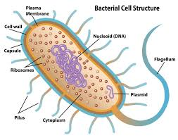
🔹 Basic Structure
- Cell Wall 🧱 – rigid layer providing shape, protection, contains peptidoglycan (main target of many antibiotics).
- Cell Membrane 🩸 – selectively permeable, controls movement of substances.
- Cytoplasm 💧 – contains enzymes, ribosomes, nutrients.
- Nucleoid 🧬 – single circular DNA; no true nucleus.
- Ribosomes (70S) 🎯 – site of protein synthesis.
- Flagella 🚩 – for motility (movement).
- Pili/Fimbriae 🌾 – attachment and conjugation.
- Capsule 🛡️ – protects from phagocytosis.
- Spores 🥚 – highly resistant dormant structures (e.g., Bacillus, Clostridium).
2️⃣ VIRUSES 🦠
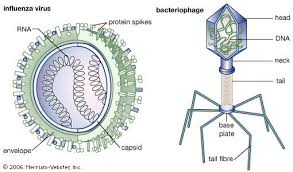
🔹 Basic Structure
- Nucleic Acid Core 🧬 – DNA or RNA (never both).
- Capsid 🛡️ – protein coat protecting genetic material.
- Envelope 🎀 (some viruses) – derived from host membrane, contains glycoproteins.
- Spikes 🌟 – help attach to host cells.
- Obligate intracellular parasites – can reproduce only inside living cells.
3️⃣ FUNGI 🍄
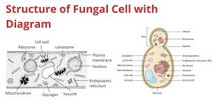
🔹 Basic Structure
- Cell Wall 🧱 – composed of chitin and polysaccharides.
- Cell Membrane 🫧 – contains ergosterol (target of antifungal drugs).
- Hyphae 🌿 – long branching filaments.
- Mycelium 🌲 – network of hyphae.
- Spores 🍃 – for reproduction.
- Nucleus 🎯 – well organized, eukaryotic.
4️⃣ PROTOZOA 🦠
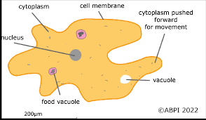
🔹 Basic Structure
- Single-celled eukaryotes 🧬
- Nucleus – well-defined.
- Pellicle – protective covering.
- Locomotory structures 🚀 – pseudopodia, cilia, or flagella.
- Contractile vacuole 💧 – osmoregulation.
- Food vacuole 🍽️ – digestion of nutrients.
5️⃣ ALGAE 🌿
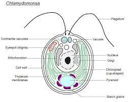
🔹 Basic Structure
- Chloroplasts 🌞 – contain chlorophyll for photosynthesis.
- Cellulose cell wall 🧱
- Nucleus, mitochondria – eukaryotic cell components.
- Flagella (in some) – for movement.
- Pigments 🎨 – chlorophyll, carotenoids, phycobilins.
6️⃣ HELMINTHS 🪱
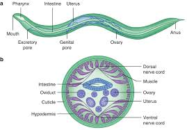
🔹 Basic Structure
- Multicellular organisms
- Complex reproductive system
- Digestive tract (varies by type)
- Cuticle or outer covering
- Organs and tissues like animals
7️⃣ PRIONS 🧬⚠️
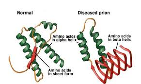
🔹 Basic Structure
- Infectious proteins only
- No DNA, no RNA
- Cause degenerative brain diseases (e.g., Creutzfeldt–Jakob)
🧩 CLASSIFICATION OF MICROBES
1️⃣ BACTERIA 🧫
Based on Shape
- Cocci ⚪ – spherical (Streptococcus, Staphylococcus)
- Bacilli 🔶 – rod-shaped (E. coli, Bacillus)
- Spirilla/Spirochetes 🌀 – spiral (Treponema)
- Vibrios ✔️ – comma-shaped (Vibrio cholerae)
Based on Gram Staining
- Gram-positive 💜 – thick peptidoglycan
- Gram-negative ❤️ – thin peptidoglycan + outer membrane
Based on Oxygen Requirement
- Aerobic 🌬️ – require oxygen
- Anaerobic 🚫🌬️ – no oxygen
- Facultative anaerobes 🔄 – both
- Microaerophilic ⚠️ – low oxygen
Based on Spore Formation
- Spore-forming 🥚 – Bacillus, Clostridium
- Non-spore-forming 😌 – others
2️⃣ VIRUSES 🦠
Based on Genetic Material
- DNA viruses
- RNA viruses
Based on Shape
- Icosahedral 🔷
- Helical 🌀
- Complex 🛸
Based on Host Range
- Animal viruses
- Plant viruses
- Bacteriophages (infect bacteria)
3️⃣ FUNGI 🍄
Based on Structure
- Yeasts 🍞 – unicellular
- Molds 🌿 – filamentous
- Dimorphic fungi ♻️ – both yeast & mold forms
4️⃣ PROTOZOA 🦠
Based on Locomotion
- Amoeboids 🤲 – pseudopodia
- Flagellates 🚩 – flagella
- Ciliates 🌾 – cilia
- Sporozoans/Apicomplexa 🧬 – non-motile (e.g., malaria parasite)
5️⃣ ALGAE 🌿
Based on Pigments
- Green algae 💚
- Brown algae 🤎
- Red algae ❤️
- Blue-green algae (cyanobacteria) 💙 (prokaryotes)
6️⃣ HELMINTHS 🪱
Based on Type
- Nematodes 🧵 – roundworms
- Cestodes ➿ – tapeworms
- Trematodes 🍃 – flukes
7️⃣ PRIONS 🧬⚠️
- Classified under proteinaceous infectious agents
- Cause transmissible spongiform encephalopathies
🌱 Morphological Types in Microbiology
Microorganisms show a wide range of shapes, sizes, and structural variations, which help in identification, classification, and diagnosis in microbiology.
🦠 1. Bacterial Morphology
🔵 Cocci (Spherical Bacteria)
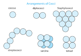
These bacteria are round or oval-shaped.
Key points:
- 🟣 Found singly or in groups
- 🟣 Non-motile generally
- 🟣 Common in skin, respiratory tract infections
Forms of Cocci:
- 🔹 Coccus – single round cell
- 🔹 Diplococci – in pairs (e.g., Neisseria)
- 🔹 Streptococci – chain-like arrangement
- 🔹 Staphylococci – grape-like clusters
- 🔹 Tetrads – groups of four
- 🔹 Sarcinae – cube-like arrangement of eight cells
🪵 Bacilli (Rod-Shaped Bacteria)
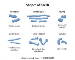
These bacteria appear as straight or slightly curved rods.
Key points:
- 🟤 May form spores
- 🟤 Can be motile due to flagella
- 🟤 Common in soil, water, and GI tract
Forms of Bacilli:
- 🔹 Single bacillus – one rod
- 🔹 Diplobacilli – rods in pairs
- 🔹 Streptobacilli – chains of rods
- 🔹 Coccobacilli – short rods resembling cocci
🌀 Spirilla (Rigid Spiral Bacteria)
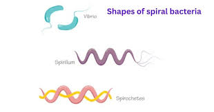
Spiral-shaped, thick, rigid organisms.
Key points:
- 🔵 Rigid spiral form
- 🔵 Have external flagella
- 🔵 Cause waterborne infections
🔍 Spirochetes (Flexible Spiral Bacteria)
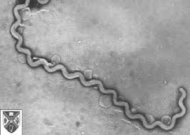
Long, thin, flexible spiral-shaped bacteria.
Key points:
- 🧬 Move in corkscrew fashion
- 🧬 Endoflagella present
- 🧬 Cause syphilis & leptospirosis (Treponema, Leptospira)
🪱 Vibrio (Comma-Shaped Bacteria)
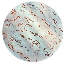
These are curved rod-shaped, like a comma.
Key points:
- 💧 Aquatic organisms
- 💧 Motile with a single polar flagellum
- 💧 Example: Vibrio cholerae (cholera)
🍄 2. Fungal Morphology
🧫 Yeasts
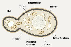
Unicellular fungi, oval or round in shape.
Key points:
- 🍞 Reproduce by budding
- 🍞 Form smooth, creamy colonies
- 🍞 Example: Candida albicans
🕸️ Molds
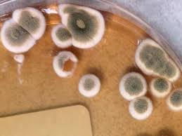
Multicellular fungi with filamentous structures called hyphae.
Key points:
- 🌿 Hyphae form mycelium
- 🌿 Produce spores (asexual/sexual)
- 🌿 Common in environment
🌗 Dimorphic Fungi
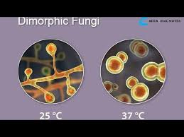
Exist in two morphological forms depending on temperature.
Key points:
- 🔥 Yeast form at body temperature (37°C)
- ❄️ Mold form at room temperature (25°C)
- 🔥 Pathogenic (e.g., Histoplasma, Blastomyces)
🧫 3. Viral Morphology
🧊 Icosahedral Viruses
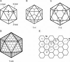
These viruses have a 20-sided symmetrical capsid.
Key points:
- 🟡 Very stable
- 🟡 Includes adenovirus, poliovirus
🧵 Helical Viruses
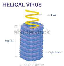
Capsid proteins are arranged in a spiral or helix around the viral genome.
Key points:
- 🟠 Tobacco mosaic virus (plants)
- 🟠 Human viruses include rabies, influenza
🧩 Complex Viruses
Have irregular shapes with additional structures.
Key points:
- 💉 Bacteriophages have head + tail
- 🟤 Pox viruses are brick-shaped
🧫 4. Protozoal Morphology
🟤 Amoeboid (Sarcodina)
Move via pseudopodia.
- Example: Entamoeba histolytica
🟢 Flagellates (Mastigophora)
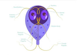
Move via flagella.
- Example: Giardia, Trypanosoma
🔵 Ciliates (Ciliophora)
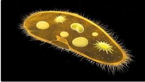
Covered with cilia.
- Example: Balantidium coli
🔴 Sporozoa (Apicomplexa)
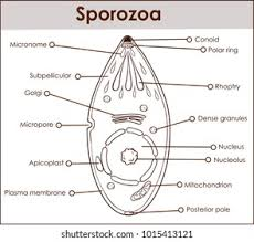
Non-motile, spore-forming parasites.
- Example: Plasmodium (malaria)
🧬 5. Algal Morphology
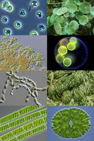
Algae show wide variety:
- 🌿 Unicellular (Chlamydomonas)
- 🌱 Filamentous (Spirogyra)
- 🌊 Colonial (Volvox)
🔍 SIZE OF BACTERIA
📏 General Size Range
- Most bacteria range between 0.2 µm to 2.0 µm in diameter and 1–10 µm in length.
- They are much smaller than human cells and can only be seen clearly under a compound microscope or electron microscope.
⭐ Important Size Variations
- Tiny bacteria (smallest):
Mycoplasma — about 0.1–0.25 µm (smallest free-living organisms) 🦠 - Large bacteria:
Bacillus anthracis — up to 1.5 µm × 10 µm 🧫 - Giant bacteria (rare):
Epulopiscium fishelsoni — up to 600 µm (visible to naked eye) 😲 - Average pathogens:
E. coli — about 1 µm × 3 µm
Staphylococcus aureus — about 1 µm diameter
🧪 Factors Affecting Bacterial Size
- Growth conditions
- Nutrition availability
- Stage of growth (lag, log, stationary)
- Environmental stress
🔷 FORM (SHAPE) OF BACTERIA
already discussed in above topics
OTHER SPECIAL BACTERIAL SHAPES 🧬
Some bacteria show unique or irregular shapes.
- Pleomorphic bacteria – shape varies due to lack of cell wall
Example: Mycoplasma - Actinomycetes – branching, filamentous bacteria
Example: Actinomyces, Nocardia 🌿 - Stellate (star-shaped) bacteria ⭐
- Square-shaped bacteria found in salty environments ⬛
🔶 ARRANGEMENT OF BACTERIA
The arrangement depends on the plane of cell division:
- One plane → Chains (strepto-)
- Multiple random planes → Clusters (staphylo-)
- Two planes → Tetrads
- Three planes → Sarcina (cube of 8 cells)
📚 IMPORTANCE OF SIZE & FORM IN MICROBIOLOGY
- Helps in identification in Gram staining
- Used for diagnosis of infectious diseases
- Determines motility, virulence, and survival
- Guides selection of antibiotics (cell wall structure depends on shape)
⭐ Motility
Microbial motility refers to the ability of microorganisms to move from one place to another using specialized structures or mechanisms. This movement is essential for survival, colonization, nutrient acquisition, escaping harmful environments, and causing infections.
🌟 Definition
Motility is the self-directed movement of microorganisms by using external or internal structures, enabling them to swim, glide, twitch, or rotate in response to various stimuli.
🔥 Importance of Motility
- ⭐ Helps organisms reach favorable environments
- ⭐ Avoids harmful conditions (e.g., toxic chemicals)
- ⭐ Assists in colonization and infection in host tissues
- ⭐ Essential for biofilm formation
- ⭐ Supports nutrient search and uptake
🧬 Types of Motility in Microorganisms
🚩 1. Flagellar Motility (Most Common)
Many bacteria use flagella—long, whip-like structures—to swim.
🔑 Key Features
- ⭐ Flagella rotate like a propeller
- ⭐ Allows swimming in liquid environments
- ⭐ Found in Bacillus, E. coli, Vibrio cholerae, Pseudomonas
- ⭐ Can move forward (run) or stop and change direction (tumble)
🌀 Flagellar Arrangement
- ⭐ Monotrichous – single flagellum (e.g., Vibrio)
- ⭐ Lophotrichous – tuft at one end
- ⭐ Amphitrichous – one on each end
- ⭐ Peritrichous – all around the cell (e.g., E. coli)
💥 2. Brownian Movement (Not True Motility)
Random vibration of cells due to water molecules.
🔑 Key Points
- ⚠️ Not actual motility
- ⚠️ No direction or energy use
- ⚠️ Seen as “shaking movement”
- ⭐ Helps differentiate motile vs non-motile bacteria
🧭 3. Chemotaxis (Movement Toward or Away From Chemicals)
Microbes move based on chemical signals in the environment.
🔑 Key Features
- ⭐ Move toward nutrients (positive chemotaxis)
- ⭐ Move away from toxins (negative chemotaxis)
- ⭐ Uses receptor proteins to sense environment
- ⭐ Highly developed in E. coli and Pseudomonas
🧷 4. Gliding Motility
Some bacteria move smoothly over surfaces without flagella.
🔑 Key Features
- ⭐ Seen in Myxobacteria, Cytophaga
- ⭐ Allows surface colonization
- ⭐ Useful in biofilm formation
- ⭐ Movement is slow and smooth
🧲 5. Twitching Motility
Movement using type IV pili that extend, attach, and pull the cell forward.
🔑 Key Features
- ⭐ “Jerky” or “twitchy” motion
- ⭐ Seen in Pseudomonas, Neisseria
- ⭐ Important in biofilm formation
- ⭐ Helps in movement across solid surfaces
🌊 6. Spirochete Motility
Spirochetes move by axial filaments (endoflagella) wrapped around the cell inside the periplasmic space.
🔑 Key Features
- ⭐ Produces corkscrew-like movement
- ⭐ Helps movement in viscous tissues
- ⭐ Found in Treponema pallidum (syphilis), Borrelia
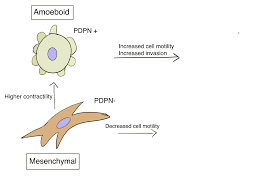
💫 7. Amoeboid Motility (Pseudopodia Formation)
Used by protozoa like Amoeba.
🔑 Key Features
- ⭐ Cell extends pseudopodia (false feet)
- ⭐ Movement is slow and crawling
- ⭐ Important for phagocytosis
🦠 8. Ciliary Motility
Protozoa like Paramecium use cilia to move.
🔑 Key Features
- ⭐ Fast movement
- ⭐ Coordinated beating of many cilia
- ⭐ Also aids in feeding by moving food particles
🧪 Methods to Detect Motility in Microbiology
1️⃣ Hanging Drop Method
- ⭐ Direct observation under microscope
- ⭐ Shows true motility vs Brownian motion
2️⃣ Motility Agar (Soft Agar 0.4%)
- ⭐ Motile bacteria spread from inoculation line
- ⭐ Non-motile remain in place
3️⃣ Flagella Staining
- ⭐ Visualizes flagella under microscope
4️⃣ Electron Microscopy
- ⭐ High-resolution view of motility structures
🚫 Non-Motile Organisms
Some bacteria do not show motility, e.g.:
- ⭐ Klebsiella
- ⭐ Shigella
- ⭐ Streptococcus
- ⭐ Corynebacterium