ENGLISH – ANATOMY & PHYSIOLOGY-2017 PAPER 3
GUJARAT NURSING COUNCIL PAPER 2017.
Que-1 Answer the following questions
(a) List out the organs of the urinary system. 02
The urinary system is the excretory system of the body. Which removes waste products from the body. It has the following organs.
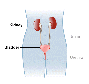
Kidney (Right and Left) 2
Ureter (right and left) 2
Urinary Bladder 1
Urethra 1
Que-1 🔸(b) Describe gross structure of kidney ..04
There are two kidneys in the human body. They are located one on both sides of the vertebral column on the poster side of the body on the right and left sides of the abdominal cavity.
Kidney is a shapeless organ. It lies from the level of the twelfth thoracic vertebra to the level of the third lumbar vertebra.
Kidney is 11 cm long by 5 to 6 cm wide. Its weight is approximately 150 grams. The right kidney is positioned slightly lower than the left kidney because the liver occupies a larger portion on the right side.
Veins around the kidney.
The kidney is an organ located in the abdominal cavity. One is located on both the right and left sides. Abdominal cavity organs like liver, small intestine, adrenal glands, stomach, spleen, pancreas etc. are located around both kidneys.
Structure of the Kidney..
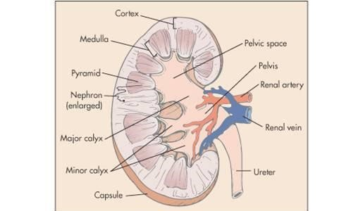
Kidney is a shapeless organ. A groove in the middle is called hilum or renal hilum. Through which the structures of renal artery, renal vein, nerves, lymph vessels and ureter enter and exit.
The inner border or hilum of the kidney is found on the side of the vertebral column. Its outer border is convex. The kidney is a hanging organ on both sides of the abdominal cavity. To hold it in position, it is surrounded by a network of fatty tissue and fibroelastic connective tissue called the renal fascia. With the help of this kidney can maintain its position and it also gets protection.
When the kidney is viewed in a longitudinal section, it is seen to be distributed into three kidney structures.
Fibrous capsule.: It is a part made of fibrous tissue surrounding the kidney. This membrane is arranged around the kidney. Which acts as a layer to protect and maintain the shape of the kidney.
Cortex.: It is a redish brown colored part of tissue. Which is located under the kidney capsule.
Medulla.
In the kidney, the inner part from the cortex is called the medulla. It also has redish brown color. The triangular shaped pyramidal structure is called renal pyramid. The base part of this renal pyramid is towards the cortex and the pointed part of the pyramid i.e. the part of the renal papilla is arranged inwards towards the hilum.
The renal papilla forms a cup-like structure anteriorly called the calyx. The part with large space is called major calyx and the part with small space is called minor calyx. The minor calyx opens into the major calyx. Beyond this calyx is the wide funnel-shaped portion called the renal pelvis.
The urine filtered by the kidney falls into the wide part of the calyx, the funnel shape, i.e. the renal pelvis. Urine collects here and then passes anteriorly from the renal pelvis through a narrow structure called the ureter that exits the kidney and reaches the urinary bladder.
Urine filtered by the kidney passes from the minor calyx to the major calyx and from the major calyx to the renal pelvis. It then reaches the urinary bladder through the ureter. This action is not controlled by any kind of nervous system. In the wall of the renal pelvis there are special muscles and pacemaker cells due to the contraction of which this urine flows forward.
Que-1🔸 (c) Write the functions of kidney in detail … 06
Kidney is mainly responsible for urine formation.
Kidneys filter the blood and remove the waste products through urine.
The function of the kidney is to maintain the normal balance of electrolytes.
It works to maintain blood pH.
The body functions to remove waste products accumulated at the end of metabolism from the body.
Kidneys secrete a hormone called erythropoietin which plays a very important role in the production of RBCs.
Kidneys secrete a hormone called renin which plays a very important role in maintaining blood pressure.
Kidneys are responsible for maintaining water balance in the body.
Kidney prevents the elements that are needed in the body from leaving the body.
Or
Que 1🔸(a) List out the components of cell. 04
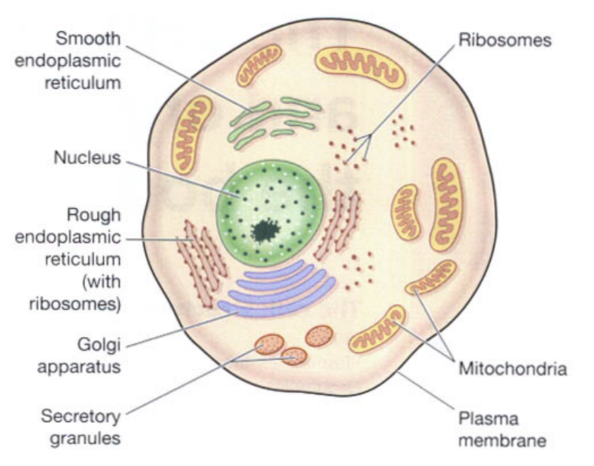
Cell is the smallest microscopic and functional unit of the human body.
The components in Cell are as follows.
cell membrane
the nucleus
Cytoplasm
protoplasm
Mitochondria
Golgi apparatus
Ribosomes
Endoplasmic Reticulum (Smooth Endoplasmic Reticulum and Rough Endoplasmic Reticulum)
Que 1🔸(b) Describe the organelles of the cell ..04 marks
Cell is the basic functional and structural unit of human body. It is the main working tax unit.
A cell is known as a mass of protoplasm. Inside the cell are organelles that are covered by a plasma membrane.
A zygote is formed by the fusion of ovum and sperm in the human body. The growth and cell division of this zygote leads to the formation of the human body.
The fluid part inside the cell is known as cytoplasm. It contains many organelles. The structure of a cell is as follows.
plasma membrane..
The membrane surrounding the cell is called the plasma membrane. This membrane has selective permeability. Due to which some substance can come inside the cell and some substance can go out of the cell. Thus the cell maintains the structure of its cytoplasm through this membrane.
The plasma membrane is a double layer membrane composed of phospholipids. It works to provide protection to the organelles of the cell and to maintain the shape of the cell.
Nucleus..
Nucleus is located in the center of the cell. It contains a liquid called protoplasm. Nucleus membrane is located around the nucleus. This membrane also has selective permeability. The nucleus membrane partially separates the cytoplasm and protoplasm. Nucleus controls all the activities inside the cell and only with its help the cell can stay alive.
Inside the nucleus are stringy proteins called chromatin. This chromatin turns into chromosomes during cell division and performs the function of cell division.
These chromosomes inside the nucleus are responsible for the inherited traits of an individual. Chromosomes are found in 23 pairs in the cells of the human body. 22 pairs of these chromosomes are called ordinary chromosomes while 1 pair is called sex chromosomes.
Mitochondria…
Mitochondria are rod-shaped structures. Which is located in the cytoplasm inside the cell. There is a double membrane around it, the structure of the membrane is similar to the plasma membrane. The outer layer of this membrane is a smooth layer and the inner layer has many folds. This series of folds is called cristae.
Within these cristae are enzymes that release ATP. This is why mitochondria are called the power house of the cell.
Ribosomes..
They are tiny granules in the cytoplasm. They are made up of proteins and RNA. It performs the function of protein synthesis from amino acids. Some ribosomes lie free in the cytoplasm and some are attached to the surface of the endoplasmic reticulum.
Endoplasmic reticulum…
It is a series of interconnecting membrane or channel like structures. which connects one structure of cytoplasm to another structure. There are two types of it.
- Smooth endoplasmic reticulum.. Its surface is smooth. It functions in steroid hormone and lipid synthesis. It also helps to detoxify certain drugs.
- Rough endoplasmic reticulum.. Its surface is rough. Ribosomes are located on its surface. These ribosomes perform the function of protein synthesis. Endoplasmic reticulum also helps transport substances from one place to another in the cytoplasm of the cell.
Golgi apparatus…
The Golgi apparatus is a bag-like structure with four to eight folds. These folds overlap each other. The end portion of this structure forms a pouch-like structure called a cisterna. Proteins synthesized by ribosomes are collected and stored in the form of secretory vesicles at the ends of these cisternae. When needed, these secretory vesicles release proteins into the cytoplasm. The Golgi apparatus is a structure located near the nucleus.
Lysosomes…
Lysosomes are a type of secretory vesicles that are secreted through the membrane of the Golgi apparatus. These lysosomes contain the content of certain enzymes that break down certain large molecules in the cytoplasm of the cell. It works to protect cells from foreign material and microorganisms. These lysosomes also work to remove the waste material accumulated inside the cell.
A sun-shaped centrosome is also present in the cytoplasm of the cell, which plays an important role in cell division.
Apart from this, the cytoplasm of the cell also contains a network of microfilaments and microtubules which function to maintain the shape of the cell and to protect and support the structure of the cell.
Que 1🔸(c) Explain the epithelium tissue in detail 04
This type of tissue is found in many places throughout the body. which are mainly scattered on the surface or on the surface lining.
Epithelial tissue is the tissue lining the inner wall of a body cavity, gland, organ or blood vessel.
Functions of the Epithelial Tissue..
Epithelial tissue is important in forming the inner wall of any organ.
Epithelial tissue acts to provide protection where it is located in the inner wall of an organ.
Epithelium is a tissue associated with making any type of secretion through secretory cells located in the tissue.
Epithelial tissue functions in the absorption of material as it is located in the inner wall of the structure.
Characteristics of Epithelium Tissue…
Epithelium TSUs are scattered over the basement membrane of any organ or structure.
The cells in this tissue are closely fitted to each other i.e. arranged close together.
The matrix in this tissue is in liquid form.
Due to the presence of special types of cells in this tissue, they are connected with the functions of secretion and absorption.
Classification of Epithelium Tissue.
- Simple Epithelium Tissue.. This type of tissue is always found in single layer.
- Stratified Epithelium Tissue .. This tissue is in multiple layers.
A. Classification of Simple Epithelium Tissue: This type of tissue is found in the inner wall of any structure or organ. They are associated with activities like absorption and secretion. There are four main types of this type of tissue.
- Simple squamous epithelium tissue… These types of tissues are scattered in the basement layer, the inner wall of any structure or organ. Its cells are close to each other and are flat and arranged in a row. Between them is the nucleus. This type of tissue is found in the lining of the inner wall of the heart, alveoli of the lungs, blood vessels and lymph vessels.
- Simple cuboidal epithelium tissue…. The shape of the cells in this tissue is cube shaped. which are closely related to each other. This type of tissue is spread over the basement membrane. Usually this tissue is found in renal tubules and thyroid gland.
- Simple columnar epithelium tissue … This cell is rectangular in shape. Which are more in length and less in width. These types of tissues are found in the lining of the respiratory tract and the lining of the alimentary tract. It also contains goblet epithelium cells which perform the action of mucus secretion.
- Ciliated Simple Epithelium Tissue …. This tissue contains cells similar to those of cuboidal and columnar tissue. In addition, this tissue has hair-like processes on the cell margin, i.e. cilia, so it is called ciliated epithelium tissue. This type of tissue is especially found in the lining of the respiratory tract and the lining of the fallopian tubes. They are here associated with specific movements.
B. Stratified epithelium tissue…
This type of tissue is composed of more than one layer. In this cell, the size of each cell is not the same. In which cells of each layer are found in irregular size and shape.
In this tissue, the cells of the bottom layer are found in large size and they decrease in size when they come to the surface.
This type of tissue is mainly concerned with protection and support of the structure. Stratified epithelium tissue is divided into two parts.
Stratified squamous epithelium tissue.
Transitional epithelium tissue.
Stratified squamous epithelium tissue.: Stratified squamous epithelium tissue is found in multiple layers. It is mainly divided into two parts.
A. Non Keratinized Stratified Epithelium Tissue..
This tissue is mainly found in moist surface areas of the body. Like conjunctiva, esophagus, vaginal cavity, fairings etc.
It is a cell with a nucleus. It has a flat shape.
B. Keratinized stratified epithelium tissue…
This type of tissue is mainly found in dry areas of the body such as skin, hair, nails etc.
This tissue mainly contains keratin substance. Which makes water resistant. So mainly evaporation cannot take place. It is the main characteristic of this tissue
Transitional epithelium tissue.. This tissue is found in more than one layer but its main characteristic is that it does not have a basement membrane. Pear shaped cells are seen in this. This type of tissue is mainly found in the inner wall of the urinary bladder.
Que 2 Answer the following questions
🔸(a) Explain the gross structure and function of liver. 08
Describe the gross structure of the liver and explain its function
Among all the glands in the body, the liver is the largest gland. which lies below the diaphragm in the right quadrant of the abdominal cavity. It weighs approximately 1.4 kilograms inside an adult. It is located below the ribs of the chest. Ribs protect it. A part of it is also located in the region of the left abdominal cavity.
The upper surface of the liver is smooth. This portion is attached to the diaphragm and has an irregular surface and margin on the back and underside of the liver.
Organs around the liver.
Organs such as the diaphragm, anterior abdominal wall muscles, stomach, duodenum, kidney, inferior vena ceva, gallbladder etc. are arranged around the liver.
Liver is mainly divided into two lobes, right lobe and left lobe.
The right lobe is larger than the left lobe and both lobes are separated by the falciform ligament.
The quadrate lobe is found on the posterior side of the liver and the quadrate lobe is found on its inferior side.
Thus four lobes of liver can be seen anatomically.
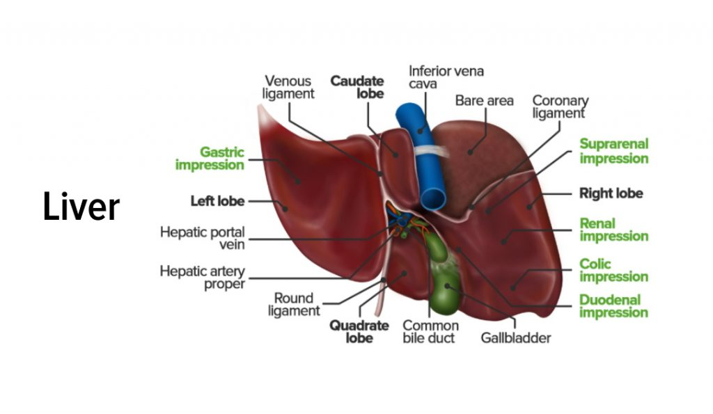
Portal fissure…
The groove on the posterior surface of the liver is called the portal fissure. From this fissure some structures enter the liver and some structures exit the liver viz.
The portal vein carries deoxygenated and nutritious blood from the intestine and enters the liver through this fissure.
The hepatic artery carries oxygenated blood and enters the liver through this fissure.
Nerve fibers of the sympathetic and parasympathetic nerves enter the liver through this fissure.
Left and right hepatic ducts exit this fissure to carry bile from the liver to the gall bladder.
The hepatic vein exits this fissure to carry deoxygenated blood from the liver.
Structure of the Liver…
The liver is an organ located in the right quadrant of the abdominal cavity. It is mainly composed of 2 lobes. These lobes are made up of many lobules. These lobules are made up of special type of epithelium cells and cells called hepatocytes. These cells are found in a hexagonal pattern in the liver.
After entering the hepatic artery and portal vein within the liver, a capillary network of arteries and veins is formed. This network of capillaries is called sinusoids. Kupffer cells are found in the wall of these sinusoids, which Kupffer cells act to protect the liver from bacteria, foreign material or toxins and perform a protective function in the liver.
Hepatocytes cells within the liver secrete bile. This bile enters the bile canaliculi. These bile canaliculi carry the bile into small bile ducts. These small bile ducts join to form the right and left hepatic ducts. which drains bile from the liver through the common hepatic duct. The common hepatic duct joins with the cystic duct from the anterior gallbladder to form the common bile duct. Bile is drained into the small interstitium and plays an important role in the digestion of fat.
Blood supply of the liver.
The liver is supplied with blood by the hepatic artery and the portal vein carries nutritious blood and enters the liver.
The hepatic vein drains deoxygenated blood from the liver and joins the inferior vena cava.
Functions of the Liver..
Liver is a very important organ of our body. It is associated with many important functions. These functions are as follows.
The liver is responsible for the metabolism of carbohydrates.
The liver functions to maintain normal carbohydrate ie blood glucose levels. When blood glucose levels fall, glucose is produced from glycogen through the action of glycogenolysis and blood glucose levels are maintained.
When the amount of glucose in the blood increases, glucose is converted into glycogen by the action of glycogenesis.
The liver converts fat into fatty acids so that it can be used in the body, this is called desaturation of fat, thus it helps in lipid metabolism.
The liver helps in protein metabolism so that amino acids are synthesized in the body.
The liver converts ammonia and makes urea so that this waste product can be excreted through urine.
The death of red blood cells releases bilirubin, which is modified by the liver to help remove excess bilirubin from the body.
Liver helps to detoxify the toxic substances, alcohol, drugs etc. introduced in the body and remove them from the body.
The liver synthesizes bile salts, which are essential for emulsification of fats and hence absorption of lipids and cholesterol.
Helps to maintain the level of vitamin D in the body.
Since Kupffer cells are located inside the liver, they perform a protective function by protecting the liver from harmful substances and performing phagocytosis.
The liver acts as a storage of vitamins and minerals and releases these vitamins and minerals into the body when needed.
Liver is responsible for heat production in the body.
Que 2🔸(b) Discuss Menstrual cycle. 04 marks
Menstruation cycle occurs after puberty phase in females. In which changes are seen in the function of ovaries and uterus.
A menstrual cycle occurs every 26 to 30 days. This is seen due to changes in the level of hormones in the blood.
The onset of the menstruation cycle is known as menarche.
Females find this cycle continuous after the age of puberty. Which stops temporarily during pregnancy and stops completely after the period of menopause.
The onset of menstruation is due to the degeneration of the corpus luteum layer in the uterus and bleeding occurs through the vaginal cavity.
Menstruation cycle has the following phases.
- Menstrual phase..
This phase occurs every 28 days and lasts for about four days. When fertilization of the egg does not take place in the female, the hormones estrogen and progesterone that support the uterine wall decrease and the hormone oxytocin increases. So the stimulation of contraction of the uterus increases and degeneration of the corpus luteum layer of the wall of the uterus starts and blood drains from the uterus through vaginal discharge. This phase lasts from 1 to 4 days.
This menstrual flow contains endometrial glands, endometrial cells, blood and unfertilized ovum. Approximately 100 to 200 ml of blood is shed during the 3 to 5 days of this phase which is called the menstrual phase.
- Proliferative phase..
The menstrual phase ends on the 5th day. After that, the proliferative phase starts from day 6 and lasts for 14 days.
In this phase follicle stimulating hormone stimulates the ovarian follicles and hence increases estrogen production. This estrogen stimulates the proliferation of the endometrium.
The endometrium of the uterus begins to develop from the sixth day.Its cells multiply and due to this increase in mucus secreting glands and blood capillaries. Thus the endometrium of the uterus becomes bulky and vascular.
At the end of this phase, the inner wall of the uterus is ready for implantation of the fertilized egg. This phase ends with ovulation. Towards the end of this phase, there is a decrease in estrogen levels.
- Secretary phase.
After the proliferation phase is completed, the secretory phase begins. The secretory phase is observed from the 15th day of the menstrual cycle to the 28th day.
As progesterone hormone is important in this phase, this phase is also called progesterone phase.
When the mature egg is released by the ovary due to ovulation, the amount of estrogen and progesterone hormones decreases, but the corpus luteum of the uterine wall maintains the pregnancy by secreting progesterone.
As this mature ovum is not fertilized by a sperm, the corpus luteum decreases progesterone and due to the decrease in progesterone hormone, there is an increase in the amount of oxytocin hormone and the uterine muscles begin to contract.
The next cycle begins at the end of this phase due to the corpus luteum not receiving a fertilized ovum and increased uterine contractions.
Que 3 Answer the following (Any Two)
(a) Describe the cardiac cycle. 2×6-12
Heart is a continuously pumping organ. The pumping action of the heart is called its cardiac cycle. A healthy person has a cardiac cycle ie pumping action of 68 to 72 times in a minute. One cardiac cycle takes 0.8 seconds to complete. This work runs continuously through the heart of a living human being.
The contraction and relaxation of the heart muscle is done by the impulses generated from the S node of the heart. Contraction is called systole and relaxation is called diastole.
The following events occur in the cardiac cycle.
Atrial systole.. Atrial systole means simultaneous contraction of both atria which takes 0.1 second. In this atrial systole, when both atria are filled with blood, impulses are generated by the S node and these impulses reach the AV node. During this time it takes 0.1 second and both atria contract simultaneously both atrioventricular valves open and both atriums empty of blood and both ventricles fill with blood. This phase is called atrial systole.
Ventricular systole.. Ventricular systole is the simultaneous contraction of both ventricles. It takes 0.3 seconds. During ventricular systole, when both ventricles are filled with blood, impulses from the AV node reach the bundle of His and Purkinje fibers, i.e., the heart. During this time it takes 0.3 seconds and both ventricles contract simultaneously. The blood of both the ventricles respectively, the blood of the right ventricle goes to the lung through the pulmonary artery and the blood of the left ventricle circulates throughout the body through the aorta. This phase is called ventricular systole.
Complete cardiac diastole.. Complete cardiac diastole means simultaneous relaxation of both the atria and the ventricles i.e. the four chambers of the heart. This action takes 0.4 seconds.
During complete cardiac diastole there is no electrical activity in the heart. Myocardium muscles are relaxed. During this time both the atria and both the ventricles dilate i.e. relax. Both atriums are refilled with blood during this time. This relaxation time is 0.4 seconds. This is called complete cardiac diastole.
Thus it takes 0.8 seconds to complete a complete cardiac cycle and blood circulation occurs due to the contraction and relaxation of the heart.
Que 3🔸(b), Explain the role of nurse in bio medical waste management.–
Nurses have a very important role in the management of bio medical waste
Disinfect biomedical waste generated on board as early as possible so that it does not become a source of infection.
To reduce as much as possible the transportation and storage of waste
According to the medical waste policy, it should be put in separate bags or containers
To discard disposable items so that they are not reused
Infectious plastic waste that can be recycled should be reused only after sterilization.
Sharp instruments should be kept in a puncture proof white container
Sharp instruments should be treated for dish effect before transport
A chemical like sodium hypochlorite should be used
If there is any mistake in segregation of bio medical waste, it should be corrected by the nursing staff
If infection occurs through bio medical waste, it should be surveyed
Clean and use should be handled carefully to prevent dusting
Bio medical waste should be segregated according to the color coding of bio medical waste
Education should be provided for buy medical waste
Proper records and reports of bio medical waste should be maintained
Bio medical waste containing harmful micro-organisms that can harm hospitalized patients, health workers working in hospitals and the general public
Bio medical waste contains sharp instruments such as syringes scalpel etc. which may cause injury.
Toxic products of pharmaceutical products especially antibiotics and released into the environment also cause mercury damage.
Chemical burns can occur during dish infection or waste treatment activities
The incineration process causes air pollution
Infection occurs by using needle etc
Due to lack of safe injection practices, infectious diseases like HIV, hepatitis B, hepatitis B etc. are caused by medical waste.
All these diseases can also be caused by needle stick injury
People handling bio medical waste can get special injuries and other diseases
🔸(c) Describe the factors affecting the growth of microbes.
1) Moisture
Like nourishing food, every bacteria needs water for growth. In fact, bacteria cannot get food in the absence of water, because every food element needs to be in a liquid state to pass through the wall of the bacteria. All types of bacteria grow well in an aqueous medium, an environment without complete moisture prevents its growth. or destroys.
Apart from this, cells cannot live in low or high humidity
2) Light
Most bacteria are destroyed by direct exposure to ultraviolet rays in sunlight.
3) Temperature :-
Temperature is a very important factor affecting the growth of bacteria. Optimal temperature with food, water is necessary for bacteria growth.
Different bacteria have different optimal temperatures.
37°C is the optimal temperature for bacteria growing in the human body.
However, many bacteria are mesophilic (meso = middle, phille = loving). The optimum temperature for it is 25 to 39* C.
Most bacteria grow this way.
Whereas psychrophilic (psychro = cold) bacteria grow better between 4°C to 10°C, some
Thermophilic (Therma – Heat) is also found. Its growth is best between 55°C to 75°C.
Temperature above 75 C is fatal for bacteria. In fact high temperatures are created to kill bacteria in different ways.
Like moist heat (steam), boiling water, pasteurization & autoclaving.
Many species can survive even at very low temperatures. Like yeast, mould, viruses & Rickettsia, spirochetes (76* C can survive for years).
(4) Oxygen
O2 also plays an important role in the life of bacteria. Many types of bacteria can only survive, or grow, in the presence of O2. They are called Aerobes (EX.Sarcina).
Conversely, Anaerobes can live or grow in the absence of 02. E.g. Closteridium tetani-
Apart from this there are also bacteria. which can survive in the presence or absence of 02. They are known as facultative anaerobes. E.g. Salmonella typhi.
Microaerophils grow more in less oxygen than is present in air.
(5)Hydrogen Ion Concentration: (Acidity and Alkalinity) PH medium
The acid or alkaline concentration of the liquid in which the bacteria grow affects the growth.
This is seen from the hydrogen ion concentration index.
PH – 0 (Zero) is the most acidic,
PH – 14 shows the lowest acidic concentration.
PH – 7.) A nutral (neutral),
pH < 7 is acidic
and alkaline at pH >7
Most bacteria grow best between pH 5.0 to 8.5. There are some exceptions to this too.
6) Osmotic pressure :-
The life of bacteria also depends on high or low osmotic pressure. If the bacteria are immersed in a liquid whose osmotic pressure is very high or very low, the bacterial cell collapses or becomes dormant due to leakage of liquid.
Carbon Dioxide is also necessary for the growth of bacteria.
Que 4 Short notes (Any Three) Write track notes. (any three)
🔸(a) Physiology of Hearing-Physiology of Hearing 3×4-12
Mechanism of hearing i.e. physiology of hearing means act of hearing. The wavelength for hearing is 20 to 20,000 Hz. The human ear is capable of frequencies between 500 and 5,000 hz. The frequency at which the sound waves vibrate is known as the pitch, as the vibration increases, the pitch increases.
Every sound produces sound waves and they strike the outer part of the auricle and from there enter through the external auditory canal, these sound waves vibrate the tympanic membrane i.e. the ear drum which is the junction between the external ear and the middle ear.
The sound waves are connected to the tympanic membrane by the malleus bone to the incus and incus to the stapes and the stapes bone is further connected to the oval window. goes and from there goes to the endolymph and the round window vibrates and the vestibule goes to the cerebrum through the cochlear nerve and the sound is recognized.
Que 4 🔸(b) Neuron-
Brain has a large number (100 billion) of neurons.
Each neuron has the following characteristics:
Cell body and its processes
Axon
Dendrites
Some thread-like nerve fibers are also present.
Neurons cannot divide and need constant oxygen for their survival. It gets its energy from glucose.
A neuron has properties of conductivity and excitability, through which it responds to stimuli from the external environment, including mechanical, electrical and chemical stimuli.
This stimulus travels from the dendrites of the nerve cell to the cell and the axon in a process known as slow forward conduction.
Cell Body: Nerve cells vary in size and shape which is not visible to the naked eye.
Gray matter is formed from the part of the body of the nerve cell which is located in the periphery of the brain and in the middle of the spinal cord.
A cell body, like any other cell, contains a nucleus, cytoplasm, and other organelles.
The body of the nerve cell unites to form nuclei in the central nervous system and ganglia in the peripheral nervous system.
Axon and dendrites:- They are processes extending from the cell body. Each neuron has an axon and a large number of dendrites called nerve fibers. Each nerve contains a bunch of sensory as well as motor nerve fibers.
Axons and dendrites make up the white matter. Which is in the central part of the brain and in the peripheral part of the spinal cord.
Structure of Axon:-
An axon is a process extending from the cell body. A neuron has one axon. The axon is a slender, cylindrical process. The length of which can be 100cm.
A volume of axon is called a track.
Axons also have organelles similar to cell organelles. The fluid inside it is called exoplasm and the membrane surrounding it is called axolyma.
Schwann cells are present in the peripheral nervous system surrounding axons.
The small gap between the Schwann cells is called the node of Ranvir. These nodes and the myelin sheath are essential for proper nerve conduction with nerve fibers.
The membrane surrounding the Schwann cell is called neurilemma.
Surrounding the axons of neurons is a multi-layered sheath of lipids and proteins called the myelin sheath.
A neuron that has such a sheath around its axon is called a myelinated neuron and one that does not have a sheath around it is called a non-myelinated or unmyelinated neuron.
Nerve transmission is reduced in unmyelinated neurons.
Dendrites :- are branches extending from the cell body. which conducts impulses towards the cell body of the neuron. Dendrites are not myelinated.
Que 4🔸(c) Thyroid Gland-…
The thyroid gland is an important gland of the endocrine system. It is located in the soft tissue of the neck. This gland is a butterfly shaped gland.
The weight of this gland is approximately 30 grams. Its length is 5 cm and width is 3 cm.
This gland is located from the level of the fifth cervical vertebra to the level of the first thoracic vertebra.
Thyroid gland has one lobe on both sides. Which is covered with fibrous tissue around it. The middle part connecting the two lobes is called the isthmus. The lobes of the thyroid gland have a pyramidal shape.
The tissue in the thyroid gland is made up of tiny structures called follicles. Each follicle is composed of simple cuboidal glandular epithelium tissue. which is connected with secretion.
The function of the thyroid gland is regulated by thyroid stimulating hormone, a hormone released from the pituitary gland.
The thyroid gland secretes the following hormones.
Triiodothyronine T3… The main function of this hormone is to maintain normal physical growth and development in the body. This hormone also plays a useful role in maintaining heart rate and certain metabolic activities.
Thyroxine T4… This hormone also performs the same function as the T3 hormone. That is, the body maintains metabolic activity and functions to maintain normal physical growth and development. This hormone increases the basal metabolic rate.
Calcitonin.. This hormone is secreted by the parafollicular cells of the thyroid gland. Which affects the metabolism of calcium in the blood.
Que 4🔸(d) Innominate bone
The innominate bone is the bone of the pelvic girdle. It is also known as hip bone.
They are two in number in the body. It is located one on both the right side and left side of the pelvic cavity. Both innominate bones articulate with the sacrum bone behind and form the pelvic cavity.
The innominate bone is the larger bone. It is a flat and irregular type of bone.
Each innominate bone consists of three bones.
Ilium
Two ischium
pubis.
Ilium…
Innominate Bone The ilium bone is a flat bone on the upper side. At its uppermost point there is a ridge called the iliac crest. It falls below the iliac crest.
Anterior superior iliac spine (top of front)
Anterior inferior iliac spine (at the bottom of the front)
Posterior superior iliac spine (upper back)
Posterior inferior iliac spine (lower back)
The ilium bone forms the sacroiliac joint where it joins the sacrum bone at the back.
There is a big groove at the bottom of this joint. The notch is called the greater sciatic notch. From where the sciatic nerve and blood vessels pass to the lower extremities.
The gluteal muscles attach to the posterior surface of the ilium bone. And that makes up the gluteal region.
The anterior surface of the ilium bone is known as the iliac fossa. In this part there is a depressed part where the muscles are attached.
ischium …
Below and behind the ilium bone lies the ischium bone.
Between the ilium bone and the ischium bone, the posterior side is an inferior pointed part. This part is called ischial spine.
Below it lies a small notch called the laser sciatic notch.
This laser has a strong thick process below the sciatic notch. Which is called ischial tuberosity. Body no weight comes on this part while sitting in sitting position. This has a stronger structure than a weight beer.
Pubis…
The pubis bone forms the most anterior part of the innominate bone. Both the innominate bones of the pubis bone join anteriorly to form the symphysis pubis joint.
There is a large foramen at the bottom of this pubis bone. It is called obturator foramen. Through which nerves and blood vessels pass downwards towards the extremity.
The three bones ilium, ischium and pubis located in the hip bone form a cavity-like structure called the acetabulum cavity. The head of the femur bone joins this KVT and the hip joint is formed there.
Que 5Define the terms (Any Six)
🔸 a. Carrier-–
A person or animal that harbors specific disease-causing micro-organisms that can spread or carry the disease but does not show any signs or symptoms of the disease is called a carrier.
E.g. Typhoid
(b) Medial
Medial terminology is used to describe the medial side of the body. Anatomically the term medial is also used to describe the middle side or midline of the body. E.g. Heart is located in the medial part of the body. Among the ulna bone and radius bone, the ulna bone is the bone on the medial side.
c. Bacteriostatic —
Agents that inhibit the growth of bacteria are called bacteriostatic
D. T :- Betadine
d. Oogenesis-
Oogenesis is the process of formation of the mature female gamete (ovum) in the ovary which further participates in fertilization.
🔸 e. Hypersensitivity-
Immunity is seen as a protective process. But it is only a small part of the process for antigen response. Sometimes the immune response can be injurious to the host. Which is also responsible for tissue damage, diseases or death. Harmful effects arising from contact with specific antigen is called hypersensitivity.
🔸f. Microbiology-
“Micro” means minute and “bio” means life and “logy” means study. Thus, microbiology means the study of microscopic organisms that are invisible to the naked eye.
Que 5🔸(g) Vital capacity –
The total capacity of the lung is called vital capacity. The capacity of maximum air inhaling and exhaling by this person in addition to the total reserve air in his lungs is called the vital capacity of the lungs.
Vital capacity = IRV+TV+ERV.
The total of tidal volume, inspiratory reserve volume and expiratory reserve volume
Vital capacity of lung is called. Which is approximately 3500 to 3800 ml.
Que 5🔸(h) Glycogenesis
The process of storing glucose in the body’s blood in the form of glycogen in the liver is called glycogenesis. This process starts when the blood glucose level rises above normal.
🔸Q.6 a. Fill in the blanks 05
a. Kleb’s Loffler bacilli is also known as__________
Kleb’s-Loeffler bacilli also called Corynebacterium diphtheriae.
b. B.C.G. vaccine is____________type of vaccine.
B.C.G. vaccine is a live type vaccine
c. Infection occure in patient after admission in hospital is known as_____
Infections that occur after a patient is admitted to a hospital are called nosocomial infections.
d. Types of retinal cells are_____and________.
Retinal cell types are rod cells and cone cells
(B) Match the “A” with “B”.
“A” “B”
a. Thyroid gland a. Prothrombin – Prothrombin
b. Adrenal gland-adrenal gland b. 0 – o
c. Universal Donor c. Thyroxin – Thyroxine
d. Universal Recipient – Universal Race, Payant d. AB-AB
e. Sesamoid bone-Sesamoid bone e. Glucocorticoid – Glucocorticoid
f Patella – Patella
A – C
B – E
C – B
D – D
E – F.
🔸(C) State whether the following statements are True or False. 05
a. Storage of bile is in liver.
Bile is stored in the liver. (false)
b. Optic nerve is a sensory nerve,
The optic nerve is a sensory nerve. (correct)
c. Prolactin releases from anterior pituitary gland
Prolactin is released from the anterior pituitary gland. (correct)
d. Dermis is the outer layer of the skin,
The dermis is the outermost layer of the skin (false)
e. A.V. node is the pace maker of the heart. The AV node is the pacemaker of the heart. (False).