ENGLISH – ANATOMY & PHYSIOLOGY-2018 PAPER 2
GUJARAT NURSING COUNCIL PAPER 2018.
Que. 1 🔸 (a) List out the organs of digestive system. 03
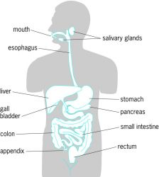
Organs of the Digestive System…
Digestive tract also called alimentary tract is a hollow tube which starts from mouth and extends to anus and its organs are as follows.
mouth
Fairings
Esophagus
Stomach
The small intestine consists of the duodenum, jejunum, and ileum.
Large intestine which includes cecum, ascending colon, transverse colon, descending colon, sigmoid colon, rectum and anal canal.
Accessory organs of the digestive tract…
These organs do not come in the main track of the digestive track but are located in the side which pours its secretion into the alimentary canal and help in the process of digestion so they are called accessory organs.
3 pairs of salivary glands
Liver and biliary tract
Goal Bladder
Pancreas.
Que. 1🔸(b) Describe gross structure of Small Intestine. 04 marks.
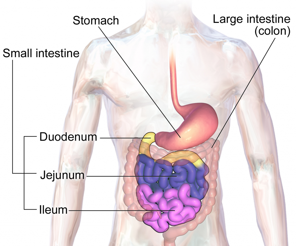
The small intestine is a tube-like structure in the gastrointestinal tract that begins in the stomach and extends to the ileocecal valve, where it joins the large intestine.
Part of the small intestine lies in the abdominal cavity around the umbilical region, with the large intestine arranged around it. Its diameter is about an inch wide and its length is about 20 ft. It is arranged like a tangle in the abdominal cavity. Small intestine is divided into three parts.
Duodenum.. Duodenum is the beginning part of small intestine. It starts at the end of the stomach i.e. the pylorus and is approximately 25 cm long and arranged in a c shape.
Jejunum…It is the post-duodenum portion of the small intestine whose upper portion joins the end portion of the duodenum. From here these tube-like structures are arranged downwards in clusters. The length of the jejunum is approximately 8 feet. The jejunum joins the ileum at the bottom.
Ileum…The ileum is the portion of the small intestine after the jejunum that joins the jejunum at the top and is approximately 12 feet in length. It connects to the large intestine through the ileosic sphincter.
Structure of the Small Intestine…
The small intestine is also composed of four tissue layers, similar to the basic structure of the digestive tract.
The outermost layer is the peritoneum. It is made up of serous membrane and is in double layer.
Below the peritoneum layer is the muscular layer. The lining of this track is composed of smooth muscles with an outer layer of longitudinal muscle fibers and an inner layer of circular muscle fibers. Due to the contraction of the muscles, peristalsis movement is observed and the content moves along the track.
Below the muscular layer is the submucosal layer which contains blood vessels, nerves etc. Some glands are also located in this layer.
The inner lining of the small intestine is made up of a mucus layer. Between this mucus layer and the submucous layer is a layer of plain muscles which is called the muscularis mucosa. Contractions of these muscle fibers empty the lactics.
Lining the mucosal layer are permanent fold-like structures called valvular cone vents. Due to this fold, the content remains there for a long time and maximum absorption can take place and the digestive juices can act on the food there for a long time so that proper digestion takes place.
The mucosal layer contains a fibrous structure that absorbs the digested food.
In this layer there are some nodules of lymphatic tissue called Peyer’s patches which perform a protective function in the intestine.
Part of the small intestine is supplied with blood by the superior mesenteric artery. and venous return via the mesenteric vein.
The nerves are supplied by the sympathetic and parasympathetic nerves.
Que. 1🔸(c) Write the functions of liver. 05 marks
Liver is a very important organ of our body. It is associated with many important functions. These functions are as follows.
The liver is responsible for the metabolism of carbohydrates.
The liver functions to maintain normal carbohydrate ie blood glucose levels. When blood glucose levels fall, glucose is produced from glycogen through the action of glycogenolysis and blood glucose levels are maintained.
When the amount of glucose in the blood increases, glucose is converted into glycogen by the action of glycogenesis.
The liver converts fat into fatty acids so that it can be used in the body, this is called desaturation of fat, thus it helps in lipid metabolism.
The liver helps in protein metabolism so that amino acids are synthesized in the body.
The liver converts ammonia and makes urea so that this waste product can be excreted through urine.
The death of red blood cells releases bilirubin, which is modified by the liver to help remove excess bilirubin from the body.
Liver helps to detoxify the toxic substances, alcohol, drugs etc. introduced in the body and remove them from the body.
The liver synthesizes bile salts, which are essential for emulsification of fats and hence absorption of lipids and cholesterol.
Helps to maintain the level of vitamin D in the body.
Since Kupffer cells are located inside the liver, they perform a protective function by protecting the liver from harmful substances and performing phagocytosis.
The liver acts as a storage of vitamins and minerals and releases these vitamins and minerals into the body when needed.
Liver is responsible for heat production in the body.
Or
Que 1🔸(a) List out the organs of Respiratory system. 03
The organs of the respiratory system are as follows.
Nose
Pharyngus
Larings
Thrace
Bronchi
Bronchioles
alveoli
Lungs are 2 in number.
Que 1🔸(b) Describe gross structure of Right Lung ..04 marks
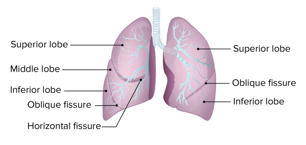
Right lung
Lung is an important organ of respiratory system. They are located on either side of the mediastinum space in the thoracic cavity in total number of two.
The lungs take in oxygen from the atmosphere and expel carbon dioxide from the body.
Structure of the Lung..
Lungs are located in the thoracic cavity in number of two. They are conical in shape.
The lungs are separated from the heart and the thoracic cavity by the mediastinum space.
The lungs are made of spongy tissue within which many air field cavities are located. Its color is brown or grey.
The weight of the right lung is approximately 625 grams and the weight of the left lung is approximately 575 grams. The right lung is heavier in weight and larger in structure than the left lung.
Lungs are divided into lobes. The right lung has three lobes, namely the superior lobe, middle lobe and inferior lobe, while the left lung has two lobes, the superior lobe and the inferior lobe. These lobes are separated by a fissure. There are two fissures in the right lung. A fissure is seen in the left lung.
Lungs are classified into the following parts.
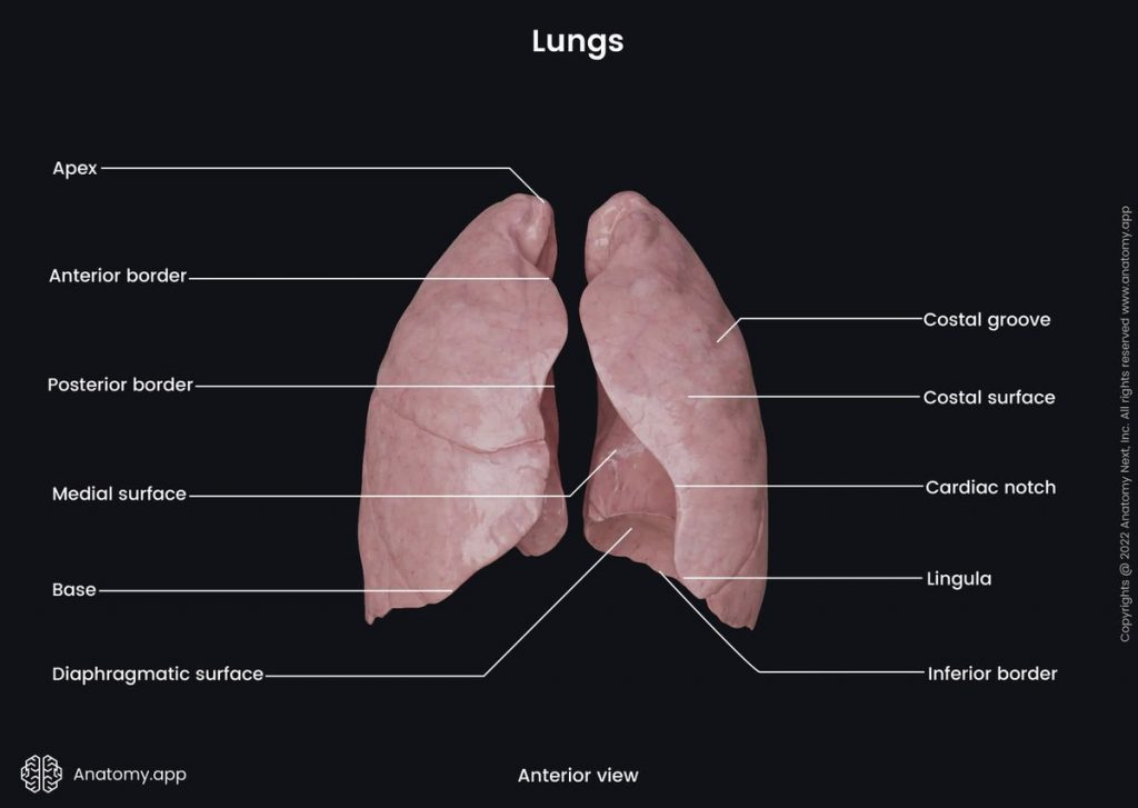
- Apex.. The upper triangular and round part of lung is called Apex. Which is seen up to the level of the clavicle bone.
- Base. The lower broad part of the lung is called the base. This base portion is attached to the diaphragm at the bottom. This part is of concave shape.
- Anterior border.. It is thin. It is shorter than the posterior border. It has a cardiac notch. In which the heart part is arranged.
- Posterior border.. It is thick. It is found from the 7th cervical vertebra to the 10th thoracic vertebra.
- Inferior border.. It is located at the bottom of the lung. It separates the costal surface and the medial surface. The costal surface is large and convex. It is in contact with the costal pleura. It is attached to the ribs and intercostal muscles by costal cartilage.
- Medial surface It is concave. There is a groove in the middle which is called hilum. The hilum lies at the level of the fifth, sixth and seventh thoracic vertebrae. Through this hilum, bronchi, pulmonary blood vessels, lymphatic vessels and nerves enter and exit each lobe of the lung.
In the middle of the medial surface lies the mediastinum space. which separates the two lungs. In this space there are structures like heart, great vessels, trachea, bronchi, esophagus etc. which separate both the lungs.
Structure of the Lobe of the Lung..
The lobes of the lungs are made up of many lobules. One lobe is separated from the other lobe by a fissure. In the center of the lung is a groove called the hilum. From this hilum the following structures are found in each lobe.
Bronchi enter from each lobe of the lung. After entering, it divides and transforms into secondary bronchus, tertiary bronchus, terminal bronchioles, alveolar shakes and small grape-like alveoli. Thus, this structure is seen in a tree-like structure in the lobes of the lung, which is called bronchial tree or respiratory tree.
Surrounding these alveoli is a network of capillaries of the pulmonary artery and pulmonary vein. Gas exchange takes place here between oxygen in the alveoli and carbon dioxide in the blood capillaries through inspiration. This is known as external respiration.
Thus, in each lobe of the lung there is a network of bronchial tree, capillaries of pulmonary vessels, lymph capillaries, nerves and parenchymal tissue of the lung.
pleura..
The pleura is the serous membrane surrounding both lungs. Which is found in double layer. The outer layer is known as parietal pleura and the inner layer is known as visceral pleura.
Between the parietal pleura and the visceral pleura lies a cavity called the pleural cavity. There is serous fluid which is also called pleural fluid.
Due to the pleural fluid in the pleural cavity, the two layers do not rub against each other and due to this, the lungs get enough space for expansion. The pleural fluid in this cavity is viscous and also acts as a lubricant.
The visceral pleura is the layer adjacent to and adjacent to the lungs.Whereas the parietal pleura is the layer attached to the ribs and muscles.
Que 1🔸(c) Explain the physiology of Respiration. 05
cycle of respiration.. or
Mechanism of Respiration ..
Respiration is gas exchange between two surfaces. In which air from the atmosphere enters the lungs. Gas exchange between lung tissue and blood is called external respiration. Gas exchange between each cell tissue of the body and blood is called internal respiration.
During the act of respiration, oxygen enters the lungs through inhalation and carbon dioxide leaves the body through exhalation.
Normally, the act of respiration is observed 16 to 18 times in a minute.
The following steps are observed in the cycle of resuspension.
Inspiration
expiration
pause.
Inspiration…
Inhalation of atmospheric air into the lungs is called inspiration.
When the brain receives nerve impulses for contraction of the diaphragm and intercostal muscles, the contraction of the diaphragm and intercostal muscles increases the size of the thoracic cavity. The air pressure inside the thoracic cavity decreases so that air from the outside atmosphere can enter the lungs through the action of inspiration. This action is called inspiration.
Contraction impulses to the diaphragm cause the diaphragm to flatten downwards and contraction of the intercostal muscles causes the ribs and intercostal muscles to move upwards and outwards. So the size of the thoracic cavity increases and negative pressure is created inside the cavity. As the air pressure in the outside environment is higher and the air pressure in the thoracic cavity is lower, the action of inspiration can take place. The act of inspiration is an active process.
Expiration…
The process of expelling air from the lungs into the atmosphere is called expiration. The action of expiration is a passive process that begins after the action of inspiration is completed.
During exhalation, the contracted diaphragm and intercostal muscles relax. So the diaphragm returns to its original position and the ribs move downwards and inwards, reducing the size of the thoracic cavity and exhaling. In which the air from the lungs is thrown out into the atmosphere.
In the act of exhalation, the air pressure in the lungs is greater than the atmospheric pressure so that the act of exhalation takes place.
Pause… This is the relaxed stage of the lung. In which no action of inspiration or expiration takes place. This period is called pause period.
Que 2 🔸(a) What is incubation period? Write the importance of incubation period in disease transmission. What is incubation period? Write the importance of incubation period in the spread of disease. 08
The time from when the micro-organism enters the body until the first signs and symptoms are observed is called the incubation period.
The incubation period is the basis of the generation time of the particular pathogen
An infectious dose of it
The portal is based on entry and personal identity
Many diseases can become infectious and communicable during this time. Da. T :- Measles, whooping cough etc
The duration of incubation period of different diseases varies from character to character.
Also it varies from person to person even within the same disease
The duration of each disease is different, many diseases have a very short incubation period such as a few hours to two to three days, medium 10 to three weeks such as typhoid chicken pox and very long hepatitis B leprosy and Many times we cannot predict it
Due to the incubation period, the main information is obtained as follows
To know the source of infection
For information on surveillance and quarantine period
To carry out immunization
To find out whether a disease is epidemic or not and its point no source
To know the prognosis of any disease i.e. how long it will take to get better
To know the gap of primary and secondary cases
🔸(b) Enlist the name of abdominal cavities in human body o4 marks
Abdominal Cavity is mainly divided into nine different regions according to its location which are as follows.
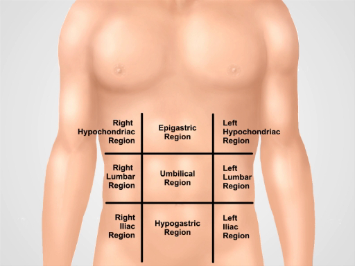
Right hypochondriac region
Epigastric region
Left hypochondriac region
Right lumbar region
Umbilical region
Left lumbar region
Right iliac fossa region
Hypo gastric region
Left iliac fossa region
Que 3🔸(a) What is the importance of Microbiology in nursing?
1) Microorganisms have the potential to cause disease in humans so their characteristics and behavior can be known through this study.
(2) Study of Microbiology how disease occurs. We can learn how it spreads and how to stop it.
(3) Need to check laboratory to identify bacteria.
(4) Understand the importance of personal health and immune measures for resistance to microbial diseases.
(5) The prevailing superstition, ignorance and confusion among the people about diseases caused by microorganisms can be removed and correct understanding can be given. Social Stigma
Precautions can be taken while treating the patient.
(7) Microbial disease can be recognized and measures can be taken to diagnose and prevent its spread.
Que 3🔸(b) Describe health hazards of Biomedical waste.
Bio medical waste containing harmful micro-organisms which can harm hospitalized patients, health workers working in hospitals and general public
Bio medical waste contains sharp instruments such as syringes, scalpels, etc. which may cause injury.
Toxic products of pharmaceutical products, especially antibiotics, are released into the environment, mercury and boxing also cause harm.
Chemical burns can occur during dish infection or waste treatment activities
The incineration process causes air pollution
Infection occurs by using needles etc
Due to lack of safe injection practices, infectious diseases like HIV, hepatitis B, hepatitis B etc. are caused by medical waste.
All these diseases can also be caused due to needle stick injury. People handling bio medical waste can suffer special injuries and other diseases.
Que 3 🔸(c) Explain the cardiac cycle..
Heart is a continuously pumping organ. The pumping action of the heart is called its cardiac cycle. A healthy person has a cardiac cycle ie pumping action of 68 to 72 times in a minute. One cardiac cycle takes 0.8 seconds to complete. This work runs continuously through the heart of a living human being.
The contraction and relaxation of the heart muscle is done by the impulses generated from the S node of the heart. Contraction is called systole and relaxation is called diastole.
The following events occur in the cardiac cycle.
- Atrial systole..
Atrial systole means simultaneous contraction of both atria which takes 0.1 second.
In this atrial systole, when both atria are filled with blood, impulses are generated by the S node and these impulses reach the AV node. During this time it takes 0.1 second and both atria contract simultaneously both atrioventricular valves open and both atriums empty of blood and both ventricles fill with blood. This phase is called atrial systole.
- Ventricular systole..
Ventricular systole is the simultaneous contraction of both ventricles. It takes 0.3 seconds.
During ventricular systole, when both ventricles are filled with blood, impulses from the AV node reach the bundle of His and Purkinje fibers, i.e., the heart.
During this time it takes 0.3 seconds and both ventricles contract simultaneously. The blood of both the ventricles respectively, the blood of the right ventricle goes to the lung through the pulmonary artery and the blood of the left ventricle circulates throughout the body through the aorta. This phase is called ventricular systole.
- Complete cardiac diastole..
Complete cardiac diastole means simultaneous relaxation of both the atria and the ventricles i.e. the four chambers of the heart. This action takes 0.4 seconds.
During complete cardiac diastole there is no electrical activity in the heart. Myocardium muscles are relaxed. During this time both the atria and both the ventricles dilate i.e. relax. Both atriums are refilled with blood during this time. This relaxation time is 0.4 seconds. This is called complete cardiac diastole.
Thus it takes 0.8 seconds to complete a complete cardiac cycle and blood circulation occurs due to the contraction and relaxation of the heart.
Que 4 🔸(a) Spleen
SPLEEN: It is an oval drop. SPLEEN is the largest lymphatic tissue in the body.
Length : 12 cm Width : 7 cm Thickness : 22.5 cm
It is located in the left hypochondriac region between the stomach and the diaphragm lateral to the liver. It weighs approximately 200 gm.
Organs Associated with the Spleen : Around the Spleen there are organs such as Diaphragm, Intestines, Stomach, Pancreas, Left Kidney, etc.
Structure:
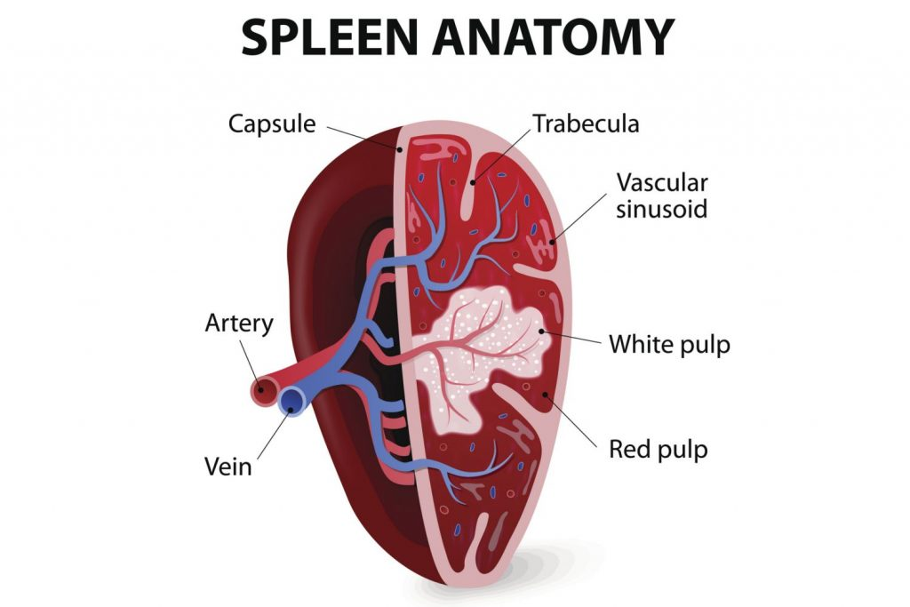
The spleen has a network of connective tissue on the inside called the splenic pulp. It contains lymphoid tissue and some blood cells. A capsule made of smooth muscle fibers and elastic tissue surrounds it. Its tissues are divided into two parts inside the spleen. White pulp and Red pulp. Whight pulp is for immunity where the number of B-cells increases to help make antibody plasma. Which is useful against infection. Red pulp is vascular. Bacteria are phagocytosed there and RBCs and platelets also act to segregate and phagocytose damaged cells.
Functions of Spleen:
Phagocytosis: Helps to destroy bacteria and RBCs that are damaged are removed from circulation by phagocytosis.
Storage of blood: Spleen stores approximately 350 ml of blood. Which sends this blood back into the circulation as soon as sympathetic stimulation is received in a situation like hemorrhage.
Immue response: T and B lymphocytes that are activated by antigens to maintain immunity.
Erythropoiesis: Spleen and liver are important for the production of cells during fetal life. Even in adults, Spleen produces cells when needed.
Que 4 🔸(b) C.S.F.
Cerebrospinal fluid is produced by the choroid plexus in the wall of the ventricle. The choroid plexus is a network of capillaries located in the wall of the lateral ventricle. Cerebrospinal fluid from this lateral ventricle drains into the third ventricle through the interventricular foramen of Munro and from there into the fourth ventricle through the cerebral aqueduct. From there, some fluid flows into the central canal of the spinal cord and some fluid circulates through the foramen of Lasca and Magendi into the subarachnoid space.
The fluid circulating in the subarachnoid space is absorbed into the blood through the arachnoid wall. CSF is formed at a rate of 20 ml per hour i.e. 480 ml per day and is absorbed at the same rate.
CSF pressure can be measured using a lumbar puncture needle which is 10 cm h2o in the lying position and 30 cm h2o in the sitting position.
CSF is a clear fluid that is slightly alkaline with a specific gravity of 1.005.
Its composition consists of water, mineral salts, glucose, plasma proteins, some amount of albumin and globulin, creatinine, urea, and some leukocytes.
Que 4 (c) Describe the mechanism of hearing
Mechanism of hearing i.e. physiology of hearing means act of hearing. The wavelength for hearing is 20 to 20,000 Hz. The human ear is capable of frequencies between 500 and 5,000 hz. The frequency at which the sound waves vibrate is known as the pitch, as the vibration increases, the pitch increases.
Every sound produces sound waves which strike the outer part of the auricle and from there enter through the external auditory canal, these sound waves vibrate the tympanic membrane i.e. the ear drum which is the junction between the external ear and the middle ear.
The sound waves are connected to the tympanic membrane by the malleus bone to the incus and incus to the stapes and the stapes bone is further connected to the oval window. Goes and from there goes to the endolymph and the round window vibrates and the vestibule goes to the cerebrum through the cochlear nerve and the sound is recognized.
Que 4🔸 (d) Reflex Action
Spinal cord acts as a connection between the brain and body parts. The length of the spinal cord is 45 cm.
- It carries sensory impulses to the brain and motor impulses from the spinal cord to different parts of the body.
- Some activities are individualized by the spinal code, which are not required for the functioning of the brain. After the operation is completed, the brain becomes aware of them. This action is completed by spinal reflexes. It is called reflex action.
•Sensory and motor neurons in the spinal cord are connected by connecting neurons found at different levels in the cord for the action of spinal reflexes.
A reflex arch is formed by the spinal cord which results in a quick action which reduces the workload of the brain.
Que 4🔸 (e) Sternum Bone …
The sternum bone is the bone of the axial skeleton. It is the anterior medial bone in the thoracic cavity.
It is the bone immediately below the skin. Sternum bone is also known as breast bone. The sternum is a flat bone.
The ribs are attached to the sternum bone with the help of costal cartilage. Hence it is a very important bone for building the thoracic cage.
Below the sternum bone is divided into three parts.
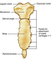
- Manubrium.
The uppermost part of the sternum bone is called the manubrium. It forms the upper broad part of the sternum. It has a rough triangular shape.
The notch at the top of the middle of the manubrium is called the suprasternal notch. The suprasternal notch is also known as the second jugular notch.
Below the suprasternal notch of the manubrium there is a small groove on both sides called the clavicular notch. At this notch, the clavicular bone joins and forms the sternoclavicular joint.
The first pair of ribs are attached to the menorium by the costal cartilage.
The second pair of ribs are joined by costal cartilages near the angle between the manubrium and the body.
- Body…
The body forms the longest part between the sternum bone no. On its upper side is the manubrium and on its lower side is the xiphoid process. The part between these two is known as the sternum body.
There are many small grooves on both sides of the body. On either side of this groove, the third, fourth, fifth, sixth and seventh pairs of ribs are joined with the help of costal cartilage.
- Xiphoid process…: The lowest part of the sternum bone is called the xiphoid process. This part is attached to the muscles of the abdominal wall as well as the diaphragm.
Que 5 Write Meaning (ANY SIX) 6X2=12
🔸 a. Aseptic technique-
Aseptic technique is a technique used to perform a procedure free from pathogenic micro-organisms using only sterile materials.
🔸(b) Tidal Volume .: Tidal volume is the volume of air entering the lungs during inspiration and the volume leaving the lungs during expiration, which is called the titular volume. Tidal volume within a normal adult is approximately 500 ml.
c. Infection: Microorganisms (bacteria, viruses, fungi) enter the human or animal body and multiply, which are not normally found in the body, which is called infection. Its signs and symptoms are visible or subclinical
d. Epidemic: If there are many cases of the same disease in a specific geographical area, it is called epidemic. For example. T. Dengue
🔸 e. Pandemic: Pandemic means any disease that spreads from one state to another state and from one country to another country and is seen all over the world is called a pandemic.
E. X. Swine flu covid 19 etc
🔸f. Vector – Vectors are living organisms that can transmit pathogenic micro-organisms between humans or from animals to humans.
E. X :-Mosquitoes, sand flies etc
🔸g. Immunity: Immunity is the barrier shown by the host against the condition caused by the micro organism and its products (toxin).
or
Immunity means when any antigen or microorganism enters our body and our body protects against it by resisting it is called immunity.
🔸 h. Histology: Histology is a branch of anatomy that deals with the scientific microscopic study of cells and tissues of the body.
🔸Que 6 (a) Fill in the blank 05 marks
The left lung has_______lobes. . (two lobes)
_____blood group is known as universal donor … (O Group)
….. is the smallest functional unit of the body. (cell)
________valve is located between left atrium and left ventricle (bicuspid valve)
Tears are produced by______ gland. Tears are produced from the ………. gland. (lacrimal gland)
Que 6 (b) State whether following statements are True or False 05 marks
- There are three main lymphatic ducts in the body. (false)
- Bile stored in gall bladder. (correct)
- Anti-Diuretic Hormone is secreted by posterior lobe of pituitary gland. (correct)
- Mitochondria is a power generating unit of cell (correct)
- Innermost layer of the heart is called pericardium. (false)
🔸Que 6 (c) Match the following pairs. 05 marks
A…………………………. B
a. Cornea Cornea I Breast Bone Breast Bone
b.Master gland Master gland ii Fertilization of ovum Fertilization of ovum
c. Fallopian tube Fallopian tube iii Transparent sclera Transparent sclera
d. Sternum Sternum iv. White blood cell
e. Fluid connective tissue v. Pituitary gland – fluid connective tissue pituitary gland
vi. Collar Bone
Answer
a – iii
b – v
c – ii
d – I
e – iv.