ENGLISH – General Nursing & Midwifery (First Year) BIO-SCIENCES-2023 PAPER NO 6
GNC BIO SCIENCE
Date: 11/10/2023
Q-1 a. List out the organs of the respiratory system. – 03 marks.
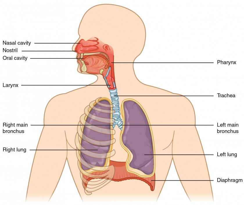
Following is the list of organs of respiratory system.
Organs located above the respiratory tract outside the thoracic cavity are called organs of the upper respiratory tract. Which includes the following organs.
Nose
Pharynx
Larynx
The organs of the respiratory tract located inside the thoracic cavity are called the organs of the lower respiratory tract. The organs are as follows.
Trachea
Bronchi (Right and Left)
Bronchioles
Terminal Bronchioles
Alveoli
Lungs (Right and left).
b. Write the gross anatomical structure of lungs. -Describe the anatomical structure of lung. 04
Lung is an important organ of respiratory system. They are located on either side of the mediastinum space in the thoracic cavity, totaling 2 in number.
The lungs take in oxygen from the atmosphere and expel carbon dioxide from the body.
Lungs are located in the thoracic cavity in number of 2. They are conical in shape.
The lungs are separated from the heart and the thoracic cavity by the mediastinum space.
The lungs are made of spongy tissue within which many air field cavities are located. Its color is brown or grey.
The weight of the right lung is approximately 625 grams and the weight of the left lung is approximately 575 grams. The right lung is heavier in weight and larger in structure than the left lung.
Lungs are divided into lobes. The right lung has 3 lobes, namely the superior lobe, middle lobe and inferior lobe, while the left lung has 2 lobes, the superior lobe and the inferior lobe. These lobes are separated by a fissure. There are two fissures in the right lung. A fissure is seen in the left lung.
Lungs are classified into the following parts.

- Apex..
The upper triangular and round part of lunge is called Apex.Which is seen up to the level of the clavicle bone.
- Bayes.
The lower broad part of the lung is called the base. This base portion is attached to the diaphragm at the bottom. This part is of concave shape.
- Anterior border..
It is thin. It is shorter than the posterior border. It has a cardiac notch. In which the heart part is arranged.
- Posterior border..
It is thick. It is found from the 7th cervical vertebra to the 10th thoracic vertebra.
- Inferior border..
It is located at the bottom of the lung. It separates the costal surface and the medial surface. The costal surface is large and convex. It is in contact with the costal pleura. It is attached to the ribs and intercostal muscles by costal cartilage.
- Medial surface
It is concave. There is a groove in the middle which is called hilum. The hilum lies at the level of the fifth, sixth and seventh thoracic vertebrae. Through this hilum, bronchi, pulmonary blood vessels, lymphatic vessels and nerves enter and exit each lobe of the lung.
In the middle of the medial surface lies the mediastinum space. which separates the two lungs. In this space there are structures like heart, great vessels, trachea, bronchi, esophagus etc. which separate both the lungs.
Structure of the Lobe of the Lung..
The lobes of the lungs are made up of many lobules. One lobe is separated from the other lobe by a fissure. In the center of the lung is a groove called the hilum. From this hilum the following structures are found in each lobe.
Bronchi enter from each lobe of the lung. After entering, it divides and transforms into secondary bronchus, tertiary bronchus, terminal bronchioles, alveolar shakes and small grape-like alveoli. Thus, this structure is seen in a tree-like structure in the lobes of the lung, which is called bronchial tree or respiratory tree.
Surrounding these alveoli is a network of capillaries of the pulmonary artery and pulmonary vein. Gas exchange takes place here between oxygen in the alveoli and carbon dioxide in the blood capillaries through inspiration. This is known as external respiration.
Thus, in each lobe of the lung there is a network of bronchial tree, capillaries of pulmonary vessels, lymph capillaries, nerves and parenchymal tissue of the lung.
pleura..
The pleura is the serous membrane surrounding both lungs. Which is found in double layer. The outer layer is known as parietal pleura and the inner layer is known as visceral pleura.
Between the parietal pleura and the visceral pleura lies a cavity called the pleural cavity. There is serous fluid which is also called pleural fluid.
Due to the pleural fluid in the pleural cavity, the two layers do not rub against each other and due to this, the lungs get enough space for expansion. The pleural fluid in this cavity is viscous and also acts as a lubricant.
The visceral pleura is the layer adjacent to and adjacent to the lungs.Whereas the parietal pleura is the layer attached to the ribs and muscles.
e. Explain the pulmonary circulation. – .05
Pulmonary Circulation..
Pulmonary circulation started from the right ventricle and the blood goes to the lungs and from there returns to the left atrium, so the circulation from the right ventricle to the left atrium is called pulmonary circulation.
In the pulmonary circulation, deoxygenated blood in the right ventricle exits the right ventricle through the pulmonary artery. As it exits, the pulmonary artery divides into a right and a left pulmonary artery and both enter the lung. In which two branches in the left lung and three branches in the right lung enter the pulmonary artery which is according to each lobe of the lung.
In the lungs, gas exchange takes place between the blood and the tissue of the lungs, and two pulmonary veins take oxygenated blood from both sides of the lungs and enter the left atrium of the heart by taking oxygenated blood from each lobe.
Pulmonary circulation converts deoxygenated blood in the heart to oxygenated blood via the lungs. This blood enters the left ventricle and supplies oxygenated blood to the whole body through the systemic circulation.
The circulation from right ventricle to left atrium is called pulmonary circulation.
Or
a. List out the components of cell. – List the components of a cell.03
Cell is the smallest microscopic structural and functional unit of the human body. The cells in it are important for the function of every organ in the body. Every organ can perform normal function only with the function of this cell. There are many different types of cells in the body.
The components in Selma are as follows.
Cell membrane
Nucleus
Cytoplasm
Protoplasm
Mitochondria
Golgi apperatus
Ribosomes
Endoplasmic Reticulum (Smooth Endoplasmic Reticulum and Rough Endoplasmic Reticulum)
B. Write different types of tissue. 04
Cells performing similar functions join together to form a particular type of tissue. It is also seen that there are more than one cells in a single tissue.
Many such types of tissues are found in the human body. Each has a different function and structure.
Types of the tissue..
Epithelial Tissue
Connective Tissue
Muscles Tissue
Nervous Tissue
Classification of Epithelium Tissue.
- Simple Epithelium Tissue..
- Stratified epithelium tissue ..
A. Classification of simple epithelium tissue.
- Simple squamous epithelium tissue…
- Simple cuboidal epithelium tissue….
- Simple columnar epithelium tissue …
- Ciliated Simple Epithelium Tissue ….
B. Stratified epithelium tissue…
- Stratified squamous epithelium tissue.
- Transitional epithelium tissue.
- Stratified squamous epithelium tissue.
A. Non Keratinized Stratified Epithelium Tissue..
B. Keratinized stratified epithelium tissue…
- Transitional epithelium tissue..
Classification of connective tissue
Areolar connective tissue
Adipose tissue.
- White adipose tissue.
- Brown adipose tissue.
Dense connective tissue..
Fibrous tissue.
Elastic tissue.
Blood.
Lymphoid tissue.
Cartilage..
- Highline cartilage..
- Fibro cartilage…
- Elastic cartilage..
Bon..
Muscle tissue…
- Skeletal muscles…
- Smooth muscles…
- Cardiac muscles…
Nervous tissue..
- Excitable Sales.
- Non excitable cell.
c. Explain the epithelial tissue in detail. -Explain epithelial tissue with area. 05
Epithelial tissue…
This type of tissue is found in many places throughout the body. which are mainly scattered on the surface or on the surface lining.
Epithelial tissue is the tissue lining the inner wall of a body cavity, gland, organ or blood vessel.
Functions of the Epithelial Tissue..
Epithelial tissue is important in forming the inner wall of any organ.
Epithelial tissue acts to provide protection where it is located in the inner wall of an organ.
Epithelium is a tissue associated with making any type of secretion through secretory cells located in the tissue.
Epithelial tissue functions in the absorption of material as it is located in the inner wall of the structure.
Characteristics of Epithelium Tissue…
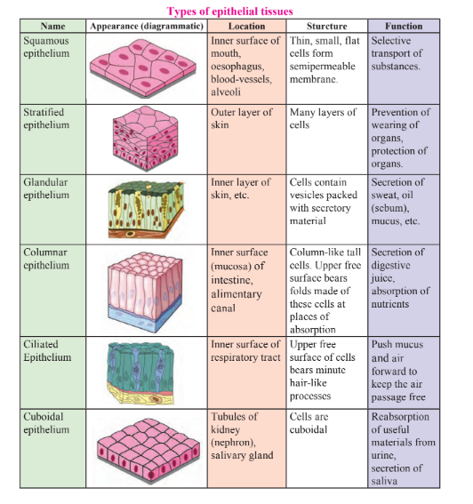
Epithelium TSUs are scattered over the basement membrane of any organ or structure.
The cells in this tissue are closely fitted to each other i.e. arranged close together.
The matrix in this tissue is in liquid form.
Due to the presence of special types of cells in this tissue, they are connected with the functions of secretion and absorption.
Classification of Epithelium Tissue.
- Simple Epithelium Tissue..
This type of tissue is always found in a single layer.
- Stratified epithelium tissue ..
These tissues are located in multiple layers.
A. Classification of simple epithelium tissue.
This type of tissue is located in the inner wall of any structure or organ. They are associated with activities like absorption and secretion. There are four main types of this type of tissue.
- Simple squamous epithelium tissue…
These types of tissues are scattered in the basement layer, the inner wall of any structure or organ. Its cells are close to each other and are flat and arranged in a row. Between them is the nucleus.
This type of tissue is found in the lining of the inner wall of the heart, alveoli of the lungs, blood vessels and lymph vessels.
- Simple cuboidal epithelium tissue….
The shape of the cells in this tissue is cube shaped. which are closely related to each other. This type of tissue is spread over the basement membrane. Usually this tissue is found in renal tubules and thyroid gland.
- Simple columnar epithelium tissue …
This cell is rectangular in shape. Which are more in length and less in width.
These types of tissues are found in the lining of the respiratory tract and the lining of the alimentary tract. It also contains goblet epithelium cells which perform the action of mucus secretion.
- Ciliated Simple Epithelium Tissue ….
This tissue contains cells similar to those of cuboidal and columnar tissue. In addition, this tissue has hair-like processes on the cell margin, i.e. cilia, so it is called ciliated epithelium tissue.
This type of tissue is especially found in the lining of the respiratory tract and the lining of the fallopian tubes. They are here associated with specific movements.
B. Stratified epithelium tissue…
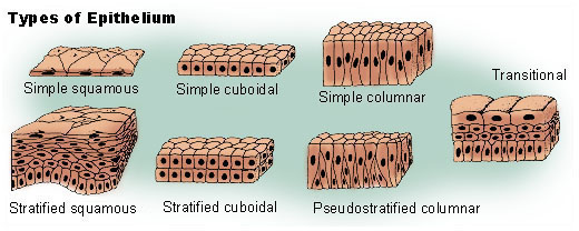
This type of tissue is composed of more than one layer. In this cell, the size of each cell is not the same. In which cells of each layer are found in irregular size and shape.
In this tissue, the cells of the bottom layer are found in large size and they decrease in size when they come to the surface.
This type of tissue is mainly concerned with protection and support of the structure. Stratified epithelium tissue is divided into two parts.
- Stratified squamous epithelium tissue.
- Transitional epithelium tissue.
- Stratified squamous epithelium tissue.
Stratified squamous epithelium tissue is found in multiple layers. It is mainly divided into two parts.
A. Non Keratinized Stratified Epithelium Tissue..
This tissue is mainly found in moist surface areas of the body. Like conjunctiva, esophagus, vaginal cavity, fairings etc.
It is a cell with a nucleus. It has a flat shape.
B. Keratinized stratified epithelium tissue…
This type of tissue is mainly found in dry areas of the body such as skin, hair, nails etc.
This tissue mainly contains keratin substance. Which makes water resistant. So mainly evaporation cannot take place. It is the main characteristic of this tissue
- Transitional epithelium tissue..
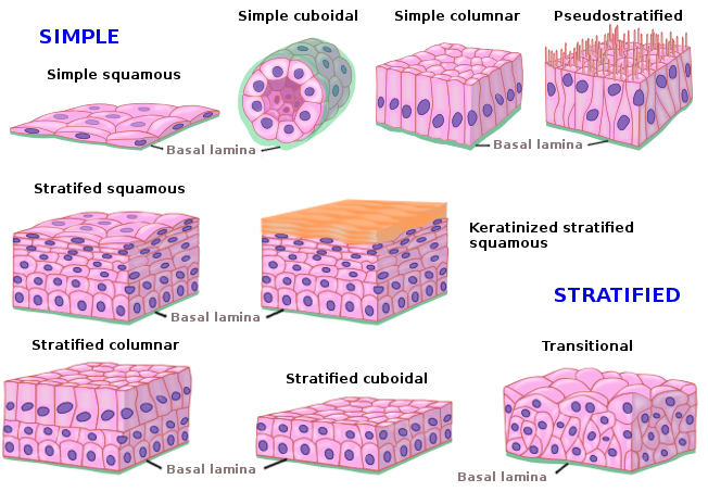
This tissue is found in more than one layer but its main characteristic is that it does not have a basement membrane.
Pear shaped cells are seen in this. This type of tissue is mainly found in the inner wall of the urinary bladder.
Q-2 a) Draw a diagram of heart and describe the cardiac cycle. 08
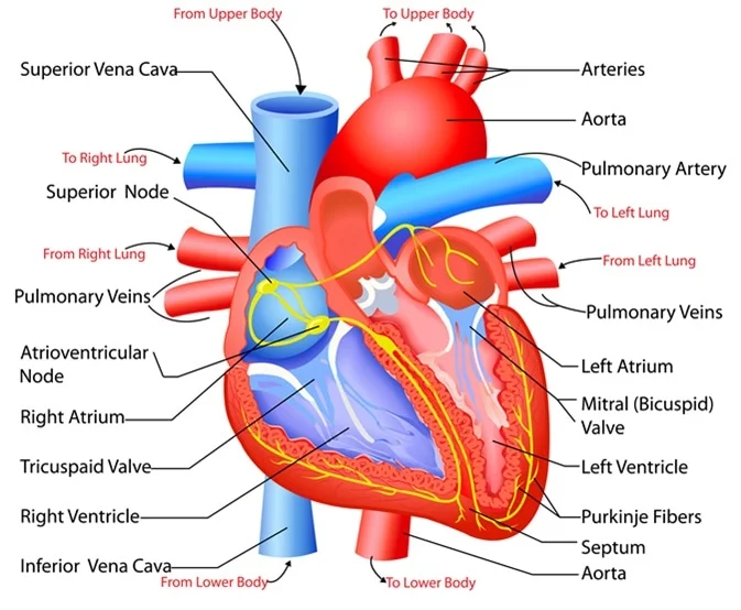
Cardiac Cycle of the Heart..
Heart is a continuously pumping organ. The pumping action of the heart is called its cardiac cycle. A healthy person has a cardiac cycle ie pumping action of 68 to 72 times in a minute.One cardiac cycle takes 0.8 seconds to complete. This work runs continuously through the heart of a living human being.
The contraction and relaxation of the heart muscle is done by the impulses generated from the S node of the heart. Contraction is called systole and relaxation is called diastole.
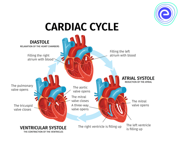
The following events occur in the cardiac cycle.
- Atrial systole..
Atrial systole means simultaneous contraction of both atria which takes 0.1 second.
In this atrial systole, when both atria are filled with blood, impulses are generated by the S node and these impulses reach the AV node. During this time it takes 0.1 second and both atria contract simultaneously both atrioventricular valves open and both atriums empty of blood and both ventricles fill with blood. This phase is called atrial systole.
- Ventricular systole..
Ventricular systole is the simultaneous contraction of both ventricles. It takes 0.3 seconds.
During ventricular systole, when both ventricles are filled with blood, impulses from the AV node reach the bundle of His and Purkinje fibers, i.e., the heart.
During this time it takes 0.3 seconds and both ventricles contract simultaneously. The blood of both the ventricles respectively, the blood of the right ventricle goes to the lung through the pulmonary artery and the blood of the left ventricle circulates throughout the body through the aorta. This phase is called ventricular systole.
- Complete cardiac diastole..
Complete cardiac diastole means simultaneous relaxation of both the atria and the ventricles i.e. the four chambers of the heart. This action takes 0.4 seconds.
During complete cardiac diastole there is no electrical activity in the heart. Myocardium muscles are relaxed. During this time both the atria and both the ventricles dilate i.e. relax. Both atriums are refilled with blood during this time. This relaxation time is 0.4 seconds. This is called complete cardiac diastole.
Thus it takes 0.8 seconds to complete a complete cardiac cycle and blood circulation occurs due to the contraction and relaxation of the heart.
b) Wine the difference between R.B.C and W.B.C Write the difference between R.B.C and W.B.C. 04
(Write the question difference as A. Both rates are given here for the sake of brevity.)
RBCs appear red in color while WBCs are white in color or colorless.
The shape of RBC is circular biconcave disk shape.. while WBC is round shape.
Nucleus is absent in RBC.. whereas nucleus is present in WBC..
RBCs are involved in the transportation of oxygen while WBCs are involved in maintaining immunity and defense mechanisms in the body.
The life span of RBC is 90 to 120 days while the life span of WBC is 5 to 21 days.
Its function is linked to the cardiovascular system while the function of wbc is linked to both the cardio vascular system and the lymphatic system.
RBCs constitute 40 to 45% of the total blood while WBCs constitute one percent of the total blood.
Only one type of RBC is found in blood whereas five types of WBC are found in blood.
RBCs have the property of circulating only in the blood circulation while WBCs can travel beyond the blood circulation to the connective tissue and lymphatic system when needed.
Decreased RBC than normal results in anemia while decreased in WBC results in leukopenia.
OR
a) List out the hormones secreted by anterior pituitary gland and describe posterior pituitary gland – List out the hormones secreted by anterior pituitary gland and describe posterior pituitary gland. 08.
Anterior Lobe
This lobe is also known as adenohypophysis. There are fibers of connective tissue in its structure.
Due to the releasing and inhibiting hormones secreted from the hypothalamus, the following hormones are secreted from the anterior lobe of the pituitary gland.
The hypothalamus is connected by blood to the anterior lobe of the pituitary gland and the hormones secreted from the anterior lobe are controlled by the hypothalamus.
The following hormones are secreted from the anterior lobe.
A. Growth Hormone
B. Thyroid Stimulating Hormone (TSH)
Ç. Adreno Corticotropic Hormone ACTH
D. Prolactin Hormone
E. Gonadotropic Hormone
- Follicle Stimulating Hormone (FSH)
- Luteinizing Hormone (LH)
Posterior Lobe
This is a lobe of the posterior pituitary gland.
It is known as neurohypophysis.
This lobe of the pituitary gland is connected to the hypothalamus by nerves and nerve fibers. Due to which it is known as neurohypophysis. The following hormones are secreted by this lobe.
A. Oxytocin
It is a hormone secreted by the posterior lobe. It is the hormone responsible for initiating normal labor pain during delivery and the contraction of the myometrium muscles in the uterus. By which the process of child birth can be done well.
Oxytocin hormone also acts to eject the milk from the breast when the baby sucks the mother’s breast milk.
This hormone is also responsible for smooth muscle contractions during sexual intercourse that allow sperm to travel from the vaginal cavity and uterus to the fallopian tubes.
B. Anti Diuretic Hormone (ADH)
This hormone is also known by another name of vasopressin. This hormone prevents large amount of urine coming out of the body which leads to absorption of water in the renal tubules due to which urine excretion is controlled.
It plays a very important role in maintaining water content in the body and maintaining fluid balance.
It causes the contraction of smooth muscles which is important for increasing blood pressure.
b) Write the difference between artery and vein. 04
(Write the question difference as A. Both ratios are given here for the sake of brevity.)
The blood vessels that carry blood from the heart to the body are called arteries, while the blood vessels that carry blood from the body to the heart are called veins.
Oxygenated blood circulates in arteries except the pulmonary artery and deoxygenated blood circulates in veins except the pulmonary vein.
Arteries do not have valves while veins do.
As the blood in the artery contains oxygen, it appears bright red in color, while the blood in the vein appears dull red or less red due to the presence of carbon dioxide.
As the artery originates directly from the heart, pulsation is heard in it, whereas no pulse occurs in the vein.
Arteries are mainly located deep in the body while veins are located superficially.
The middle layer of an artery is more elastic so that its diameter can change more while the middle layer of a vein is not as elastic as an artery.
The lumen of an artery is normally narrow while the lumen of a vein is narrow compared to that of an artery.
Blood no force is high in artery while blood no force is low in vein.
When the amount of blood in the artery is less, its wall does not shrink (collapse) due to its strength, while when the amount of blood in the vein is less, it shrinks.
Q.3 Write short answer (any two) 6×2-12
a) Draw a diagram of skin and describe its structure. Draw a diagram of the skin and describe its structure.

A skin is a covering that completely covers the body from the outside. It is also called integumentary system. It is the largest organ or part of our body.
The total surface area of the skin is about 2 meters square and its thickness is approximately 1 to 2 mm.
Structure Of the Skin (Structure of the Skin).
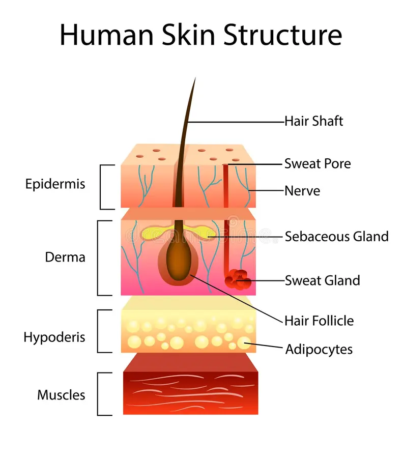
The structure of the skin consists of layers of skin, glands, nails and hair.
Layers of the Skin (Layers of the Skin).
There are mainly three layers of skin.
- Epidermis
- Dermish
- Hypodermis
- Epidermis.
It is the most superficial and outermost layer of the skin. Its structure consists of stratified columnar epithelium tissue. This layer does not contain blood vessels.
The epidermis is not evenly distributed throughout the body. Somewhere it is thicker like the soles of the feet and the palms of the hands. Somewhere its thickness is also less, like the part of the cornea of the eye.
In the epidermis layer, cells grow from the basement layer and up to the superficial layer.It takes about 35 to 40 days for the entire epidermis to be replaced.
The epidermal layer consists of the following layers.
A. Stratum Corneum
This is the outermost layer among all the layers. Dead cells are arranged in a line. These cells are flat and very thin.
Keratin is present in this layer. It is a protein. It works to protect the cells inside and prevents them from drying out.
It maintains the elasticity of the skin and helps to keep it soft.
This layer is constantly worn away due to external wear and tear.
B. Stratum Lucidum.
This layer is also made up of dead and flattened cells. It is also called block layer because the cells in this layer do not contain water and nucleus.
This layer contains a protein called LEDN. It works to protect the skin from the ultraviolet rays coming from the sun.
Ç. Stratum Granulosum
Because these cells contain granules, it is called the granulosum layer.
This layer is 2 to 4 cell rows thick.
D. Stratum Germinative.
This layer is the innermost layer of the epidermis.
This layer produces new cells from time to time and they rise towards the cell surface. Two types of cells are found here, precal cells and basal cells.
- Dermis Layer
This layer of skin is below the epidermis. It contains connective tissue. This layer contains collagen fibers, elastic fibers and reticular fibers. Due to which the elasticity of the skin is maintained. These fibers are important in providing strength and support to the skin.
Cells in the dermis include fat cells, fibroblasts and macrophages cells.
The dermis layer is composed of a papillary layer and a reticular layer. Both of which are connected to each other, cannot be separated.
The structure of the dermis layer consists of blood vessels, lymph vessels, hair follicles, sensory nerve endings,
Sweat gland, sebaceous gland etc. structures are present.
- Hypodermis Layer.
This layer is below the dermis layer. It is also called the subcutaneous layer.
This layer is made up of loose fibrous connective tissue. It is thicker than the dermis. It contains blood vessels, lymph vessels and nerves.
b) List out the organs of lymphatic system and describe structure and functions of lymph node. List the organs of the lymphatic system. Describe the structure and functions of lymph nodes.
The Lymphatic System consists of a fluid called Lymph that flows into lymph vessels. Lymphatic tissue is a part of reticular connective tissue that contains a large number of lymphocytes.
Lymph flows in lymph vessels and drains into lymph nodes and mixes with blood.
Lymphatic System includes the following organs
lymph, lymph vessels, lymph nodes, lymph organs (spleen, thymus gland), lymphoid tissue (tonsils), bone marrow.
structure and functions of lymph nodes.
Lymph Nodes: Lymph nodes are small oval or non-sap structures. Which lies in the way of lymph vessels. Which does the job of filtering lymph and lymphocytes are formed in it. Lymph nodes are mainly located in the joints and axilla, thorax, abdomen and groin.
Structure of lymph nodes:
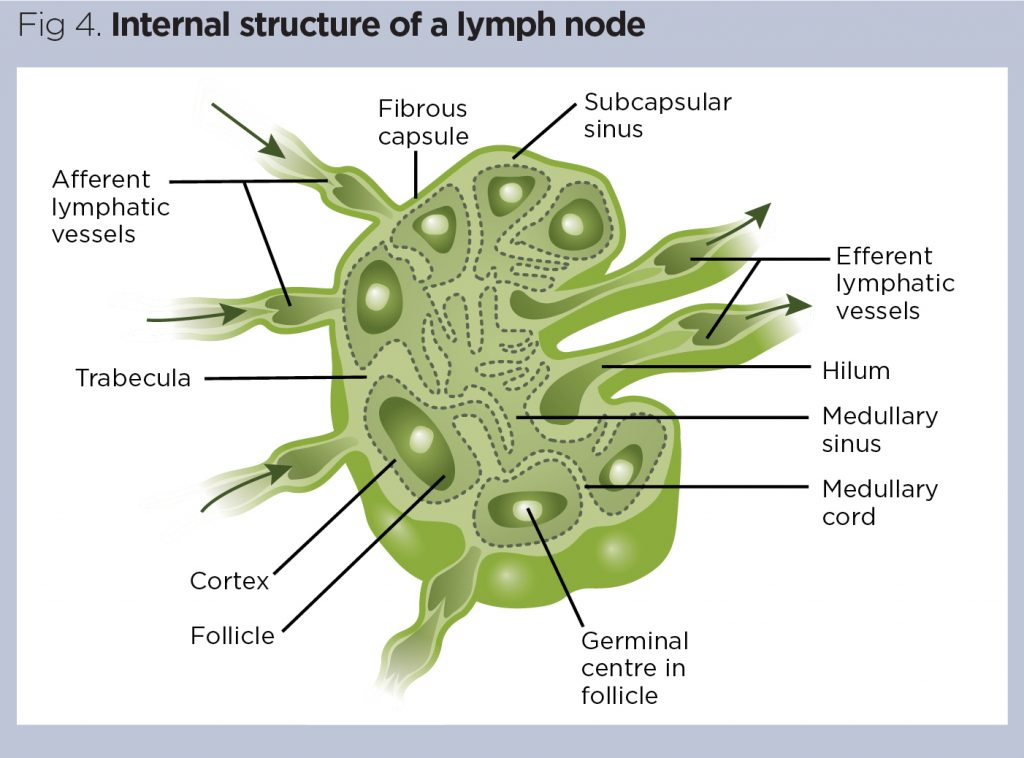
Each lymph node has an outer covering made of dense connective tissue called a capsule and an inward extension called a trabeculae. which divides the lymph node into several parts.
The lymph node portion is divided into two parts. The part towards the outer surface is called the cortex and the inner part is called the medulla.
Lymph is carried in one direction in a lymph node, the vessels entering the lymph node are called afferent vessels and the vessels leaving the lymph node are called efferent vessels. The depressed part of the entering and exiting vessels is called the hilum. Blood vessels also go in and out from here.
Functions of lymph nodes:
Filtering and Phagocytosis: The lymph node filters the lymph in which the lymph enters from one side and is filtered out from the other side. The macrophages in it destroy the foreign substance in it by the action of phagocytosis and the lymph mixes with the blood.
Proliferation of Lymphocytes: Plasma cells and T cells may increase in number in the lymph nodes and circulate from the lymph nodes to other body parts to sustain the immune response.
Hematopoiesis: Some lymphocytes and monocytes from the bone marrow enter the lymph nodes and undergo their final maturation there.
c) List out the bones present in middle ear and describe about long bone (Femur).
Auditory ossicles in the middle ear…
The bones in the middle ear are called auditory ossicles which include the malleus, incus and stapes bones. Its number is one in each ear, that is, there are total 6 auditory ossicles in the body.
Melus 02
Incus 02
Step 02
describe about long bone Femur.
Femur Bone
The femur bone is the bone in the lower extremity. It is the longest and strongest bone among all the bones in the body.
This bone has two extremities and a smooth part
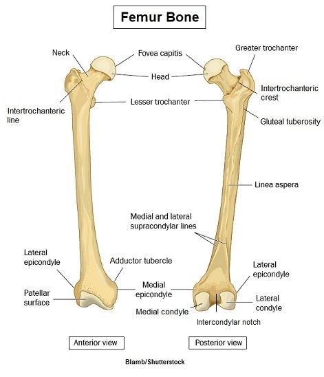
Upper extremity.
The uppermost one-third of the femur bone is called the upper extremity. It has the following structure.
head..
The most anterior part of the femur bone is the round bone part.The part which is called head of femur.
This round part joins with the acetabulum cavity of the innominate bone to form the hip joint.
Hip joint is a synovial joint so all the characteristics of synovial joint can be seen here.
Neck..
The narrow part after the head is called the neck. Which is found in four to five centimeters long and round shape.
Greater trochanter and lesser trochanter..
Where the neck ends, two large rough and raised portions of bone are seen. Which is called trochanter.
The larger part that is raised on the outside is called the greater trochanter.While the small portion raised on the inner side is called the lesser trochanter. The line connecting these parts of the trochanter is called the intertrochanteric line. This part is where the muscles are attached.
After the upper extremity is completed, the middle part of the bone, i.e. the middle part of the bone, is known as the saft. Its structure is as follows.
Linea Aspera..
A bony ridge is raised on the posterior side of the face.It is called linea aspera. This part is where the muscles are attached. The smooth part of the femur bone is cylindrical and round in shape.
Lower Extremity..
The lower one-third of the femur bone is called the lower activity. In which the lower side is seen as two rounded bony parts. Which is called condyle. The condyle on the medial side is the medial condyle and the lateral side is the lateral condyle. The part separating these two condyles is called the intercondylar notch.
The anterior surface of this condyle is a smooth surface called the patellar surface where the patella bone articulates.
A triangular surface is formed at the back of the lower extremity of the femur bone. It is called the popliteal surface. Popliteal vessels and nerves are seen in this part.
Q.4 Write short notes. Write a short note. (Any three) 12
a) Difference between active immunity and passive immunity.
Write the difference between active immunity and passive immunity.
The difference between active immunity and passive immunity is that both systems differ in the way they provide protection against diseases:
- Active Immunity
Definition: Active immunity is the condition in which the body itself makes immune proteins, such as antibodies or cells, against itself.
Cause: This type of immunity is matured when a person is infected or scientifically processed through immunization (lymph, vaccine).
Duration: Active immunity can last for a long time and provides protection against all types of infections.
Example: Getting a seasonal flu vaccine, naturally contracting the disease.
- Passive immunity
Definition: Passive immunity is the condition in which the body receives antibodies from external sources, which it does not make itself.
Cause: This type of immunity is provided by various sources, such as mother in breast milk or special antibodies, which are introduced from outside.
Duration: Passive immunity may provide immediate protection but is not durable; It usually works for a while.
Example: Medically administered antibodies (such as rabies immunoglobulin), antibodies given to the baby in breast milk after birth.
As a whole, active immunity induces the body’s natural defense mechanisms, while passive immunity provides immediate protection from external sources, but for a short period of time.
Describe the health hazards of bio medical waste
b) Write the hazards of Bio-Medical waste.
Bio medical waste containing harmful micro-organisms which can harm hospitalized patients, health workers working in hospitals and general public
Bio medical waste contains sharp instruments such as syringes, scalpels, etc. which may cause injury.
Toxic products of pharmaceutical products, especially antibiotics, are released into the environment, mercury and boxing also cause harm.
Chemical burns can occur during dish infection or waste treatment activities
The incineration process causes air pollution
Infection occurs by using needles etc
Due to lack of safe injection practices, infectious diseases like HIV, hepatitis B, hepatitis B etc. are caused by medical waste.
All these diseases can also be caused due to needle stick injury. People handling bio medical waste can suffer special injuries and other diseases.
c) Importance of microbiology in nursing
1) Microorganisms have the potential to cause disease in humans so their characteristics and behavior can be known through this study.
(2) Study of Microbiology how disease occurs. We can learn how it spreads and how to stop it.
(3) Need to check laboratory to identify bacteria.
(4) Understand the importance of personal health and immune measures for resistance to microbial diseases.
(5) The prevailing superstition, ignorance and confusion among the people about diseases caused by microorganisms can be removed and correct understanding can be given. Social Stigma
Precautions can be taken while treating the patient.
(7) Microbial disease can be recognized and measures can be taken to diagnose and prevent its spread.
d) Factors affecting on growth of microorganism
1) Moisture
Like nourishing food, every bacteria needs water for growth. In fact, bacteria cannot get food in the absence of water, because every food element needs to be in a liquid state to pass through the wall of the bacteria. All types of bacteria grow well in an aqueous medium, an environment without complete moisture prevents its growth. or destroys.
Apart from this, cells cannot live in low or high humidity
2) Light
Most bacteria are destroyed by direct exposure to ultraviolet rays in sunlight.
3) Temperature :-
Temperature is a very important factor affecting the growth of bacteria. Optimal temperature with food, water is necessary for bacteria growth.
Different bacteria have different optimal temperatures.
37°C is the optimal temperature for bacteria growing in the human body.
However, many bacteria are mesophilic (meso = middle, phille = loving). The optimum temperature for it is 25 to 39* C.
Most bacteria grow this way.
Whereas psychrophilic (psychro = cold) bacteria grow better between 4°C to 10°C, some
Thermophilic (Therma – Heat) is also found. Its growth is best between 55°C to 75°C.
Temperature above 75 C is fatal for bacteria. In fact high temperatures are created to kill bacteria in different ways.
Like moist heat (steam), boiling water, pasteurization & autoclaving.
Many species can survive even at very low temperatures. Like yeast, mould, viruses & Rickettsia, spirochetes (76* C can survive for years).
(4) Oxygen
O2 also plays an important role in the life of bacteria. Many types of bacteria can only survive, or grow, in the presence of O2. They are called Aerobes (EX.Sarcina).
Conversely, Anaerobes can live or grow in the absence of 02. E.g. Closteridium tetani-
Apart from this there are also bacteria. which can survive in the presence or absence of 02. They are known as facultative anaerobes. E.g. Salmonella typhi.
Microaerophils grow more in less oxygen than is present in air.
(5)Hydrogen Ion Concentration: (Acidity and Alkalinity) PH medium
The acid or alkaline concentration of the liquid in which the bacteria grow affects the growth.
This is seen from the hydrogen ion concentration index.
PH – 0 (Zero) is the most acidic,
PH – 14 shows the lowest acidic concentration.
PH – 7.) A nutral (neutral),
pH < 7 is acidic
and alkaline at pH >7
Most bacteria grow best between pH 5.0 to 8.5. There are some exceptions to this too.
6) Osmotic pressure :-
The life of bacteria also depends on high or low osmotic pressure. If the bacteria are immersed in a liquid whose osmotic pressure is very high or very low, the bacterial cell collapses or becomes dormant due to leakage of liquid.
Carbon Dioxide is also necessary for the growth of bacteria.
Q.5 Define following (any six) 12
a) External Respiration –
Respiration is gas exchange between two surfaces. In which air from the atmosphere enters the lungs. Gas exchange between lung tissue and blood is called external respiration.
b) Nasocomial infection –
A nosocomial infection, also called a hospital-acquired infection or healthcare-associated infection (HAI), is an infection that a patient acquires after admission to a hospital and that was not present at the time of admission. These infections usually occur in healthcare facilities and are spread through contact with patients, healthcare staff, visitors, medical devices, or the surrounding environment.
Some common types of nosocomial infections are:
Urinary tract infection (UTI): Caused by catheter use.
Surgical Site Infection: Occurs at the surgical site after surgery.
Respiratory Infection: Occurs in patients using ventilators.
Bloodstream Infection: Occurs when using an IV catheter.
C) Cross infection
Cross infection is a situation in which an infectious agent (such as bacteria, virus, or fungus) is spread from one person to another. This usually occurs in health care facilities where infections are spread through contact with patients, medical instruments, or healthcare staff.
Cross-infection can occur as a result of poor hygiene, inadequate disinfection practices, and inappropriate infection control policies.
d) Pandemic–
Pandemic means any disease that spreads from one state to another state and from one country to another country and is seen all over the world is called a pandemic.
D. T. Swine flu covid 19 etc
e) Pathogen –
Any harmful living micro-organism that produces a disease or micro-organism capable of producing a disease is called a pathogen.
Da. T. virus
F) Hypersensitivity
Immunity is seen as a protective process. But it is only a small part of the process for antigen response.Sometimes the immune response can be injurious to the host. Which is also responsible for tissue damage, diseases or death.Harmful effects arising from contact with specific antigen is called hypersensitivity.
G) Incubation period-Incubation period
The incubation period is the period between the time of infection and the appearance of the first symptoms of the disease. During this period the infectious agent (such as a virus or bacteria) replicates in the host, but the host shows no symptoms of disease. The length of the incubation period depends on the specific infectious agent and can range from a few hours to several months.
H) Histology –
A branch of anatomy that deals with the scientific microscopic study of the cells and tissues of the body.
Q-6(A) Fill in the blanks.05
1……….is a power generating unit of a cell.
……… is the power generating unit of the cell. MITOCHONDRIA
2……….blood group is known as universal donor.
………..blood group is known as universal donor. GROUP O
- There are ………pairs of chromosomes in human cells.
A human body cell has…….pairs of chromosomes. 23 PAIRS
4.Sketetal system divided into …….and……
Skeletal system……. and is divided into…. AXIAL AND APPENDICULAR
5.Process of development of R.B.C is called ……..
The process of making RBCs is……. says HEMATOPOESIS
B) True or False – State true or false. 05
- Alpha cells of pancreas secretes insulin.
Pancreas’s Alfacel insulin directs. wrong
2.A.V node is the pace maker of the heart.
The AV node is the pacemaker of the heart. wrong
- Cerebrospinal fluid is secreted by cerebellum.
Cerebrospinal fluid is secreted by the cerebellum. wrong
4.Joseph Lister is known as father of microbiology. Joseph Lister is known as the father of microbiology. wrong
- There are three main lymphatic ducts in the body.
There are mainly three lymphatic ducts in the body. wrong
C) Match the following – Jodka Jodka.05
(A) Widal test (A) SARS-Covid 19 – Good Covid 19
Vidal test
(B) Schick test (B) Tuberculosis – Tuberculosis
Shake test
(C) Western blot test (C) Typhoid-Typhoid western blot test
D) RTPCR test (D) AIDS-Aidreds ATPCr test
(E) Montoux test (E) Diphtheria-Diphtheria Montoux test
A – C
B – E
C – D
D – A
E – B.
ALL THE BEST FROM MY NURSING APP FAMILY.