ENGLISH PEDIATRIC UNIT 6 LOCOMOTOR DISORDERS
Problem with the locomotion
- explain the Kyphosis (hunchback).
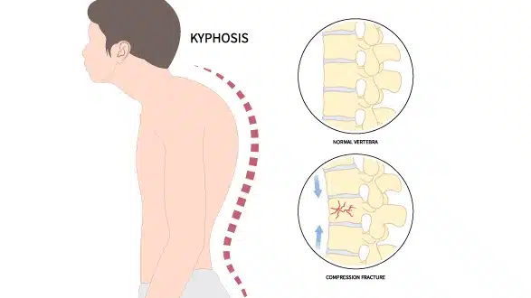
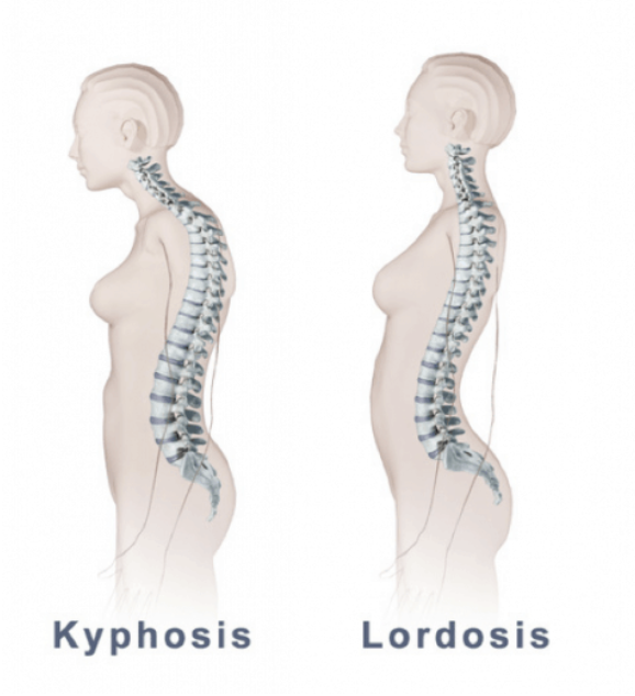
Kyphosis is an abnormality of the spine in which there is an outward curvature of the spinal cord.
Spinal coda is a rounding or hunchback.
In kyphosis the convexity of the spine increases outward.
Explain the type of kyphosis. Describe the types of kyphosis
1) Angular
Copy
Gibbs
2) Round
Other type
1) Postural kyphosis
2) Suermanus kyphophis
3) Congenital kyphosis
1) Angular
The vertebrae of the spinal cord in the kyphosis form a curved curvature and form an angle.
Copy
In this, only one vertebra in the spinal cord is curved.
Gibbs
It involves the involvement of more than two vertebrae in the curvature of the spinal cord.
2) Round
This involves the involvement of more than three vertebrae in the curvature of the spinal cord.
It has many vertebrae fused together and forms a round shape curvature.
Another type of kyphosis
1) Postural kyphosis
Postural kyphosis is the most common type of kyphosis.
Postural kyphosis is mainly due to abnormal posture.
2) Kyphosis in Scheuer
Kyphosis is primarily named after a Danish radiologist.
Kyphosis in Scheuer mainly affects the thoracic spine.
This is mainly found in the (lower) lumbar back area.
3) Congenital kyphosis
In some infants, the spinal cord does not develop properly during intrauterine life.
Explain the Etiology of Kyphosis.
- Due to degenerative diseases of the spine.
- Due to injury.
- Due to trauma.
- scoliosis.
- Due to one vertebra being forward over the other.
- Marfan syndrome.
- Due to infection.
- Muscular dystrophy.
- Neurofibromatosis.
- Paget Diss.
- Polio.
- Poor posture.
- Edge.
- Osteoporosis (weakening of bone).
- Due to injury to the spinal cord.
- Congenital
- Due to abnormality.
- Ankylosis due to spondylosis.
- Spina bifida.
- Tumors and Endocrine Disorders
Explain the clinical manifestation/sign and symptoms of kyphosis.
- Poor posture.
- “Hunchback”.
- Round back appearance.
- Mild back pain.
- Difficulty breathing.
- Burning sensation in upper back and neck area.
- Muscle fatigue.
- Pulmonary and heart failure.
- Stiffness in the spine.
- Loss of bowel and bladder control.
Explain the diagnostic evaluation of the Kyphosis.
- history taking and physical examination.
- Neurological examination
- X Ray.
- ct scan.
- MRI.
- Pulmonary function test
Explain the management of the kyphosis
Explain the medical management.
Providing bone strengthening drugs to strengthen the bones of the spinal cord and prevent fractures.
Asking a child with Scheuermann’s disease to wear body braces to prevent progression of kyphosis to prevent bone growth.
Perform stretching exercises that increase the flexibility of the spinal cord.
Exercise strengthens muscles and improves body posture.
Exercise strengthens the abdominal muscles and helps improve body posture, so the child should be asked to exercise.
If the kyphosis is more severe, surgery may be needed to reduce the spinal curvature.
A “spinal fusion” is performed in surgery.
Provide analgesic medicine to the child.
Explain the nursing management of Kyphosis.
Elevate the affected leg.
Ask the child to take adequate rest.
Ask the child to involve in maximum activity.
Ask the child to adopt relaxation techniques.
Reducing muscle tension.
Inspect the skin daily for any redness, wartiness, pressure sores.
Assess the child’s circulation.
Ask the child to exercise regularly.
Ask the child to do range of motion exercises.
Ask the child to do daily routine exercises in small amounts.
To provide all the information about the treatment and disease to the child in a proper manner.
To clear all the doubts of the child and his family members.
Providing counseling to the child to improve his coping ability.
- Explain the Lordosis.
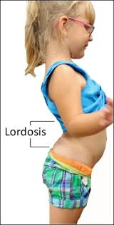
In lordosis, the curvature of the lumbar spine increases with an inward curvature. So swayback is seen in lordosis (swayback means pelvis tilts forward and abdomen protrudes).
Explain the type of Lordosis.
1) Cervical lordosis.
This lordosis is seen in the cervical region.
2) Lumbar lordosis
This lordosis is seen in the lumbar region.
3) Hyper lordosis
This results in an inward curvature in the lumbar region.
4) Hypo lordosis
This causes inward curvature in the lumbar region to a lesser extent.
Explain the Etiology Of Lordosis
- Because of obesity. Rickets.
- Pregnancy.
- Because of too much fat.
- Due to inflammation in the intervertebral disc.
- Due to developmental abnormality at birth.
- Spondylolithiasis (in which the vertebrae in the lumbar region are pushed forward)
- Osteoporosis (bones become brittle in this).
- Enchondroplasia (in this the bones do not grow normally and instead remain short.)
- Due to abnormal posture.
- Muscular imbalance.
Explain the Clinical manifestation/sign and symptoms of Lordosis.
- Symptoms in lordosis depend on its severity.
- Back pain.
- Muscle pain.
- “Sway back” appearance.
- Discomfort in the lower back.
- Movement problems.
- Numbness, tingling sensation.
- Loss of bowel bladder control.
- Difficulty standing up.
- Compression of the spinal canal.
- Weakness in legs.
explain Diagnostic evaluation of Lordosis (
- history taking and physical examination.
- X Ray.
- ct scan.
- MRI.
- Neurological examination
Explain the management of Lordosis.
Treatment of lordosis depends on its severity.
If mild cases
Asking a child to do yoga increases body strength, flexibility and range of motion.
Asking the child to undergo physical therapy.
Ask the child to get enough exercise.
Use Braces in Children and Teens.
Provide the child with analgesic medicine and take measures to reduce swallowing.
explain surgery
1) Spinal fusion
In this, more than two vertebrae of the spinal cord are joined together and moments are prevented.
2) Discectomy
This involves removing damaged discs in the spinal cord.
3) Laminectomy
In this the lamina in the vertebra is removed.
Explain the prevention of Lordosis.
Weight should be limited to prevent lordosis.
Good posture should be maintained.
Ask the child to do regular exercise.
Ask the child to do regular physical activity.
Explain/Define Scoliosis.
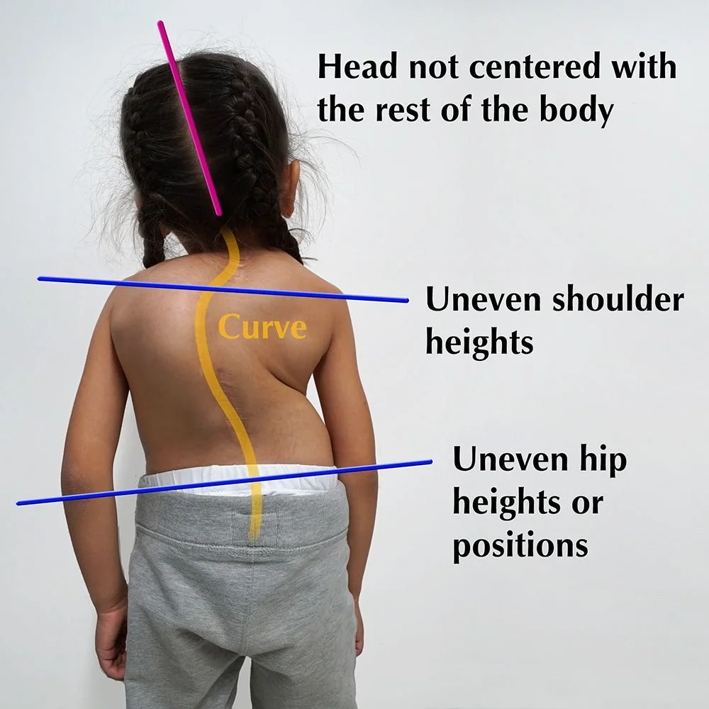
Scoliosis is a condition in which lateral curvature of the spine occurs, either on the left or right side.
The spine of a normal child is straight but the curvature of the spine of a child with scoliosis is lateral.
In scoliosis the spine becomes C-shaped or S-shaped.
Explain the types of Scoliosis.
1)Temporary:= for some time
Postural scoliosis
Compensatory scoliosis
Sciatic scoliosis
•••>
Postural scoliosis
Postural scoliosis is a common form of scoliosis mainly due to abnormal posture of the body.
In which mainly the left side curvature of the spine occurs.
Compensatory scoliosis
Compensatory means when one thing gets better, another thing gets worse.
That is, if a child has one leg long and one leg is short, the person on the side whose leg is short tries to walk from the same side, so scoliosis develops in the spine.
Sciatic scoliosis
Scoliosis develops due to spasm of the three paraspinal muscles on the back side.
2) Permanent
idiopathic scoliosis,
Congenital scoliosis
Paralytic scoliosis
••>
Idiopathic scoliosis
About 8 in 10 people develop scoliosis because the cause is unknown, so it is called idiopathic scoliosis.
Congenital scoliosis
Congenital scoliosis mainly occurs during the development of the spine during the intrauterine life of the fetus.
Paralytic scoliosis
This is a condition that affects the muscles and tendons of the back.
Ex:= Cerebral Palsy,
Muscular dystrophy
explain Etiology/cause of the Scoliosis
Marfan syndrome
(This is an inherited connective tissue disorder that affects the connective tissues so the curvature of the spine is abnormal.)
Rheumatoid arthritis
(This is an autoimmune inflammatory disorder that affects the joints.)
Stylus Disease
(This is an inflammatory condition that affects the joint.)
Osteogenesis im perfecta
(This is an inherited disorder in which bones become brittle and the curvature of the spine becomes abnormal.)
Spine tumor.
Explain the Clinical manifestation/sign and symptoms of the Scoliosis.
- Uneven shoulder (shoulder on one side going down and shoulder on one side going up.).
- Spinal curvature.
- Uneven pelvis.
- One leg is down and one leg is up.
- Back pain.
- Tingling and numbness.
- Permanent deformities.
- feeling tired
- Difficulty breathing.
- Mitral valve prolapse.
Explain the diagnostic evaluation of the scoliosis
history taking and physical examination.
A Neurological Examination.
X Ray.
CT SCAN.
MRI.
Bone scan.
Explain the management of Scoliosis.
Treatment of scoliosis depends on the age and sex of the patient.
Observation
If scoliosis is mild, it does not affect normal function, but regular check-ups by a doctor are necessary.
2) Braces
Braces are provided when there is mild scoliosis.
Surgery depends on the curvature of the spine.
Ex:=spinal fusion.
Other Treatment
Providing physiotherapy to the child.
Providing medicine to the child in an adequate amount.
Explain the nursing management of scoliosis.
Assess the child’s respiratory status every four hours.
To assess the type intensity and location of the child’s pain.
Ask the child to breathe slowly.
Providing semi-founder position to child.
Checking the child’s vital signs every hour.
Assess the child’s circulatory status.
Preparing the child for immobilization.
To maintain the comfort of children.
Assessing the child’s skin integrity.
Asking the child to wear a cotton shirt.
Asking the child to do a previous activity.
Ask the child to adopt relaxation techniques.
Referring the child to diversional therapy.
Providing Mind Diversional Therapy for Child Pain Management.
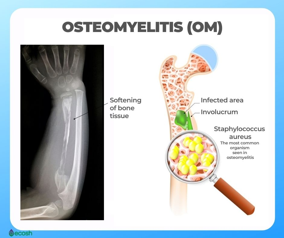
- Explain osteomyelitis.
Osteomyelitis is a pyogenic infection of bone and surrounding tissues.
Osteomyelitis is an infection of the bone and mainly involves the cortex and medullary bone.
Osteomyelitis is an acute infection of bone. It is mainly in acute, subacute and chronic process.
explain Etiology/cause of Osteomyelitis
- Staphylococcus auris,
- E coli,
- pseudomonas,
- Proteas,
- salmonella,
- rheumatoid arthritis,
- Sickle cell disease.
- Obese or
- A malnourished child.
- Whose immune system is impaired.
- Because of the post-operative wound.
- Chronic disease ie (Diabetes, Rheumatoid Arthritis).
- alcoholism,
- Intravenous drug users or drug abusers.
explain clinical manifestation/ sign and symptoms of Osteomyelitis
- bone pain,
- fever,
- General Dishcompleta.
- feeling tired
- Swelling and warmth in the affected area.
- Swelling in the local area.
- Redness and warmth.
- Loss of range of motion.
- Feeling cold.
- Sweat profusely.
- Low back pain.
- Swelling of ankles, feet and legs,
- Drainage of pus from the skin.
- General discomfort.
- Nozia.
- Sweat profusely.
- Swelling in ankles, feet and legs.
- Changes in gait.
Explain Diagnostic evaluation of the Osteomyelitis.
- history taking and physical examination,
- Bone X Ray,
- ct scan,
- MRI,
- blood test,
- blood culture,
- needle aspiration,
- biopsy,
- bone scan,
- bone biopsy,
- Bone X Ray,
- Complete blood count,
- C reactive protein,
- Erythrocyte Sedimentation Rate (ESR),
- MRI of Bone,
- needle aspiration,
Explain the medical management of osteomyelitis.
Provide proper antibiotic medication to the child
cefriaxone,
Ciprofloxacine,
clindamycin,
vancomycin,
lenezolid
Provide analgesic medication if the child is in pain
surgical management
Sequestrectomy
In this the dead bone is removed.
Debridement
This involves removing as much of the dissected bone as possible.
Drainage
In this, open wound and access are drained by needle aspiration.
Nursing management
If the child is in pain, provide opioids.
Examining the body area for any tenderness, warmth and swelling.
Ask the child to explain his feelings.
Asking the child to self-care.
Maintain strict aseptic technique when changing dressings and irrigating the wound.
Providing full information about the disease condition to the child.
Tell the child to rest.
Ask the child to adopt relaxation techniques.
Ask the child to adopt non-pharmacological techniques including relaxation techniques, guided imagery, and deep breathing.
Provide support with pillows to the affected limb.
Elevate the affected area to reduce swelling.
Check the child for any pressure ulcers present.
Assess the vascular status of the affected area.
Ask the child to take complete bed rest.
To check vital sign of child.
Ask the child to take complete bed rest.
Provide the child with a high protein, vitamin C rich diet, and a well balanced diet for proper healing.
Splint should be provided in the affected area to reduce pain and muscle spasm.
Provide the child with the prescribed antibiotic.
Ask the child to do range of motion exercises every four hours.
Provide support to the affected extremities.
Explain /define fracture.
A fracture means a breakdown in the continuity of a bone is called a fracture.
In a fracture, there is a break down in the structure of the bone. The fracture also includes the bone, its tissues, bone marrow and periosteum.
Bone fracture is either partial or complete.
Explain Etiology/cause of the Fracture.
- Due to trauma,
- Due to road traffic accident,
- due to falling,
- Due to injury,
- Due to any disease condition,
- osteoporosis,
- osteomalacia,
- cancer,
- Other Bone Infections
- Due to prolonged use of corticosteroids.
- Due to direct injury.
- Crushing force.
- Torson
- Due to excessive muscle contraction.
- Bending force.
- Due to compression force applied.
- Due to an accident.
- Due to the occurrence of bondiasis.
- Occupation.
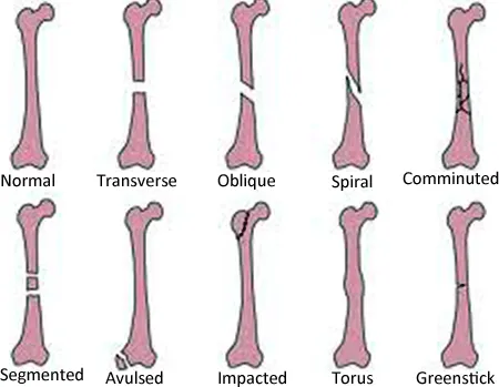
Explain the Classification of fracture.
1) Complete fracture
In this, the bone breaks down in the cross section. In a complete fracture, the bone is divided into two parts.
2) Incomplete fracture
The bone does not break down completely. In an incomplete fracture, the bone cracks but does not break down completely.
3) Closed fracture
A close fracture is also called a simple fracture. In this, the bone is the background.
But it remains inside the skin, that is, the skin is intact, so the wound is not open or visible, and the skin is intact on the fracture side.
4) Open fracture
An open fracture is also called a compound fracture. In this, the bone breaks down from above the skin and is visible outside.
The fracture site is the interrupted skin.
If there is an open fracture, bacteria can enter through the open site and create an infection.
According to grade
Grade 1
In this the wound is clear and smaller than one centimeter.
Grade 2
In this the wound is of moderate amount and one centimeter in size.
Grade 3
These wounds are highly contaminated with extensive soft tissue, nerve, and tendon involvement and the wound is larger than 6-8 cm.
5) Displaced fracture
This fracture is one in which the broken bone separates from each other and is mainly caused by falls.
6)Communicated fracture
This includes bonefragment a crush and break down into many parts.
This is mainly due to the fall down of elderly people.
Classification by fracture pattern
1) Linear fracture
In this, the fracture is parallel to the long axis of the bone and this is mainly due to direct force on the bone.
2) Transverse fracture
In this the fracture is seen at 90 degrees.
Ex:= paget’s disease,
Osteomalacia.
3) Oblique fracture
In this fracture is seen at an angle of 45° degree. Fracture is mainly seen due to twisting force.
4) Spiral fracture
A spiral fracture is called a torsion fracture. This is called bona facture.
This is mainly due to the application of twisting force.
5) Depressed fracture
This facture is mainly seen due to depression in skull bone and facial.
6) Longitudinal fracture
It is mainly a fracture that occurs in the long axis of the bone.
In this the facture line is longitudinally.
Classification by type of fracture
1) Avulsion fracture
This is a fracture in which a segment of bone breaks down from a ligament and tendon.
2) Compression fracture
A compression fracture is also called a cross fracture and is mainly seen due to any compression applied to bone.
3) Green stick fracture
In this, the bone breaks down from the part on one side and the bone bends on the other side.
4) Impact fracture
Continuity of the bone is lost in an impacted fracture.
5) Pathological fracture
This fracture is mainly when there is a breakdown of the bone where there is a bone and the fracture is seen.
6) Stress fracture
Stress fractures are often caused by repeated loading on the bone.
Classification by Eponym.
1) Cullis fracture
A Cullis fracture is also called a broken wrist. The radius bone is fractured about a centimeter from the wrist to the articular surface.
2) Potts fracture
Potts fractures occur mainly in the medial malleolus of the tibia and fibula.
Classification by anatomical location
1) Articular fracture
In this fracture there is involvement of the articular surface of the joint.
These fractures mainly damage the articular cartilage and also damage the subchondral bone.
2) Extracapsular fracture
These fractures are mainly near the capsule of the joint but do not involve the joint capsule and this type of fracture is mainly in the hip (waist).
3) Intracellular fracture
These fractures are mainly found within the joint capsule and are mainly found above the neck level and above the head of the femur bone.
4) Epiphyseal fracture
This structure consists primarily of the epiphysial plate of the long ball.
This fracture is also called a Salter fracture.
explain clinical manifestation/sign and symptoms
- pain,
- Tenderness at site of fracture.
- Swelling.
- Increase in body temperature.
- Loss of function.
- Deformity.
- Blood loss.
- Deformity.
- Swelling.
- pain
- Impairment in functioning.
- simply climb.
- Crepitus.
- Hypovolemic
- mourning
- Shortening of extremities.
- Discoloration.
- Impaired Sensation.
- Abnormal mobility.
- Shock.
- Diminished capillary refill.
- Paler.
Explain the diagnostic evaluation of fracture.
history taking and physical examination.
Clinical examination.
Radiographic examination.
ct scan.
MRI.
Explain the management of the fracture.
emergency care of fracture
Immobilization of the fractured part immediately after fracture.
To provide proper support to the fabricated part.
Taking measures to reduce the severity if it occurs in trow.
If there is an open fracture, provide immediate sterile dressing.
This includes applying a dressing over the fracture to control the bleeding.
Apply pressure if bleeding occurs.
Covering the child to preserve body heat.
Movement of the extremity at the fracture site, warmth, circulation, color check.
Adequate application of splint to the affected joint.
Immobilization of the affected joint.
Make the affected limb move a little bit.
Complete physical assessment to rule out any further injuries.
medical management
1) Reduction
Reduction restores the fractured part back to its anatomical alignment.
1) Closed reduction
In closed reduction, the fabricated part is properly positioned on its anatomical site and a splint is applied.
2) Open reduction
In this, internal fixation is used to fix the bone fragment.
In which metal, pin, wire, screen, rod, etc. are used.
2) Immobilization
Proper immobilization of bone fragments after fracture.
This immobilization is done by external fixator and internal fixator.
External fixator included
In this external fixator bandage, cast, spleen are used.
Internal fixation
Metal pin wire screen rod etc. are used in internal fixation.
3) Maintaining and restoring function
Elevating the affected extremity to reduce swelling.
Provide ice application to the child.
Assess child’s neurovascular status.
Asking the child to express his feelings.
Changing the position frequently to reduce the child’s pain level.
Administering tetanus injection to the child as a prophylactic.
Provide antibiotic medicine to the child.
Provide analgesic medicine to the child.
Provide calcium and iron supplementation to the child.
Provide cold application on the affected extremity of the child.
Providing education to children about alternative treatments for pain management like relaxation and guided imagery.
Asking to exercise to reduce muscle wasting.
4) Pharmacological management
Providing narcotic medicine to the child.
Provide analgesic medicine to the child.
Provide non-steroidal anti-inflammatory medicine (NSAID) to the child.
Administering antibiotic medicine to the child.
Administering anticoagulant medicine to the child.
Stool softener to the child.
Nursing management
Elevate the affected extremity to reduce swelling.
Provide a comfortable position to the child.
Maintain aseptic technique when handling the child.
Assessing child’s neurovascular status.
To check vital sign of child.
Provide prescribed antibiotic, analgesic, calcium supplement.
Maintaining Child No Intake Output Chart.
Provide protein and calcium rich diet to the child.
To provide reassurance to the client.
Assess the child frequently for any infections.
Ask the child to do daily routine activities and exercises in small amounts.
To check vital sign of child.
Check the child’s pain level using a pain scale.
Elevate the affected limb gradually.
Ask the client to do deep breathing.
Ask the child to adopt relaxation techniques.
Check the child’s capillary refill time frequently.
Aspirate the limb to see if there is any swelling or swelling.
Keep checking the tightness of the cast.
Keep the affected limb above heart level.
Maintain aseptic technique while dressing the child.
Ask to do range of motion exercises in the affected extremity.
Asking the child for early ambulation.
Providing education about child’s assistive devices like crutches, walkers, canes, slings etc.
Keep changing the position of the child every two hours.
Complications
grief,
fat embolism,
compartment syndrome,
Volkmans Contracts,
deep vein thrombosis,
infection,
Delayed Union,
Avascular necrosis of bone.
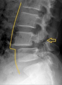
Explain/define spinal fracture.
A spinal fracture occurs when the bones of the spine, called vertebrae, break or collapse. This can occur mainly from any trauma or fall, and can also be caused by a car accident.
Explain Etiology/cause of the Spinal fracture
- Due to abnormal curvature of the spine (kyphosis,
- scoliosis, lordosis).
- Due to a tumor in the spinal cord.
- Due to any injury.
- Due to an accident.
- due to falling.
- Due to being assaulted.
- Due to sports injuries.
- Due to any injury in the spinal cord.
Explain clinical manifestation/sign and symptoms of Spinal injury (Spinal
- Symptoms and signs of spinal fracture depend on its severity and location.
- Severe pain.
- simply climb.
- Tingling and numbness sensation.
- Spasm in the muscles.
- Weakness.
- Bowel and bladder changes.
- Decreased movement.
- Paralysis.
Explain diagnostic evaluation of the Spinal fracture.
history taking and physical examination.
X Ray.
ct scan.
MRI.
Explain management of fracture of spinal cord.
There are three things to keep in mind in the management of spinal cord.
To maintain the alignment of the spinal cord.
Immobilization of the spinal cord should be maintained during healing.
Pain should be controlled by restricting movement.
Instrumentation and fusion are surgical procedures used to correct unstable fractures.
Plates for joining two fractured bones,
Rhodes,
hooks,
pedicels,
screws,
End cages are used.
It takes a few months for the bone to fuse with this implant.
Vertebroplasty and Kyphoplasty is an invasive procedure. Is done when the spine is fractured.
In Vertebroplasty
In vertebroplasty, bone cement is inserted through a hollow needle into the fractured vertebra.
In Kyphoplasty
In kyphoplasty, a balloon is inserted first and then the balloon is inflated so that the compressed vertebrae can be in a normal position.
Explain Nursing management of fracture of spinal cord.
Complete assessment of the child.
Properly monitoring the neurological status of the child.
Properly monitoring the child’s bowel and bladder function.
Properly monitoring the child for signs and symptoms of increased intracranial pressure.
Taking proper measures to prevent child complications.
To prevent any respiratory complications, urinary tract infection, pressure ulcer, contracture, I should provide proper respiratory support to the child, provide proper skin care to the child, and advise the child to do proper range of motion exercises.
Provide proper analgesic medication for pain management of child.
Provide proper physical and occupational therapy to the child to improve the child’s mobility and rehabilitation.
To provide proper psychological support to the child and his family members.
To provide complete education to the child’s parents about the child’s condition, its causes, symptoms and signs, and its treatment.
Advising the child’s parents to follow up regularly.
Explain/Define Poliomyelitis in children
Poliomyelitis is also commonly called polio. Poliomyelitis is a highly contagious, acute viral and infectious disease. which is usually caused by an RNA enterovirus called poliovirus.
Polio virus usually destroys the anterior born cells of the spinal cord and the nuclei of the cranial nerves and causes paralysis.
Polio usually affects children under the age of five. Polio virus is usually caused by coming into contact with contaminated food, contaminated water, and the feces of an infected person.
Polio usually causes paralysis with involvement of the lower legs, along with the involvement of the alimentary tract, and also involvement of the central nervous system if the respiratory muscles are involved, a life threatening condition arises.
Explain the Etiology /causes of Poliomyelitis in children
- Caused by poliovirus (type I,II,III)/ RNA enterovirus,
- Due to coming in contact with infected person’s physis,
- If an infected person coughs and sneezes, due to coming in contact with its droplets,
- By fico oral route,
- Due to intake of contaminated food and contaminated water,
- Due to poor sanitation and unhygienic conditions,
- In unimmunized children,
- Due to a weakened immune system due to malnutrition,
- In over crowded areas,
- Due to non-immunization of polio.
- Due to traveling to an area where polio is an outbreak.
Explain the mode of transmission of Poliomyelitis in children
- Mainly by the oropharyngeal route,
- Due to coming into contact with infected person’s physis,
- Due to contact of contaminated surfaces with impacted bodies,
- By fico oral route,
- Due to ingestion of contaminated food and water,
- Due to coming into contact with its droplets due to coughing and sneezing of an infected person,
Explain the incubation period of Poliomyelitis in children
The incubation period of poliomyelitis in children is 7 to 14 days.
Explain the Clinical manifestation/ Sign and symptoms of Poliomyelitis in children
The symptoms and signs of poliomyelitis depend on the different types such as,
1) Asymptomatic poliomyelitis (silent/inapparent/subclinical infection),
2) Abortive poliomyelitis
(Minor Illness),
3) Non Paralytic Polio Myelitis,
4) Paralytic polio myelitis
subtype:=
A) Spinal form,
B) Bulbar form,
C) Bulbospinal form,
D) encephalitis form
1) Asymptomatic poliomyelitis (silent/inapparent/subclinical infection)
Approximately in asymptomatic poliomyelitis
90 to 95% of infected persons are infected with the virus and the infection is asymptomatic.
2) Abortive poliomyelitis (minor illness),
Abortive poliomyelitis is a mild and self-limiting illness and accounts for 4-8% of cases. These viruses usually invade the blood stream and cause the condition of viremia. Its signs and symptoms typically include,
Fever, sore throat, headache, nosia, vomiting, loss of appetite, abdominal pain, body pain are seen.
3) Non Paralytic Polio Myelitis,
In nonparalytic poliomyelitis, the poliovirus usually enters the urinary system. And its 1% cases are seen.
Its symptoms include neck stiffness, headache, back pain, pain in legs, neck pain and nosia and vomiting. Paralysis does not occur in this. The symptoms are
Occurs for 1 to 2 weeks and then resolves.
4) Paralytic polio myelitis
Paralytic polio myelitis is the most severe form of polio. In which paralysis occurs in one or more limbs, the virus enters the central nervous system (CNS) and subsequently creates a condition of paralysis. Its most important feature is acute asymmetric flaccid paralysis (AFP). There is muscle stiffness, muscle pain, stiffness in a child with the condition of Chinese lighting polio.
A) Spinal form
Paralytic poliomyelitis of the spinal form involves the extremities, neck, abdomen, diaphragm and intercostal muscles.
Then there are symptoms like fever, muscle pain, tremors, deep tendon reflex being demineralized.
Flaccid paralysis occurs predominantly in the lower limb followed by the upper limb. And large muscles are more affected than small muscles. And due to the involvement of bladder and bowel, the condition of urinary retention and constipation is seen. Respiratory difficulty is seen due to involvement of diaphragm and intercostal muscles in spinal form.
B) Bulbar form
The bulbar form is a less common but more severe form because it involves the vital medullary center and
It involves paralysis in the muscles supplied by the cranial nervous and vital respiratory and circulatory centers. In its symptoms and signs
dysphagia,
dyspnea,
nasalspeech,
Facial paralysis occurs.
Paralysis of the vagus nerve causes weakness in the soft palate, pharyns and vocal cords, resulting in nasal speech and hoarseness of voice.
Difficulty occurs in bridging and swallowing.
There is a chance of aspirating due to regurgitation
Atelectasis and pneumonia develop. Involvement of the respiratory center leads to cellular irregular breathing, and oxygen saturation is demineralized.
Child Mother
restlessness,
Confusion and unconsciousness are seen.
C) Bulbospinal form,
Bulbar form and spinal form are seen in this. And 25% of paralytic cases are seen.
D) encephalitis form
A less common form of encephalitis presents in patients with irritability, tremors, drowsiness, convulsions, unconsciousness.
Other symptoms
feel tired,
fever,
one head,
vomiting,
Diarrhea and constipation,
sore throat,
Neck Stephenish,
Sensitivity to light,
muscle pain,
wickness,
paralysis,
Difficulty in breathing, swallowing and talking.
Explain the Diagnostic evaluation of the child with the Poliomyelitis
- History taking and physical examination
- Clinical assessment,
- laboratory test,
- stool examination,
- throat swab,
- viral culture,
- Polymerase Chain Reaction Testing (PCR Test),
- throat swab,
- Cerebrospinal fluid (CSF) assessment,
- serological test,
- imaging studies,
- x ray,
- M.R.I. testing,
- Lumbar puncture (spinal tap),
- electromyography,
Explain the management of the child with the Poliomyelitis
Providing properly supportive treatment to the child.
Advise the child to take proper bed rest.
Giving advice to maintain proper nutritional status of child.
Advise the child to have adequate fluid intake. So that the child’s fluid and electrolyte levels can be properly maintained.
Proper medication should be provided if the child has symptoms like fever, pain and discomfort.
Provide proper physical therapy if the child has muscle weakness, paralysis.
Provide proper rehabilitation therapy to strengthen muscles, improve flexibility.
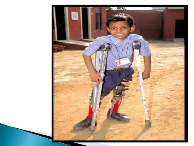
Provide supportive devices such as braces, orthoses, crutches, and wheelchairs to improve the child’s mobility and independence.
Provide proper respiratory support if the child has respiratory muscle weakness.
Closely monitor the child’s respiratory function.
If the child has joint pain, muscle pain, joint stiffness, then analgesic medicine should be provided.
Provide proper relaxation techniques to the child.
To provide adequate care to prevent the child from secondary complications.
Providing education to the child to maintain good hygiene practices.
Proper polio vaccination should be provided to the child so that the condition of poliomyelitis can be prevented.
Provide proper polio vaccination to child as per immunization.
Provide mild sedative to child for pain relief.
Any child
To assess whether the condition of respiratory distress is present or not.
To provide proper emotional support to the child and his family members.