ENGLISH PEDIATRIC UNIT 6 NERVOUS SYSTEM
Neurological system
- Explain/Define Meningitis.
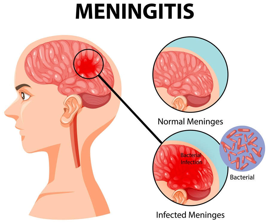
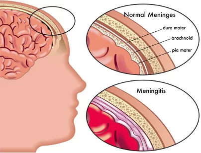
Meninges: Meninges is the protective membrane of the brain and spinal cord that covers the brain and spinal cord.
There are three other 3 layers in this meninges.
1) Durometer
(outer most layer),
2) Arachnoid Matter
(intermediate layer),
3) Pia mater
(Innermost layer)
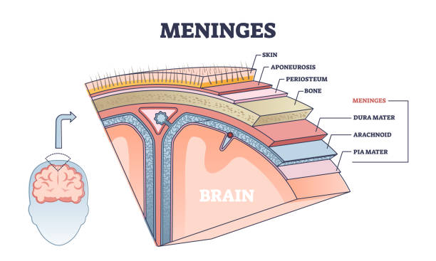
Thus, there are three layers of meninges that cover and protect the brain and spinal cord.
Meningitis: If there is infection and inflammation in the meninges layer surrounding the brain and spinal cord, the condition is called meningitis. This infection can be caused by bacteria, viruses, and microorganisms.
Explain the Etiology/cause of the meningitis.
Due to bacterial infection.
Ex:=
Mycobacterium Tuberculosis,
Streptococcus pneumoniae,
Neisseria meningitidis,
Haemophilus influenzae,
Listeria monocytogenes.
Due to viral infection.
mumps,
Herpes simplex virus,
Epstein barr viral,
Varicella-zoster viral,
Measles,
Influenza.
Due to fungal infection.
candida,
Due to systemic lupus erythematosis (SLE),
Due to certain types of medication.
Due to head injury.
Due to trauma in the head and spinal cord.
Cancer.
Due to tobacco use.
Due to impaired immune system.
Due to over crowding.
Due to brain surgery.
Explain the clinical manifestation / sign and symptoms of the patient with the Meningitis.
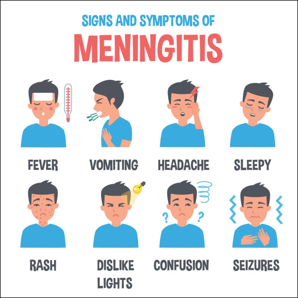
- one head,
- very high fever,
- Alteration of maintain status. Confusion.
- Altered consciousness.
- Vomiting.
- Photophobia (an inability to tolerate light).
- Irritability.
- Drowsiness.
- Confusion.
- Altered Consciousness.
- Vomiting.
- Sign of meningeal irritation.
- Neck stiffness (neck stiffness).
- headache.
- Altered mental status.
- to be startled
- Joint pain.
- Muscle union.
Positive Brudzinski sign
Brudzinski’s sign When a child is placed in a supine position and the neck is flexed towards the chest, the hip and ankle automatically flex is called Brudzinski’s sign.
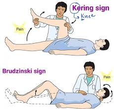
Kerning sign
In kerning sign, when the patient is placed in supine position and then the knee and hip are flexed, if the patient re-extends the knee, it is called kerning sign.
K:= Knee,
E:= Extent
R:= Resistance
That is, directly extending the knee causes pain to the patient.
Explain the Diagnostic evaluation of the Child with the Meningitis.
- History taking and physical examination.
- Blood culture.
- Lumbar puncture.
- Chest x-ray.
- CSF Examination.
- CT scan.
- Gram Stein.
- CSF culture.
- MRI test.
Explain the Management of Children with Meningitis.
Antibiotic medicine should be provided if the child has any bacterial infection.
Ex:=
Rifampicin,
Cefotaxime,
Vancomycin.
Antiviral medicine should be provided if the child has any viral infection.
If the child is in pain, provide analgesic medicine.
Ex:= Acetaminophen,
NSAID (Non steroidal anti inflammatory drug).
Provide intravenous fluid to the child.
If the child has fever, then give antipyretic medicine.
If the child has fever, provide anticonvulsant medication.
Continuous close monitoring of the child.
Continuously close monitoring of child’s vital signs.
If the child has inflammation, provide corticosteroid medicine.
If the child has the condition of meningitis, keep him properly isolated.
Properly monitor the child’s hydration status.
Providing proper nutritional support to the child.
Provide proper intravenous fluid to the child.
Explain the Nursing management of Children with the Meningitis.
To properly assess the child.
To continuously monitor the child’s vital signs.
Properly assess the child’s hydration status.
Maintain proper fluid and electrolyte levels of the child.
If the child is in pain, provide analgesic medicine.
Assess the child’s neurological status properly.
If the child has seizures, provide anti-convulsion medicine.
Providing proper nutritional support to the child.
Providing proper psychological support to the child.
To provide proper education to the child about his disease, its causes, and its symptoms and signs.
Continuously monitor the child’s intake output.
Continuous monitoring of child’s vital signs and neurological status.
Continuously monitor the child’s level of consciousness.
Advise the child to take medicine regularly.
Advise the child to follow up regularly.
Providing proper psychological support to the child.
- Define/Explain Encephalitis.
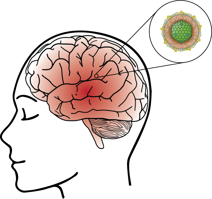
If there is infection and inflammation in the brain, the condition is called encephalitis. Encephalitis is a condition that causes infection and inflammation in the white and gray matter of the brain and mainly affects the cerebrum, brain stem and cerebellum.
Encephalitis is caused by any viral infection, bacterial infection and allergic condition.
The condition of encephalitis is mainly seen in young age and old age people.
Explain the Etiology/cause of the children with the Encephalitis.
Caused by bacteria.
Ex:=
Mycobacterium Tuberculosis, Streptococcus, due to viral infection.
Ex:=
Due to herpes simplex virus,
Due to Epstein Barr virus,
Varicellazoster
due to virus,
Caused by enterovirus
(polio virus and coxsackie virus),
Due to Arab virus,
Arthropod – Bony virus,
tick-borne virus,
Mumps, Measles and Rubella (MMR)
Rabies virus.
Due to parasitic infection.
Ex:=
roundworm,
cysticercosis,
Toxoplasmosis. Due to a weakened immune system.
Due to fungal infection.
Ex:=
Aspergillus species, Cryptococcus neoformans,
Due to autoimmune diseases,
Due to any allergic condition.
Explain the types of the Encephalitis.
There are total six types of encephalitis.
1) infectious encephalitis,
2) Postinfectious encephalitis,
3) Autoimmune encephalitis,
4) Japanese encephalitis,
5) Allergic encephalitis,
6) Chronic encephalitis
A) Subacute Sclerosing Panencephalitis B) Progressive Multifocal Leukodystrophy (PML)
•••>
1) infectious encephalitis,
Infectious encephalitis is caused by any type of infection. The condition of infectious encephalitis arises due to microorganisms like bacteria, viruses, fungal parasites etc.
2) Postinfectious encephalitis,
Postinfectious encephalitis The condition of infectious encephalitis occurs two to three weeks after the initial infection. Postinfectious encephalitis is also seen as a complication of vaccination of any viral infection.
3) Autoimmune encephalitis,
In autoimmune encephalitis, if our body’s own immune system damages our brain, it is called autoimmune encephalitis. In this condition, if the antibody of our body crosses the blood brain barrier and then damages the brain tissues and causes encephalitis, it is called autoimmune encephalitis.
4) Japanese encephalitis,
Japanese encephalitis is mainly spread by mosquitoes. This condition is mainly found in south, east, Asia, the far east and the Pacific island. And this condition is more common among people who do farming.
5) Allergic encephalitis
Allergic encephalitis is mainly caused by any allergic reaction.
6) Chronic encephalitis
Chronic encephalitis develops slowly and gradually over several months. If the condition of encephalitis persists for a long period of time, it is called chronic encephalitis.
The condition of chronic encephalitis is mainly seen in patients with HIV when the immunity is down.
There are two other types of chronic encephalitis.
A) Subacute sclerosing panencephalitis
Subacute sclerosing panencephalitis inflammation is seen as a complication of measles infection.
B) Progressive Multifocal Leukodystrophy (PML)
Progressive multifocal leukodystrophy (PML) is mainly caused by John Cunningham viral (JC viral) inflammation.
Explain the Clinical manifestation/ sign and symptoms of the Children with the Encephalitis.
- Fever,
- headache,
- Confusion and altered mental status,
- startle,
- Neurological deficits.
- Behavior changes.
- Nausea and vomiting.
- Steph Neck.
- Photophobia (sensitivity to the bright lights).
- Hallucinations (false sensory perception).
- Difficulty in speech and language.
- Motor disturbance.
- Body aches.
- Caesar is coming.
- Aphasia (inability in speaking).
- Aphagia
- (Inability in Swallowing).
- Disorientation.
- Joint pain.
- Loss of sensation in some part of the body.
- In a person’s personality and behavior
- Changes happen.
- Muscle weakness.
- Vision can be partial or total loss.
- Loss of eye movement.
- Involuntary loss of Face, Arm, and Legs.
- Anxiety.
- Unusable behavior.
Explain the diagnostic evaluation of the children with the encephalitis.
- History taking and physical examination.
- Brain MRI.
- Ct scan.
- Brain biopsy.
- Culture of the cerebrospinal fluid (CSF).
- Serology test.
- Lumbar puncture.
- CSF Examination.
- Polymerase chain reaction.
Explain the medical management of children with Encephalitis.
To provide properly supportive care to the child.
Advise the child to take proper rest.
Advising the child to take proper nutrition.
Advise the child to take proper fluids.
Antiviral medicine should be given if the child has a viral infection.
Ex:=
Acyclovir
(Zovirax),
Foscavir
(foscarnet).
Provide proper anti-inflammatory drug to the child.
Ex:= Acetaminophen,
Ibuprofen,
Naproxen.
If the child has a bacterial infection, provide antibiotic medicine to the child.
If the child has any parasitic infection, give antiparasitic medicine.
Provide anticonvulsant medicine if the child has seizure condition.
To provide properly supportive care to the child.
Provide analgesic medicine if the child has any pain condition.
Monitor the child’s neurological status.
To monitor the child’s mental status and consciousness level.
To provide proper rehabilitation services to the child.
Providing proper psychological support to the child.
Provide oxygen to the child properly.
Provide proper intravenous fluids to the child.
Explain the Nursing management of Children with the Encephalitis.
To provide properly comprehensive care to the child.
Proper assessment of the child.
Properly assess the child’s neurological status, mental status and consciousness level.
To properly assess the child’s vital signs.
Continuously monitor the child’s fluid and electrolyte levels.
Continuously monitor the child’s electrolyte levels.
Provide anticonvulsant medicine if the child has seizure condition.
If the child has a viral infection, provide antiviral medicine.
Antibiotic medicine should be provided if the child has any bacterial infection.
Antifungal medicine should be provided if the child has any fungal infection.
If the child has any parasitic infection, provide antiparasitic medicine.
Provide proper respiratory support to the child.
Providing proper nutritional support to the child.
Maintaining proper hydration status of the child.
If the child has any kind of complication, properly assess it.
To provide proper education to the child and his family members.
Providing emotional support to the child.
To monitor the child’s vital signs frequently.
Properly monitoring the child’s intake output chart.
To properly monitor the child’s vital signs.
Closely monitor the child’s neurological status.
Keeping the child’s airway properly patent.
To provide proper work and comfortable environment to the child.
Keeping the child away from noisy environment.
Properly monitor the child’s skin integrity.
Advise the child to perform proper range of motion exercises.
Provide proper standard precautions and isolation to the child.
Provide proper antipyretic medicine to the child.
Providing proper psychological support to the child.
Advising the child to take medication properly.
Advise the child to follow up regularly.
- Explain/Define seizures.
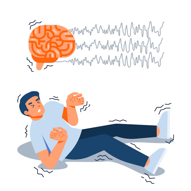
If the child has repeated episodes of seizures, the condition converts to epilepsy.
Epilepsy is a mental abnormality in which the electrical impulses of the neurons in the child’s brain are greatly disturbed due to which abnormal electrical activity is observed in the neurons and the condition of epilepsy is due to which a jerking moment arises in the body.
Explain the Etiology/cause of the seizures.
- Due to genetic factors.
- Due to head trauma.
- Due to brain tumor.
- Stroke.
- Due to brain infection.ex:= Meningitis.
- Due to drug abuse and withdrawal.
- due to hypoglycemia. due to hypoxia.
- Due to dehydration.
- Due to fever.
- Due to high blood pressure.
- Due to septicemia.
- Due to diabetes mellitus.
- Due to electric shock.
- Due to birth injury.
- Due to developmental disorder.
- Due to traumatic brain injury.
- Due to brain tumor.
- Due to exposure to any poisonous substance.
- Ex:= lead, carbon monoxide.
Explain the classification of the seizures. (
Seizures are mainly classified into three types.
1) General Caesars
2) Partial and focal onset seizures.
3) Psychogenic seizures
••••>
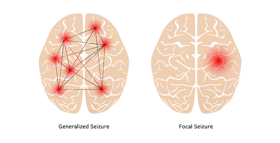
1) General Caesars
There are six types of General Caesars.
1) Tonic – clonic seizures.
2) Absent Caesar.
3) Myoclonic seizures.
4) Tonic Caesar.
5) Atonic Caesar.
6) Clonic seizures.
2) Partial and focal onset seizures.
There are two types of partial and focal onset seizures.
1) Simple focal seizure.
2) Complex focal seizures.
3) Psychogenic seizures
•••>
1) General Caesars
There are six types of General Caesars.
In generalized seizures, both the right and left hemispheres of the brain are involved.
That is, uncontrolled electrical discharge occurs in both hemispheres of the brain.
In this type of seizure, the patient becomes unconscious.
These seizures last from a few seconds to a few minutes.
1) Tonic – clonic seizures.
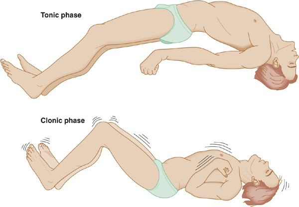
Tonic clonic seizures are also called Grandmal seizures.
In this seizure, the patient’s consciousness is completely lost. And the patient also falls on the ground.
In the tonic stage, the patient’s muscles stiffen and muscle tone increases.
In the clonic phase, the patient’s muscle tone is lost.
2) Absent seizures.
Absence seizures are also called petitmal seizures.
This type of seizure is mainly seen in children.
These seizures are seen only for a few seconds.
In these seizures, the patient has episodes lasting only a few seconds and the patient does not even know that he has any kind of issue.
3) Myoclonic seizures.
In myoclonic seizures there is involvement of muscles and excessively jerking moments of the body.
So in myoclonic seizures the body jerks excessively.
4) Tonic Caesar.
In this seizure, the muscle tone increases suddenly and the body becomes stiff and the muscle tone increases greatly.
5) Atonic Caesar.
In this seizure, the muscle tone is lost, so a drop attack is seen in this, i.e. the patient falls down standing up because in this seizure, the patient’s muscle tone stops suddenly.
In this seizure, the patient suddenly regains consciousness after having a drop attack. In this seizure, the patient has a high risk of head injury.
6) Clonic seizures.
In this seizure, the muscle mass of the patient is suddenly lost.
In this the person is found unconscious and his muscle stone is also lost.
In this seizure, jerking moment of limb and jerking moment of extremities are seen.
2) Partial and focal onset seizures.
These seizures are also called partial focal focal seizures and partial seizures.
These seizures have unilateral hemisphere involvement.
Affection is therefore seen in whatever bodily activity is regulated by the hemisphere involved.
There are two types of partial and focal onset seizures.
1) Simple focal seizure.
In this type of seizure, the patient seems to be conscious, but there is an unusual feeling and sensation.
In this, the usual feeling is that the person is very happy, very angry, or the person is suddenly sad or the feelings of nozia are seen suddenly and without any reason.
This seizure involves the involvement of only one hemisphere, so the person experiences feelings of things that do not exist in reality.
2) Complex focal seizures.
In these seizures, the patient’s level of consciousness is altered or lost.
In this seizure, a person has a dream-like experience.
In this type of seizure, the person does not even properly remember what he did.
It is called a complex focal seizure.
3) Psychogenic seizures
These seizures are also called pseudo seizures.
These seizures are mainly seen due to psychiatric conditions.
In this seizure, the person feels like they are having a seizure but they are not actually having one.
Explain the clinical manifestation / sign and symptoms of seizure.
- Temporary confusion.
- Uncontrollable jerking moment of arms and legs.
- Loss of consciousness.
- Loss of awareness.
- Temporary confusion may occur.
- Numbness in body parts.
- Loss of memory.
- Visual changes occur.
- Dizziness.
- Tingling sensation in the chest.
Explain the diagnostic evaluation of the children with the seizures.
- History taking and physical examination.
- Blood test.
- Lumbar puncture.
- Electroencephalogram.
- Computerized tomography (CT scan).
- Magnetic resonance imaging (MRI).
- Positron emission tomography (PET test).
- Single Photo Emission Computerized Tomography ( SPECT ).
- Neuropsychological test.
- Brain Mapping.
Explain the medical management of the children with the seizures.
If the child has a condition of seizures, then provide anticonvulsant medication.
Provide Phenytoin sodium medication to the child.
If the child has partial and generalized seizures, provide phenytoin medication.
This meditation is mainly used when there is a condition of cardiac dysrhythmia or some kind of nerve pain.
Providing carbamazepine meditation to the child.
Carbamazepine is mainly used for partial and generalized seizures.
Carbamazepine decreases synaptic transmission in the central nervous system.
Provide valporic acid medication to the child.
Valporic acid is used for partial and generalized seizures.
Valporic acid increases GABA.
Ethosuxamide :=
This meditation is mainly used for Absense Seizures.
Diazepam
Diazepam medication works mainly as a sedative which is mainly used in generalized seizures (Absence, Atonic, Myoclonic).
Explain the surgical management of patients with seizures.
Surgery is mainly done for the child who has any intracranial tumor, cyst, abscess and vascular abnormality.
1) Resective and palliative operations:=
If there is increased electrical activity within the brain and to treat its signs and symptoms, resection of the part of the brain causing increased electrical activity is performed.
2) Temporal lobectomy.
If the seizure is caused primarily by the temporal lobes, the temporal lobe is removed.
3) Corpus collosum.
If the seizure is due to coppice collosum, the affected party is removed.
4) Extra temporal resection
In this mainly if the seizure is caused by the temporal lobe then resection of the temporal lobe is done.
5) Hemispherectomy :=
If the seizure is primarily due to either hemisphere, no part is removed.
Explain the nursing management of children with seizures.
Maintaining a properly safe environment to prevent injury to the child.
Keep the side rails up to prevent injury to the child.
Do not put anything in the child’s mouth.
If the child is wearing anything around the neck, loosen it.
Not providing restraint to the child due to which injuries cannot be prevented.
Provide a small pillow under the child’s head.
If the child is vomiting, take proper care not to aspirate.
Keep suction readily available.
Keeping the child’s airway patent so that breathing can be done properly.
Advise the child to take proper bed rest.
Advising the child to take a specially ketogenic diet, mainly low in carbohydrates and high in fiber, minerals, and protein.
Providing intra-venous fluid to the child.
Provide oxygen therapy to the child.
To provide proper work and comfortable environment to the child.
Advising the child to take medication properly.
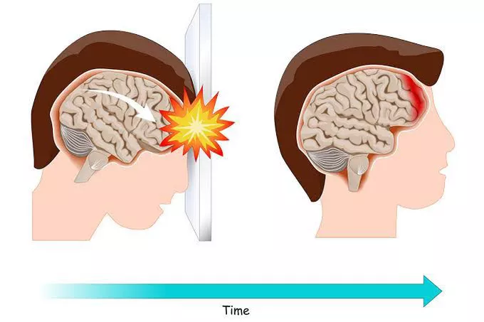
Explain/Define the Brain injury.
Brain injury is a condition in which brain tissues are damaged. It is mainly caused by brain trauma, stroke and various other medical conditions.
Brain injury is a condition in which brain function is temporarily or permanently impaired when any external mechanical force is applied to the brain.
At the same time, the dishability has also been seen in the function of the brain, it is called brain injury.
Explain the Etiology/cause of the brain injury.
- Due to head injury.
- Due to header traversal.
- Due to fall down.
- Due to penetrating injury.
- due to having a stroke.
- Due to infection.
- Due to tumor formation.
- Due to the occurrence of neurological diseases.
- Due to intracranial hemorrhage.
- due to hypoxia.
- Due to skull fracture.
- Due to neurological disease.
- Because of Alzheimer’s disease.
- Due to multiple sclerosis.
- Due to degenerative diseases.
- Because of the toxic effect.
Explain the types of brain injury.
There are mainly three types of brain injury.
1) Contusions,
2) Concussions,
3) Hematoma
•> Subdural hematoma,
•>Epidural hematoma
•••>
1) Contusions,
Contusions are a major type of head injury, the most common of which is brain injury caused by hitting the head with a movable object, this brain tissue is damaged, causing brain drain and internal bleeding, i.e. internal haemorrhage and Blood is absorbed into the cerebrospinal fluid (CSF) causing permanent brain damage. And the person becomes unconscious.
2) Concussions,
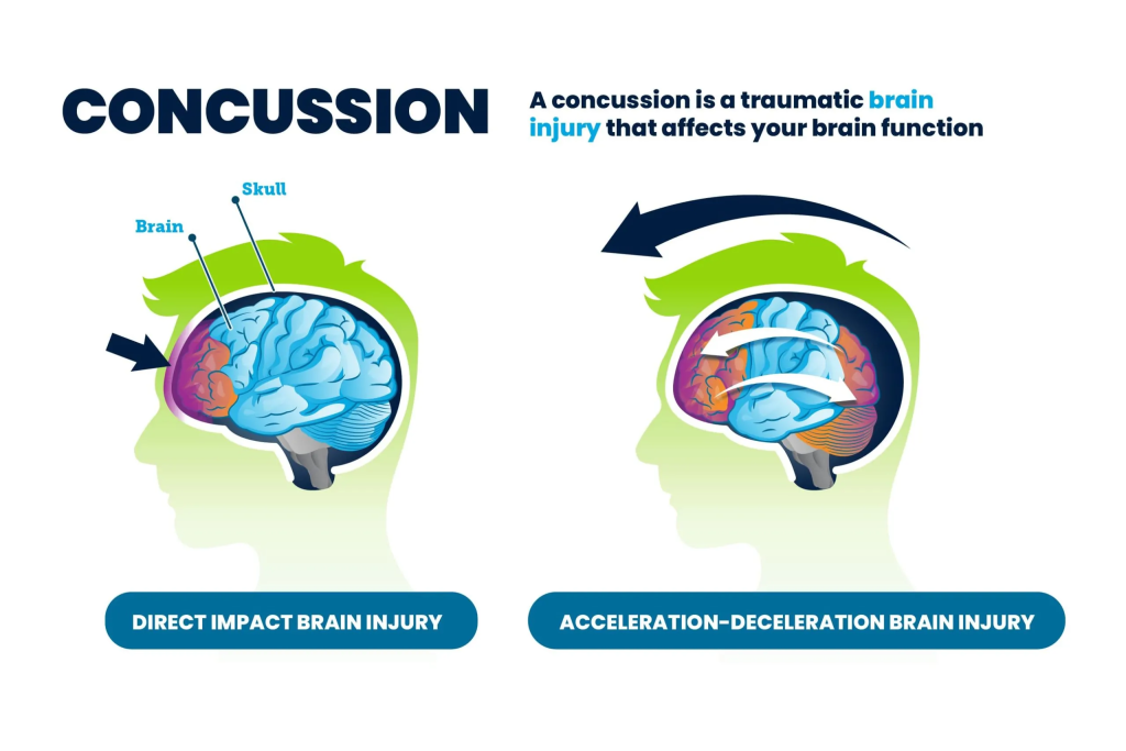
Contusions are a mild type of brain injury in which no structural damage is seen, no brain tissues are injured, mild brain damage results in temporary loss of neurological function and in this injury the person becomes unconscious for only five minutes.
3) Hematoma.
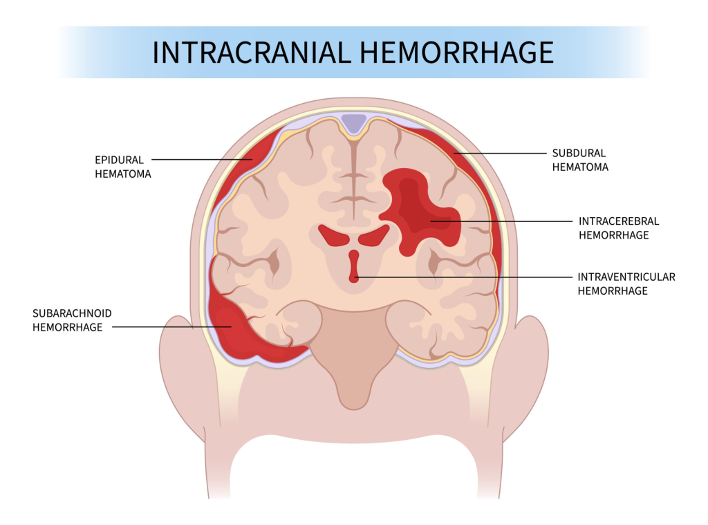
Internal bleeding is seen in hematoma due to intracranial haemorrhage and the blood becomes clotted. This clotted blood is called hematoma. There are two other types of hematoma.
•>Subdural hematoma,
If a blood clot occurs in the subdural space of the brain, it is called a subdural hematoma.
•>Epidural hematoma
If blood clots in the epidural space, it is called an epidural hematoma.
Explain the clinical manifestation/ sign and symptoms of the children with the head injury.
- Disturbing the child’s consciousness.
- Increased intracranial pressure.
- headache.
- Dizziness.
- to be startled
- Nausea and vomiting.
- Pupillary abnormality.
- Changes in vital signs.
- Tachycardia,
- tachypnea.
- Alteration of the gag reflex.
- Sensory, visual and hearing impairment.
- Disturbance of mental function.
- becoming paralyzed.
- Sleep disturbance.
- Personality change.
- Hemiplegia.
- Difficulty in concentration.
- Increases mood swings.
- feeling tired
- Slip pattern disturbance.
- Internal bleeding.
- Bleeding from ears and nose.
“Halo sign (leakage of cerebrospinal fluid into the linen and blood around it)” may be observed.
Explain the diagnostic evaluation of the children with the brain injury.
History taking and physical examination.
x ray,
CTScan
M.R.I.
PET (Positron Emission Tomography).
Angiography.
Neuropsychological test.
LabInvestigation.
Explain the management of the children with the head injury.
Keep the child’s airway properly patent.
Provide oxygen in adequate amount to the child.
Provide proper ventilatory support to the child.
Keep the child’s head elevated at a 30° angle.
Keep the child’s head and neck in a neutral position.
Keeping the child’s body temperature properly maintained.
Provide adequate amount of fluid to the child.
Provide sedation if the child is agitated.
Provide osmotic diuretic medicine to the child.
Ex:=
Inj.mannitol,
Syrup glycerol and glucocorticoid.
If the child has an infection, provide antibiotic medicine.
If the child has seizures, provide antiepileptic medicine.
Maintain child’s fluid and electrolyte levels.
Providing nutritional support to the child.
If the child is in pain, provide analgesic medicine.
To monitor the child’s vital signs properly.
Advise the child to take complete bed rest.
If the patient has inflammation in the brain, provide corticosteroid medicine.
Perform arterial blood gas analysis (ABG analysis) of the child properly.
Adequate amount of fluids to the child.
Properly monitor the child’s serum electrolyte level.
Provide proper skin care to the child.
Assessing the cognitive level of the child.
To provide proper psychological support to the child.
To provide proper work and comfortable environment to the child.
Provide proper medication to the child.