ENGLISH PEDIATRIC UNIT 5 CVS
Congenital Disorder of the Cardiovascular System (CVS)
Explain/Define Atrial Septal Defect (ASD)

Atrial septal defect is a congenital heart disease that usually results in a defect in the wall (septum) of the upper chamber of the heart i.e. the left atria and the right atria in which an abnormal opening is present at birth in the wall (septum) separating the left atria and right atria. .
Due to this abnormal opening in the atrial wall, the pressure of oxygenated blood flow in the left side atria is higher, the blood from the left side goes from the abnormal opening in the atrial septal to the atria of the right side due to which the left to right shunt is formed. .
Explain the types of the Atrial Septal Defect
There are mainly 3 types of atrial septal defect.
1) Ostium primum,
2) Osteum secundum,
3) Sinus venous defect
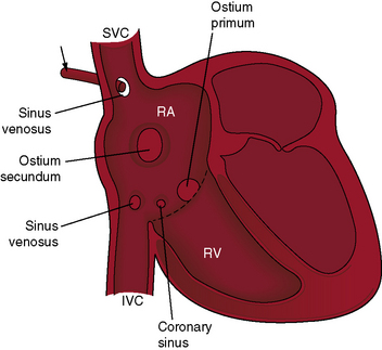
1) Ostium primum,
This type is less common and is usually found low in the atrial septum, near the valve that separates the atria from the ventricles.
2) Osteum secundum,
This is the most common type, accounting for about 70-80% of all ASDs. An abnormal opening develops in the center of the atrial septum.
3) Sinus venous defect
A sinus venous defect is an abnormal opening that usually develops near the entrance of the superior vena cava and inferior vena cava of the right atrium.
Explain the Etiology/cause of the Atrial Septal Defect.
- The exact cause is unknown,
- Due to genetic factors such as Down syndrome and other genetic abnormalities,
- Due to chromosomal abnormalities,
- Due to environmental factors like,
- Due to the mother taking certain types of toxic substances during pregnancy such as alcohol consumption, smoking, and taking certain types of medicine,
- Due to advanced maternal age,
- Due to maternal diseases like diabetes mellitus,
- Due to family history,
- Due to the condition of other congenital heart diseases.
- Due to the abnormal opening of the atrial septal during the development time of the heart.
Explain the Clinical manifestation/ Sign and symptoms of the Atrial Septal Defect.
- Difficulty breathing,
- Dyspnea especially during exercise,
- A bulging chest,
- No weight gain,
- cardiac enlargement,
- A systolic murmur sound is present,
- feel tired,
- feeding difficulties,
- Poor growth of the infant,
- Frequent respiratory infections such as pneumonia,
- heart palpitations,
- Irregular heart beat,
- Swelling in leg and abdomen,
- Congestive heart failure,
- Excessive sweating,
- Stroke and Transient Ischemic Attack (TIA).
Explain the Diagnostic evaluation of the Atrial septal Defect.
History taking and physical examination,
imaging studies,
echocardiogram,
Electrocardiogram
cardiac catheterization,
chest x-ray,
Trans esophageal echocardiogram (TEE),
Explain the medical management of the Atrial Septal Defect.
If the child has a small opening, then only the need for continuous monitoring of the child remains.
Proper medication is provided to manage the child’s symptoms.
Proper diuretic medicine is provided to prevent fluid buildup in the body.
Proper antibiotic medicine is provided to prevent the child from infection.
Digoxin medicine is provided to prevent congestive heart failure and dysrhythmias and to improve the pumping action of the heart.
Giving advice to the child’s parents for modification of the child’s lifestyle.
Explain the Surgical management of the Atrial Septal Defect.
1 .Open heart surgery
Atrial septal defect is repaired in open heart surgery.
In this procedure, the surgeon makes an incision in the chest and temporarily stops the heart while repairing the atrial septal defect with Dacron patches and sutures that are made of synthetic materials and pericardial tissues.
2 .Minimally invasive surgery
In many cases, minimally invasive surgery is used to treat atrial septal defect. In this surgery, a small incision is made on the chest. Then the defect is treated using a specialized instrument and a thoracoscope. The recovery time is short in this minimally invasive surgery. And it is less painful.
3 .Catheter based closure
Catheter-based closure is usually used for small to medium-sized openings. In this procedure, a flexible catheter is inserted from the groin area to the heart’s blood vessels, then the catheter is placed there to correct the wall defect. Because of this, tissues form in the opening and the opening can be closed.
Explain the Nursing management of the Atrial septal Defect.
Monitoring the child properly.
China To Assess Vital Signs Properly.
Assess the child’s cardiac function properly.
Properly assess the child’s symptoms like breathing difficulty, sweating, tiredness.
To provide complete education to the child’s parents about the child’s condition, its causes, symptoms and signs, its treatment and lifestyle modification.
Preoperative Nursing Management
To provide proper position to the child.
Provide oxygen to the child.
To provide proper psychological support to the child.
Monitoring the child’s blood oxygen level regularly.
Provide proper protection to prevent infection and trauma to the child.
To provide complete education to the child’s parents and caregivers about the child’s condition, its causes, symptoms and signs.
Provide care by maintaining proper aseptic technique of the child.
Provide intravenous fluids to maintain the nutritional status of the child.
Keeping the child properly clothed to prevent hypothermia.
Provide proper antibiotic medicine to prevent the child from infection.
Postoperative nursing management
Proper close monitoring of the child.
Continuously monitoring the child’s vital signs.
To continuously monitor the intake output chart of the child.
Provide adequate intravenous fluids to maintain the nutritional status of the child.
Provide adequate respiratory support to the child.
Administer oxygen properly to the child.
Proper ventilation to keep the child’s air passage clear.
Maintaining body temperature of child continuously Avoiding exposure of child to external environment.
Providing a nutritious diet to the child.
Maintain proper hygienic condition to prevent child from infection.
Provide dressings maintaining proper aseptic technique on the operative side.
Daily weight monitoring of child.
Continuously monitoring the child for any complications.
To provide education to the parents to provide adequate care to the child.
To provide complete education to the parents about the child’s condition.
Provide proper psychological support to reduce the anxiety of the child and his family members.
Advising the child’s parents to provide the prescribed medication to the child.
To provide proper psychological support to the parents of the child.
Advising the child’s parents to follow up regularly.
Explain/Define ventricular septal defect (VSD)
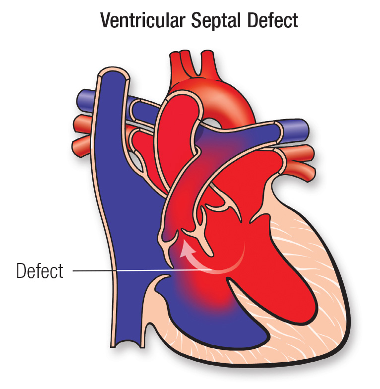
Ventricular septal defect is a congenital heart disease that usually results in a defect in the wall (septum) of the lower chamber of the heart i.e. the right ventricles and the left ventricles in which an abnormal opening is present at birth in the wall (septum) separating the right ventricles and the left ventricles. .
Because of this abnormal opening in the ventricular wall, the pressure of oxygenated blood flow in the left side ventricles is high, the blood from the left side flows through the abnormal opening in the ventricular septal into the ventricles of the right side, resulting in the formation of a left to right shunt. .
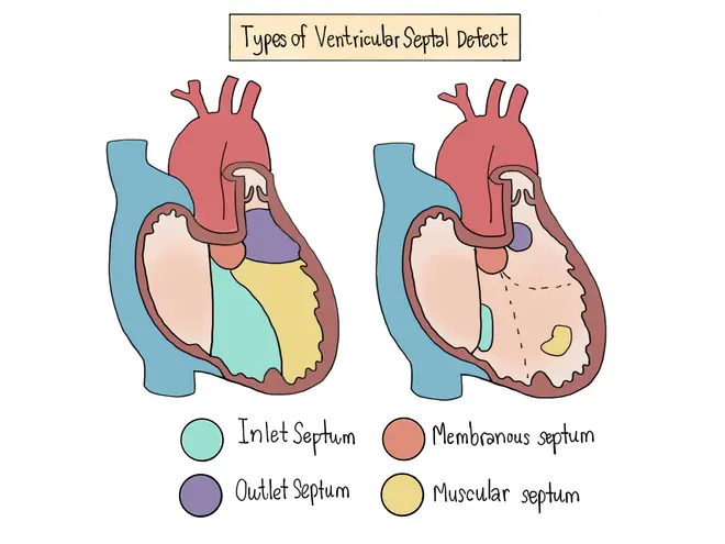
Explain the types of the ventricular septal defect
There are mainly 2 types of atrial septal defect.
1) Perimembranous Ventricular Septal Defect (VSD),
2) Muscular Ventricular Septal Defect (VSD),
••>
1) Perimembranous Ventricular Septal Defect (VSD),
This is the most common type of VSD, accounting for about 70-80% of cases. It involves an abnormal opening in the membranous portion of the ventricular septum, i.e. the upper part, which is the part of the septum near the valves of the heart.
2) Muscular Ventricular Septal Defect (VSD),
A muscular ventricular septal defect (VSD) is an abnormal opening in the muscular or lower part of the ventricular septum.

Explain the Etiology/cause of the ventricular septal defect (ventricular septal
- The exact cause is unknown,
- Due to genetic factors such as Down syndrome and other genetic abnormalities,
- Due to chromosomal abnormalities,
- Due to environmental factors like,
- Due to the mother taking certain types of toxic substances during pregnancy such as alcohol consumption, smoking, and taking certain types of medicine,
- Due to advanced maternal age,
- Due to maternal diseases like diabetes mellitus,
- Due to family history,
- Due to the condition of other congenital heart diseases.
- Due to the abnormal opening of the ventricular septal during the development time of the heart.
Explain the Clinical manifestation/ Sign and symptoms of the Ventricular septal Defect.
- Difficulty breathing,
- dyspnea,
- tachypnea,
- A bulging chest,
- No weight gain,
- cardiac enlargement,
- A systolic murmur sound is present,
- feel tired,
- feeding difficulties,
- Lack of interest in feeding,
- Poor growth of the infant,
- Frequent respiratory infections such as pneumonia,
- heart palpitations,
- Irregular heart beat,
- Tachycardia,
- Swelling in leg and abdomen,
- Congestive heart failure,
- Excessive sweating,
- Stroke and Transient Ischemic Attack (TIA).
- Palness to come.
- Biventricular hypertrophy occurs.
Explain the Diagnostic evaluation of the ventricular septal defect.
- History taking and physical examination,
- imaging studies,
- echocardiogram,
- Electrocardiogram
- cardiac catheterization,
- chest x-ray,
- Trans esophageal echocardiogram (TEE),
- Doppler studies.
Explain the medical management of the ventricular septal defect.
If the child has a small opening, then only the need for continuous monitoring of the child remains.
Proper medication is provided to manage the child’s symptoms.
Proper diuretic medicine is provided to prevent fluid buildup in the body.
Proper antibiotic medicine is provided to prevent the child from infection.
Digoxin medicine is provided to prevent congestive heart failure and dysrhythmias and to improve the pumping action of the heart.
Giving advice to the child’s parents for modification of the child’s lifestyle.
Advising the parents to provide an adequate nutritious diet to the child.
Provide proper oxygen therapy to the child.
Explain the Surgical management of the ventricular septal defect.
- Open heart surgery
Ventricular septal defect is repaired in open heart surgery.
In this procedure, the surgeon makes an incision in the chest and temporarily stops the heart while repairing the ventricular septal defect with Dacron patches and sutures that are made of synthetic materials and pericardial tissue.
- Minimally invasive surgery
In many cases, minimally invasive surgery is used to treat ventricular septal defects. In this surgery, a small incision is made on the chest. Then the defect is treated using a specialized instrument and a thoracoscope. The recovery time is short in this minimally invasive surgery. And it is less painful.
3) Catheter based closure
Catheter-based closure is usually used for small to medium-sized abnormal openings. In this procedure, a flexible catheter is inserted from the groin area to the blood vessels of the heart, and then the catheter is placed there to correct the wall defect. Because of this, the tissues in the opening are formed and the opening can be closed. This procedure is generally used for small and medium size openings.
Explain the Nursing management of the ventricular septal defect.
Monitoring the child properly.
China To Assess Vital Signs Properly.
Assess the child’s cardiac function properly.
Properly assess the child’s symptoms like breathing difficulty, sweating, tiredness.
To provide complete education to the child’s parents about the child’s condition, its causes, symptoms and signs, its treatment and lifestyle modification.
Preoperative Nursing Management
To provide proper position to the child.
Provide oxygen to the child.
To provide proper psychological support to the child.
Monitoring the child’s blood oxygen level regularly.
Provide proper protection to the child to prevent infection and trauma.
To provide complete education to the child’s parents and caregivers about the child’s condition, its causes, symptoms and signs.
Provide care by maintaining proper aseptic technique of the child.
Provide intravenous fluids to maintain the nutritional status of the child.
Keep the child properly clothed to prevent hypothermia.
Provide proper antibiotic medicine to prevent the child from infection.
Postoperative nursing management
Proper close monitoring of the child.
Continuously monitoring the child’s vital signs.
To continuously monitor the child’s intake output chart.
Provide adequate intravenous fluids to maintain the nutritional status of the child.
Provide adequate respiratory support to the child.
Administer oxygen properly to the child.
Proper ventilation to keep the child’s air passage clear.
Maintaining body temperature of child continuously Avoiding exposure of child to external environment.
Providing a nutritious diet to the child.
Maintain proper hygienic condition to prevent child from infection.
Maintain proper aseptic technique and provide dressing on the operative side.
Daily weight monitoring of child.
Continuously monitoring the child for any complications.
To provide education to the parents to provide adequate care to the child.
Providing complete education to the parents about the child’s condition.
Provide proper psychological support to reduce the anxiety of the child and his family members.
Advising the child’s parents to provide the prescribed medication to the child.
To provide proper psychological support to the parents of the child.
Advising the child’s parents to follow up regularly.
Explain/Define Patent Ductus Arteriosus (PDA)
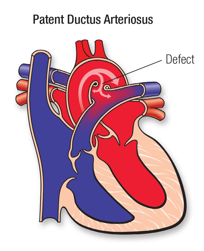
Patent doctors arteriosus is a congenital heart effect. This condition occurs when blood vessels i.e. ductor arterioses are connected to the pulmonary artery and aorta in the fetal circulation during intrauterine life. But after the birth of the newborn, it normally gets closed in the first week of life, but the condition of doctor’s arteriosis remains patent or open even after birth, i.e. if it fails to close, then the condition is called patent doctor’s arteriosis.
This condition of patent doctors arteriosis is more common in premature babies whose weight is less than 1.5 kg.
Explain the Etiology/cause of the patent ductus Arteriosus.
- The exact cause is unknown,
- Due to doctors not closing arteriosus after birth,
- Due to any infection of the mother during pregnancy,
- Due to premature birth,
- Due to genetic factors such as Down syndrome and other genetic abnormalities,
- Due to chromosomal abnormalities,
- Due to environmental factors like,
- Due to the mother taking certain types of toxic substances during pregnancy such as alcohol consumption, smoking, and taking certain types of medicine,
- Due to advanced maternal age,
- Due to maternal diseases like diabetes mellitus,
- Due to family history,
- Due to the condition of other congenital heart diseases.
- Due to the abnormal opening of the ventricular septal during the development time of the heart.
Explain the Clinical manifestation/ Sign and symptoms of the patent ductus arteriosus.
- Difficulty breathing,
- strong pulse,
- Increase in systolic pressure and decrease in diastolic pressure,
- dyspnea,
- tachypnea,
- A bulging chest,
- No weight gain,
- growth failure,
- cardiac enlargement,
- A machinery like murmur sound is present,
- feel tired,
- feeding difficulties,
- Lack of interest in feeding,
- Hosannas of Voice,
- Poor growth of the infant,
- Frequent respiratory infections such as pneumonia,
- heart palpitations,
- Irregular heart beat,
- Tachycardia,
- Congestive heart failure,
- Excessive sweating,
- Palness to come.
Explain the Diagnostic evaluation of the Patent ductus Arteriosus.
- History taking and physical examination,
- To perform heart sound auscultation,
- imaging studies,
- echocardiogram,
- Electrocardiogram
- cardiac catheterization,
- chest x-ray,
Explain the medical management of the Patent ductus Arteriosus.
There is a need for continuous monitoring of the child.
Proper medication is provided to manage the child’s symptoms.
If the child is premature and the condition is patent doctors provide indomethacin 0.1 to 0.25 mg/kg/dose intravenously to treat the condition.
Providing the child with a proper NSAID (Non Steroidal Anti-Inflammatory Drug) patent doctors to close the arteriosus.
Provide limited amount of fluid to the child to maintain the fluid level in the body.
Proper antibiotic medicine is provided to prevent the child from infection.
Proper medicine is provided to prevent congestive heart failure and dysrhythmias and to improve the pumping action of the heart.
Giving advice to the child’s parents for modification of the child’s lifestyle.
Advising the parents to provide an adequate nutritious diet to the child.
Provide proper oxygen therapy to the child.
Explain the Surgical management of the Patent ductus Arteriosus.
Open heart surgery
In open heart surgery, patent doctors divide the arteriosus and then close it with sutures.
In this procedure, the surgeon makes an incision on the chest and patent doctors repair the arteriosus with sutures.
Visual Assisted Thoracoscopic Surgery (VATS)
In this procedure, two to three incisions are made on the chest to treat patent ductus arteriosus, after which the patent ductus arteriosus is treated using a thoracoscope and instruments.
Minimally Invasive Surgery
In many cases, patent doctors use minimally invasive surgery to treat arteriosus. In this surgery, a small incision is made on the chest. The patent ductus arteriosus is then treated using specialized instruments and a thoracoscope. The recovery time is short in this minimally invasive surgery. And it is less painful.
Catheter Based Closure
Catheter-based closure is commonly used to treat patent ductus arteriosus. In this procedure, a flexible catheter is inserted from the groin area to the blood vessels of the heart, then coils and umbrella-like dividers are placed there to close the patent ductus arteriosus. Is performed.
Explain the Nursing management of Patent ductus Arteriosus.
Monitoring the child properly.
To properly assess the child’s vital signs.
Assess the child’s cardiac function properly.
Properly assess the child’s symptoms like breathing difficulty, sweating, tiredness.
To provide complete education to the child’s parents about the child’s condition, its causes, symptoms and signs, its treatment and lifestyle modification.
Preoperative Nursing Management
To provide proper position to the child.
Provide oxygen to the child.
To provide proper psychological support to the child.
Monitoring the child’s blood oxygen level regularly.
Provide proper protection to the child to prevent infection and trauma.
To provide complete education to the child’s parents and caregivers about the child’s condition, its causes, symptoms and signs.
Provide care by maintaining proper aseptic technique of the child.
Intravenous to maintain the nutritional status of the child
Provide fluids.
Dress the child properly to prevent hypothermia
Keep a stock.
Provide proper antibiotic medicine to prevent the child from infection.
Postoperative nursing management
Proper close monitoring of the child.
Continuously monitoring the child’s vital signs.
To continuously monitor the child’s intake output chart.
Provide adequate intravenous fluids to maintain the nutritional status of the child.
Provide adequate respiratory support to the child.
Administer oxygen properly to the child.
Proper ventilation to keep the child’s air passage clear.
Maintaining body temperature of child continuously Avoiding exposure of child to external environment.
Providing a nutritious diet to the child.
Maintain proper hygienic condition to prevent child from infection.
Maintain proper aseptic technique and provide dressing on the operative side.
Daily weight monitoring of child.
Continuously monitoring the child for any complications.
To provide education to the parents to provide adequate care to the child.
Providing complete education to the parents about the child’s condition.
Provide proper psychological support to reduce the anxiety of the child and his family members.
Advising the child’s parents to provide the prescribed medication to the child.
To provide proper psychological support to the parents of the child.
Advising the child’s parents to follow up regularly.
Explain/Define Coarctation of the Aorta (COA)
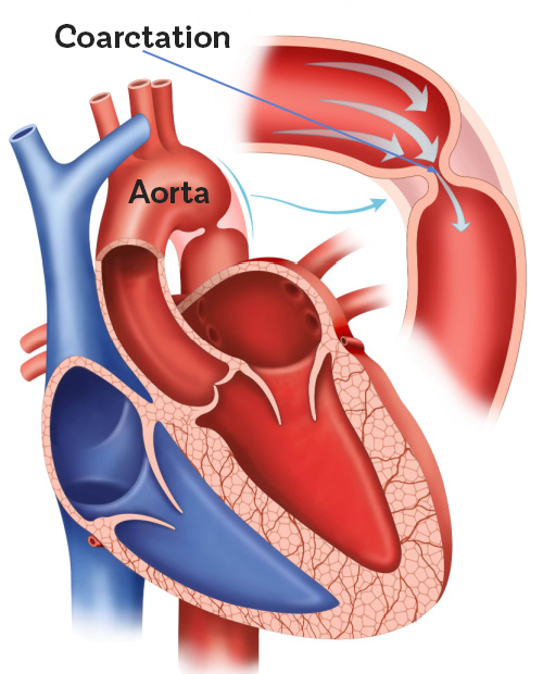
Correction := Means Narrowing,
Aorta := Min Laj blood vessel that supplies oxygenated blood from the heart to the entire body.
Correction of aorta means “narrowing of the aorta”.
Coarctation of the aorta is a congenital heart defect in which the aorta (a blood vessel that carries oxygenated blood from the heart and supplies oxygenated blood to all parts of the body) is narrowed. This narrowing occurs in close proximity to the doctor’s RTOS condition.
Due to this narrowing, the blood flow in the lower part of the body (abdomen, pelvis and legs) is restricted and the blood pressure in the lower extremities is also reduced. When the blood pressure increases in the upper extremities and head.

Explain the Etiology/cause of the Coarctation of Aorta.
- The exact cause is unknown,
- Due to any infection of the mother during pregnancy,
- Due to premature birth,
- Due to genetic factors such as Turner syndrome and other genetic abnormalities,
- Due to chromosomal abnormalities,
- Due to environmental factors like,
- Due to the mother taking certain types of toxic substances during pregnancy such as alcohol consumption, smoking, and taking certain types of medicine,
- Due to advanced maternal age,
- Due to maternal diseases like diabetes mellitus,
- Due to family history,
- Due to the condition of other congenital heart diseases.
- Due to the abnormal opening of the ventricular septal during the development time of the heart.
Explain the Clinical manifestation/ Sign and symptoms of the Coarctation of Aorta.
- Difficulty breathing,
- Bounding pulse and
- Increase in blood pressure,
- Low blood pressure and extremity are total,
- acidosis,
- dyspnea,
- tachypnea,
- No weight gain,
- growth failure,
- cardiac enlargement,
- Murmur sound being present,
- feel tired,
- feeding difficulties,
- Lack of interest in feeding,
- Poor growth of the infant,
- Frequent respiratory infections such as pneumonia,
- heart palpitations,
- Irregular heart beat,
- Tachycardia,
- Congestive heart failure,
- Excessive sweating,
- coming to palness,
- headache,
- Epistaxis, (nose bleeds),
- coming to wickness,
- Dizziness.
- Explain the Diagnostic evaluation of the Coarctation of Aorta (Coarctation
- History taking and physical examination,
- To perform heart sound auscultation,
- imaging studies,
- echocardiogram,
- Electrocardiogram
- cardiac catheterization,
- chest x-ray,
- Angiography.
Explain the medical management of the Coarctation of Aorta.
There is a need for continuous monitoring of the child.
Proper medication is provided to manage the child’s symptoms.
If the child has high blood pressure, provide antihypertensive medicine.
If the child has fluid buildup in the body and edema, then provide diuretic medicine.
If the child is experiencing pain and discomfort, provide proper pain killers and NSAID (Non-Steroidal Anti-Inflammatory Drug).
Provide limited amount of fluid to the child to maintain the fluid level in the body.
Proper antibiotic medicine is provided to prevent the child from infection.
Proper medicine is provided to prevent congestive heart failure and dysrhythmias and to improve the pumping action of the heart.
Giving advice to the child’s parents for modification of the child’s lifestyle.
Advising the parents to provide an adequate nutritious diet to the child.
Provide proper oxygen therapy to the child.
Explain the Surgical management of the Coarctation of Aorta.
Open heart surgery
Open Heart Surgery In this procedure, the surgeon makes an incision on the chest and after cutting the part of the coarctation of the aorta, the remaining part of the aorta is anastomosed end-to-end with sutures. If necessary, additional tissues are also used. is coming.
Minimally Invasive Surgery
In many cases, minimally invasive surgery is used to treat coarctation of the aorta. In this surgery, a small incision is placed on the chest. Then the corrected aorta is treated using specialized instruments and thoracoscope. In this minimally invasive surgery, the incision is also small and the recovery time is short. And it is less painful.
Catheter based
Catheter-based is commonly used to treat correction of aorta. In this procedure, a flexible catheter is inserted from the groin area to the aorta followed by placement of special devices such as balloons, angioplasty, and stents. and correction of aorta in dilatation
is coming.
Balloon angioplasty is performed to correct the dilatation of the aorta.
Explain the Nursing management of Coarctation of Aorta.
Monitoring the child properly.
To properly assess the child’s vital signs.
Assess the child’s cardiac function properly.
Properly assess the child’s symptoms like breathing difficulty, sweating, tiredness.
To provide complete education to the child’s parents about the child’s condition, its causes, symptoms and signs, its treatment and lifestyle modification.
Preoperative Nursing Management
To provide proper position to the child.
Provide oxygen to the child.
To provide proper psychological support to the child.
Monitoring the child’s blood oxygen level regularly.
Provide proper protection to the child to prevent infection and trauma.
To provide complete education to the child’s parents and caregivers about the child’s condition, its causes, symptoms and signs.
Provide care by maintaining proper aseptic technique of the child.
Intravenous to maintain the nutritional status of the child
Provide fluids.
Properly to prevent the child from hypothermia
Keeping the cloth in the bag.
Provide proper antibiotic medicine to prevent the child from infection
Postoperative nursing management
Proper close monitoring of the child.
Continuously monitoring the child’s vital signs.
To continuously monitor the intake output chart of the child.
Provide adequate intravenous fluids to maintain the nutritional status of the child.
Provide adequate respiratory support to the child.
Administer oxygen properly to the child.
Proper ventilation to keep the child’s air passage clear.
Maintaining body temperature of child continuously Avoiding exposure of child to external environment.
Providing a nutritious diet to the child.
Maintain proper hygienic condition to prevent child from infection.
Provide dressings maintaining proper aseptic technique on the operative side.
Daily weight monitoring of child.
Continuously monitoring the child for any complications.
To provide education to the parents to provide adequate care to the child.
To provide complete education to the parents about the child’s condition.
Provide proper psychological support to reduce the anxiety of the child and his family members.
Advising the child’s parents to provide the prescribed medication to the child.
To provide proper psychological support to the parents of the child.
Advising the child’s parents to follow up regularly.
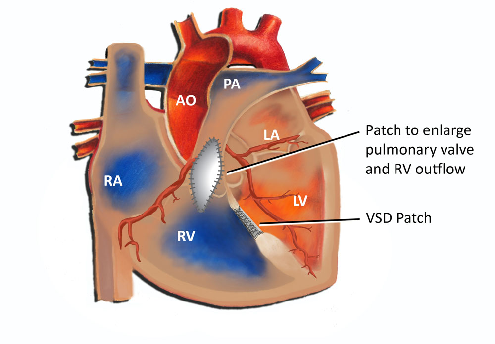
Explain/Define Tetralogy of Fallot (Define Tetralogy of Fallot).

Tetralogy of Fallot is a congenital heart defect. In which four heart defects are seen simultaneously in the child.
1) Ventricular septal defect,
2) pulmonary stenosis,
3)overriding aorta,
4) Right ventricular hypertrophy.
••>
1) Ventricular septal defect,
In a ventricular septal defect, an abnormal opening is present in the septum separating the right and left ventricles.
2) pulmonary stenosis,
In pulmonary stenosis, there is a narrowing of the pulmonary artery (the blood vessel that carries deoxygenated blood from the right ventricle to the lungs) that obstructs blood flow from the right ventricle.
3) Overriding of aorta,
Overriding of aorta is a condition in which normally the aorta (the main blood vessel that carries and transports oxygenated blood from the heart to the wall body) originates from the left ventricle of the heart but overriding of the aorta in the aorta over the left ventricles. On the side, the origin is directly above the ventricular septal defect
4) Right ventricular hypertrophy.
When the pulmonary artery (the blood vessel that carries deoxygenated blood from the right ventricle and carries blood to the lungs for oxygenation in the lungs) narrows and stenoses, the heart’s right ventricle contracts to deliver blood from the right ventricle to the lungs. There is a need to do extra work (pumping) because of this extra pumping, the muscles of the right ventricle of the heart become thickened which is called hypertrophy.
Explain the Etiology/cause of the Tetralogy of Fallot
- Due to genetic factors,
- Due to environmental factors during pregnancy like,
- maternal age,
- Maternal health conditions such as diabetes.
- Due to the mother coming in contact with certain types of toxic substances during pregnancy.
- Due to certain types of medication taken by the mother during pregnancy.
- Due to improper nutrition intake by the mother during pregnancy.
- Due to chromosomal abnormalities.
- Due to failure of proper heart development during intrauterine life of fetus.
Explain the Clinical manifestation/ Sign and symptoms of the Tetralogy of Fallot.
Cyanosis is observed,
Bluish discoloration of lips, skin and nails,
Shortness of breath,
go clubbing,
feel tired,
Growth is poor,
Breathing difficulty,
Abnormal murmur sounds,
Irritability,
dyspnea,
Episodes of cyanotic spells (tet spells) in which cyanosis, hypoxia and breathing difficulties occur in the child after feeding and after any painful procedure,
polycythemia,
The child’s skin is rough and clammy,
Explain the diagnostic evaluation of the Tetralogy of F
- History taking and physical examination,
- Heart sound auscultation,
- Electrocardiogram (ECG),
- echocardiography,
- chest x-ray,
- Cardiac Magnetic Resonance Imaging (MRI),
- cardiac catheterization,
Explain the medical management of the Tetralogy of Fallot.
To properly provide oxygen to treat the hypoxic condition of the child and his condition of cyanosis.
Provide proper sedative medicine to the child.
Provide proper intra venous fluid to the child.
Treating Child Dehydration Properly
If the child has the condition of anemia, it should be treated properly.
Provide proper chest position to treat child’s hypoxic spell.
Administer an intravenous vasopressor such as methoxamine to the child.
If the child has a severe technology of Fallot condition, provide the child with prostaglandin E1 intravenously. Due to this, ducts dilate and pulmonary blood flow increases.
If the child has a condition of acidosis, treat it properly.
Explain the Surgical management of the Tetralogy of Fallot.
Surgical management of Technology of Fallot usually consists of correcting the defect and providing palliative procedures. Performing this procedure increases the blood flow in the lungs and treats the cyanosis condition.
Most common surgeries involve intracardiac repair and complete repair.
- Complete intracardiac repair
Complete intracardiac repair is the preferred surgical procedure to treat most tetralogy of Fallot conditions. In this procedure, the ventricular septal defect is treated with a patch. Then, the obstruction of the pulmonary artery which is stenosis is relieved so that the blood can be transported from the right ventricle to the lungs properly, after which repositioning of the overriding aorta is provided. The aim of this procedure is to improve the blood flow in the lungs and treat the child’s condition properly.
- Blalock Tossing or Modified Blalock Tossing Sunt.
When it is impossible to completely repair the condition of tetralogy of fallot, blactosing sunt is not used. In this procedure, a connection is made between the subclavian artery, innominate artery and pulmonary artery, due to which the blood flow in the lungs can be improved.
- Pulmonary valve stenosis
In pulmonary valve stenosis, the stenotic valve is properly repaired.
Explain the Nursing management of the Tetralogy of Fallot.
Monitoring the child properly.
To properly assess the child’s vital signs.
Assess the child’s cardiac function properly.
Properly assess the child’s symptoms like breathing difficulty, sweating, tiredness.
To provide complete education to the child’s parents about the child’s condition, its causes, symptoms and signs, its treatment and lifestyle modification.
Preoperative Nursing Management
To provide proper position to the child.
Provide oxygen to the child.
To provide proper psychological support to the child.
Monitoring the child’s blood oxygen level regularly.
Provide proper protection to the child to prevent infection and trauma.
To provide complete education to the child’s parents and caregivers about the child’s condition, its causes, symptoms and signs.
Provide care by maintaining proper aseptic technique of the child.
Provide intravenous fluids to maintain the nutritional status of the child.
Keep the child properly clothed to prevent hypothermia.
Provide proper antibiotic medicine to prevent the child from infection.
Postoperative nursing management
Proper close monitoring of the child.
Continuously monitoring the child’s vital signs.
To continuously monitor the child’s intake output chart.
Provide adequate intravenous fluids to maintain the nutritional status of the child.
Provide adequate respiratory support to the child.
Administer oxygen properly to the child.
Proper ventilation to keep the child’s air passage clear.
Maintaining body temperature of child continuously Avoiding exposure of child to external environment.
Providing a nutritious diet to the child.
Maintain proper hygienic condition to prevent child from infection.
Maintain proper aseptic technique and provide dressing on the operative side.
Daily weight monitoring of child.
Continuously monitoring the child for any complications.
To provide education to the parents to provide adequate care to the child.
Providing complete education to the parents about the child’s condition.
Provide proper psychological support to reduce the anxiety of the child and his family members.
Advising the child’s parents to provide the prescribed medication to the child.
To provide proper psychological support to the parents of the child.
Advising the child’s parents to follow up regularly.
- Explain/Define Transposition of Great Arteries (TGA) OR Transposition of Great Vessels (TGV). (
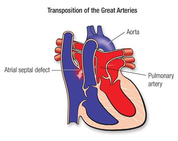
Normally the aorta (the main blood vessel that transports oxygenated blood from the heart to the body wall) originates from the left ventricle of the heart while the pulmonary artery (the blood vessel that carries deoxygenated blood from the heart to the lungs) originates from the right ventricle. .but,
Transposition := Means any thing being transported from one side (its normal place) to another.
Great Artery := Mins Largest Blood Vessels of Body (Aorta and Pulmonary Artery).
Transposition of great arteries is a condition in which the great vessels of the body (aorta and pulmonary artery) originate from the other side than their normal place.
Transposition of great arteries is a congenital heart condition in which the aorta originates from the right ventricle of the heart while the pulmonary artery originates from the left ventricle of the heart.
This condition is more common in males, in children whose birth weight is high, and in children whose grandparents have a history of diabetes.
Explain the Etiology/cause of Transposition of Great Arteries
- Due to genetic factors,
- Due to environmental factors during pregnancy like,
- maternal age,
- Maternal health conditions such as diabetes.
- Due to developmental factors,
- Due to the mother coming in contact with certain types of toxic substances during pregnancy.
- Due to certain medications taken by the mother during pregnancy.
- Due to improper nutrition intake by the mother during pregnancy.
- Due to chromosomal abnormalities.
- Due to failure of proper heart development during intrauterine life of fetus.
Explain the Clinical manifestation/ Sign and symptoms of the Transposition of Great Arteries.
- Cyanosis is observed,
- Bluish discoloration of lips, skin and nails,
- Shortness of breath,
- go clubbing,
- feel tired,
- Growth is poor,
- Breathing difficulty,
- Metabolic acidosis
- Irritability,
- dyspnea,
- Hypoxia and breathing difficulties,
- The child’s skin is rough and clammy,
- severe hypoxia,
- Symptoms of congestive heart failure,
- cardiomyopathy,
- Tachycardia.
Explain the Diagnostic evaluation of the Transposition of Great Arteries.
- History taking and physical examination,
- Electrocardiogram (ECG),
- echocardiography,
- chest x-ray,
- Cardiac Magnetic Resonance Imaging (MRI),
- cardiac catheterization,
Explain the medical management of the Transposition of Great Arteries.
To properly provide oxygen to treat the hypoxic condition of the child and his condition of cyanosis.
Provide proper intra venous fluid to the child.
Treating Child Dehydration Properly
Provide prostaglandin E to the child intravenously.
If the child has a condition of acidosis, treat it properly.
Continuously monitoring the child.
Explain the Surgical management of the Transposition of Great Arteries.
- Arterial Switch Procedure
An arterial switch procedure is usually performed during the first week of life. In this procedure, the transposed great artery is surgically repaired to its normal place and the normal anatomical function of the heart is reestablished. Because of this, this surgical procedure is performed so that the oxygenated blood can be transported to the wall body and the deoxygenated blood can be properly transported from the right ventricle to the lungs.
2.Intraarterial baffle repair
In this procedure, the surgeon creates an intra-arterial baffle or tunnel inside the heart to redirect blood flow from the ventricle (usually the left ventricle) to the appropriate great artery (aorta) and the pulmonary ventricle (usually the right ventricle). to the other great artery (pulmonary artery). This leads to proper oxygenation of the blood and improves circulation.
- Rastelli Procedure
During this procedure, the surgeon creates a tube to bypass the pulmonary stenosis and connect the right ventricle to the pulmonary artery. Additionally, a patch is placed to redirect blood flow from the left ventricle to the aorta. Due to which normal physiological circulation can be created and oxygenated blood can be transported in the wall body.
Explain the Nursing management of the Transposition of Great Arteries.
Monitoring the child properly.
To properly assess the child’s vital signs.
Assess the child’s cardiac function properly.
Properly assess the child’s symptoms like breathing difficulty, sweating, tiredness.
To provide complete education to the child’s parents about the child’s condition, its causes, symptoms and signs, its treatment and lifestyle modification.
Preoperative Nursing Management
To provide proper position to the child.
Provide oxygen to the child.
To provide proper psychological support to the child.
Monitoring the child’s blood oxygen level regularly.
Provide proper protection to prevent infection and trauma to the child.
To provide complete education to the child’s parents and caregivers about the child’s condition, its causes, symptoms and signs.
Provide care by maintaining proper aseptic technique of the child.
Provide intravenous fluids to maintain the nutritional status of the child.
Keeping the child properly clothed to prevent hypothermia.
Provide proper antibiotic medicine to prevent the child from infection.
Postoperative nursing management
Proper close monitoring of the child.
Continuously monitoring the child’s vital signs.
To continuously monitor the intake output chart of the child.
Provide adequate intravenous fluids to maintain the nutritional status of the child.
Provide adequate respiratory support to the child.
Administer oxygen properly to the child.
Proper ventilation to keep the child’s air passage clear.
Maintaining body temperature of child continuously Avoiding exposure of child to external environment.
Providing a nutritious diet to the child.
Maintain proper hygienic condition to prevent child from infection.
Provide dressings maintaining proper aseptic technique on the operative side.
Daily weight monitoring of child.
Continuously monitoring the child for any complications.
To provide education to the parents to provide adequate care to the child.
To provide complete education to the parents about the child’s condition.
Provide proper psychological support to reduce the anxiety of the child and his family members.
Advising the child’s parents to provide the prescribed medication to the child.
To provide proper psychological support to the parents of the child.
Advising the child’s parents to follow up regularly.
Explain/Define Total Anomalous Pulmonary Venous Connection
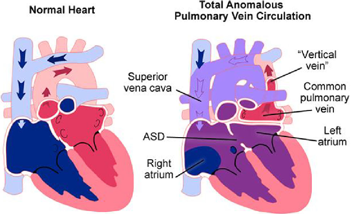
Total anomalous pulmonary venous connection (TAPVC) is a congenital heart defect where all four pulmonary veins, which normally carry oxygenated blood from the lungs to the left atrium of the heart, do not connect normally to the heart. Instead, it connects abnormally to the right atrium or to another vein draining into the right atrium. This results in mixing of oxygenated and deoxygenated blood, which leads to cyanosis (bluish discoloration of the skin) and inadequate oxygen supply to the body.
Explain the Etiology/cause of Total Anomalous Pulmonary Venous Connection
- Due to genetic factors,
- Due to environmental factors during pregnancy like,
- maternal age,
- Maternal health conditions such as diabetes.
- Due to developmental factors,
- Due to the mother coming in contact with certain types of toxic substances during pregnancy.
- Due to certain types of medication taken by the mother during pregnancy.
- Due to improper nutrition intake by the mother during pregnancy.
- Due to chromosomal abnormalities.
- Due to failure of proper heart development during intrauterine life of fetus.
Explain the Clinical manifestation/ Sign and symptoms of the Total Anomalous Pulmonary Venous Connection.
- Cyanosis is observed,
- Bluish discoloration of lips, skin and nails,
- Shortness of breath,
- go clubbing,
- feel tired,
- Growth is poor,
- Breathing difficulty,
- Irritability,
- dyspnea,
- Hypoxia and breathing difficulties,
- The child’s skin is rough and clammy,
- severe hypoxia,
- Symptoms of congestive heart failure,
- cardiomyopathy,
- Tachycardia.
Explain the Diagnostic evaluation of the Total Anomalous Pulmonary Venous Connection.
- History taking and physical examination,
- Electrocardiogram (ECG),
- echocardiography,
- chest x-ray,
- Cardiac Magnetic Resonance Imaging (MRI),
- cardiac catheterization,
Explain the medical management of Total Anomalous Pulmonary Venous Connection
To properly provide oxygen to treat the hypoxic condition of the child and his condition of cyanosis.
Provide proper intravenous fluids to maintain child’s hydration status.
Treating Child Dehydration Properly
Provide intra-venous prostaglandin to the child.
Continuously monitoring the child.
Complete assessment of the child for any other complication or not.
Properly maintaining the nutritional status of the child by providing adequate amount of breast milk, formula milk and adequate calorie rich food.
If there is fluid overload in the child’s body, provide adequate diuretic medicine to treat the condition.
Explain the surgical management of the Total Anomalous Pulmonary Venous Connection.
Surgical management of total anomalous pulmonary venous connection (TAPVC) involves an anastomosis between the pulmonary vein and the left atrium of the heart. The surgeon carefully divides the abnormal communication of the pulmonary vein with the right atrium or another vein and then sutures it to the opening made in the left atrium. This returns oxygenated blood from the lungs to the left side of the heart and is then transported to the wall body.
Explain the Nursing management of the Total Anomalous Pulmonary Venous Connection.
Monitoring the child properly.
To properly assess the child’s vital signs.
Assess the child’s cardiac function properly.
Properly assess the child’s symptoms like breathing difficulty, sweating, tiredness.
To provide complete education to the child’s parents about the child’s condition, its causes, symptoms and signs, its treatment and lifestyle modification.
Preoperative Nursing Management
To provide proper position to the child.
Provide oxygen to the child.
To provide proper psychological support to the child.
Monitoring the child’s blood oxygen level regularly.
Provide proper protection to the child to prevent infection and trauma.
To provide complete education to the child’s parents and caregivers about the child’s condition, its causes, symptoms and signs.
Provide care by maintaining proper aseptic technique of the child.
Provide intravenous fluids to maintain the nutritional status of the child.
Keep the child properly clothed to prevent hypothermia.
Provide proper antibiotic medicine to prevent the child from infection.
Postoperative nursing management
Proper close monitoring of the child.
Continuously monitoring the child’s vital signs.
To continuously monitor the child’s intake output chart.
Provide adequate intravenous fluids to maintain the nutritional status of the child.
Provide adequate respiratory support to the child.
Administer oxygen properly to the child.
Proper ventilation to keep the child’s air passage clear.
Maintaining body temperature of child continuously Avoiding exposure of child to external environment.
Providing a nutritious diet to the child.
Maintain proper hygienic condition to prevent child from infection.
Maintain proper aseptic technique and provide dressing on the operative side.
Daily weight monitoring of child.
Continuously monitoring the child for any complications.
To provide education to the parents to provide adequate care to the child.
Providing complete education to the parents about the child’s condition.
Provide proper psychological support to reduce the anxiety of the child and his family members.
Advising the child’s parents to provide the prescribed medication to the child.
To provide proper psychological support to the parents of the child.
Advising the child’s parents to follow up regularly.