ENGLISH integumentary -PART-1 SKIN DISORDERS (juhi)
INTEGUMENTARY SYSTEM DISEASES AND DISORDERS (SKIN)
Important Terminology for disorders of integumentary
system
Alopecia :– Loss of hair
Acantholysis :- Epidermal cell separation from each other (which occurs due to intracellular substrate damage or abnormality)
Carbuncle :- Carbuncle is a bacterial skin infection affecting a group of hair follicles.
Cellulitis :– Cellulitis is a bacterial skin infection. In which redness, swelling and pain are seen in the affected skin area.
Comdones :-Comdones are papule-like formations of skin color. which is found in one’s vulgaris. Which is seen due to sebum blockage in the hair follicle.
Cryosurgery:– Cryosurgery is a process in which extremely cold liquid nitrogen is produced to destroy cancerous cells and abnormal tissue.
Cyanosis :- Bluish discoloration of skin and mucus membrane.
Cytotoxic:- Cytotoxic is a substance or procedure that causes cell damage and cell death.
Dermatitis :- Inflammation of skin
Dermatosis :– Abnormal skin lesions
Debriment :– Procedure to remove necrotic and infected tissue.
Dermatophytes :– Fungal infection of skin
Erythema :- Skin red color due to congestion of capillaries.
Erysipelas: Erysipelas is a bacterial skin infection. In which the superficial dermis layer of the skin is affected.
Folliculitis (Folliculitis):- Bacterial infection of hair follicle is called folliculitis. (Infection of hair follicle)
Furuncle :– A furuncle is a bacterial hair follicle infection. In which a painful, pus-filled bump is seen under the skin.
Hydrophilic :– Hydrophilic is a type of material that absorbs moisture.
Hydrophobic :- Hydrophobic is a type of material that repels moisture.
Hygroscopic :– Hygroscopic is a type of material that absorbs moisture from the air.
Hirsutism:– Hirsutism is excessive hair growth in women.
Impetigo :– Impetigo is a common bacterial infection that causes sores and blisters on the skin.
Keratin :- Keratin is a fibrous protein that forms the outer layer of the skin.
Keloids :– Keloids are irregular, thick scars. which is seen due to abnormal wound healing process.
Melanin :- Melanin is a substance. Which is responsible for skin color.
Melanocyte :- Melanocyte is a skin cell that produces melanin.
Patechia (Petechia) :– Petechia means red color spots found on the skin. Which is seen due to blood leakage in the skin.
Plasmapharesis :- Plasmapheresis is a process in which bad plasma is removed from the blood by centrifugation and reinfusion and new plasma is added in its place.
Psoriasis:- Psoriasis is an autoimmune disorder in which the epidermal cells in the skin overproduce.
Pruritus (Pruriters) :– Pruritus is the term used for itching, due to which scratches are seen.
Paronychia :- An infection of the soft tissue around the fingernails and toenails is known as paronychia.
Scabies :– Scabies is a parasitic infestation. which is caused by Sarcoptes scabiei mite.
Sebum :- The fatty secretion produced by the sebaceous gland is known as sebum.
Telangiectasia :- Dilation of superficial and small veins in the skin.
Tinea:- Fungal infection of skin and scalp.
Ulticaria :(Ulticaria) :– Ulticaria is a type of itchy race. Which occurs due to a reaction to food, medicine or any other substance.
vitilogo:– In vitiligo, cells that produce pigmentation become dead or stop working so that the skin loses its color and the skin is excessively white.
Warts :- Warts are a condition caused by a virus in the human papilloma. In which a small bump is seen on the skin.
Wood light:- Wood light is a blue type of light which is used for skin assessment. So that abnormal skin conditions can be identified.
Xerosis:- Dry skin
a) Nursing Assessment :-
History :-
(A) Present health history :
Asking the patient about their current complaints.
To know whether the patient has itching, dryness, reses, lesion lump, swelling present in any area.
Ask about any kind of symptoms in skin, hair, nails, scalp.
Ask about when the symptoms started and their duration, intensity, and location. Past health history:
Collect information about previous sun and radiation exposure, if any. (B) Past surgical history : If any cosmetic surgery or any other type of surgery has been done in the previous years to collect information about it.•
(C) Personal history :-
Inquiring whether the patient has any type of skin allergy.
Asking about allergic reactions to any food medicine or chemical.
Knowing about cosmetic items, shops, shampoos and personal hygiene products used by patients.
To know about patient’s elimination pattern, sleep rest pattern, sexuality pattern, exercise and activity pattern.
Also know about the type of material the patient uses in the garment. (D) Family history :
Collecting the patient’s family history.
Knowing if anyone has a history of skin allergies, skin cancer, alopecia, xerosis, psoriasis, dermatitis, lupus erythematosus in the family.
Also ask if anyone in the family has vitiligo or sexually transmitted diseases. (E) Occupational history :
To collect the occupational history of the patient.
People working in metal industry, automobile industry, construction industry, X-ray department, manufacturing department, printing industry have higher skin disease level ◼️Physical assessment (objective data)
Physical assessment should include entire skin, mucus membrane, scalp, hair and nails.
Skin examination uses inspection and palpation techniques.
The examination room should have adequate lighting.
Use gloves when palpating lesions and lesions. Inspection
Checking skin color in skin inspection.
Check whether redness, cyanosis, pallor, pigmentation, vitiligo, erythema is present in the skin.
Assessing whether any type of race or lesion is present in the skin.
If a lesion is present, assess its type, size, shape, location and color.
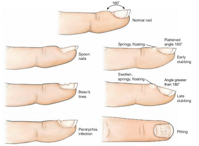
Nail examination to check nail color, shape, curvature, consistency, surface and clubbing.
A pitted surface is seen in the nails in patients with psoriasis.
Clubbing is seen in people with respiratory diseases.
Checking hair color, texture, lice, dandruff in hair inspection. In which to check what type of hair is oily, silky, dry, straight, curly.
Checking whether conditions like alopecia and hirsutism are present.
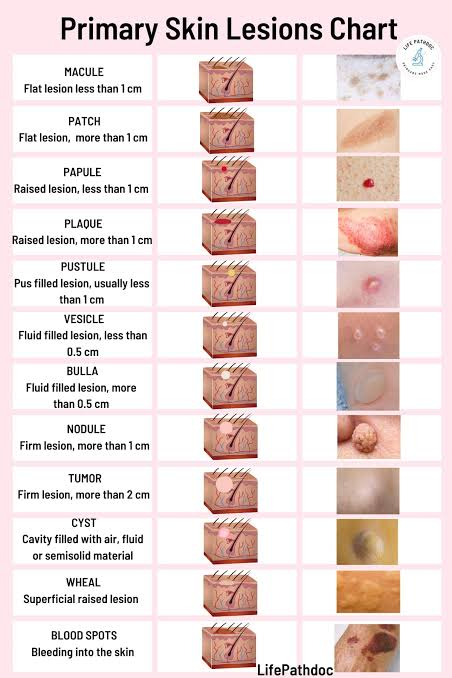
Primary skin lesions:
(Primary skin lesion)
A primary skin lesion occurs as a direct result of a disease condition.
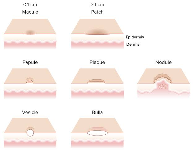
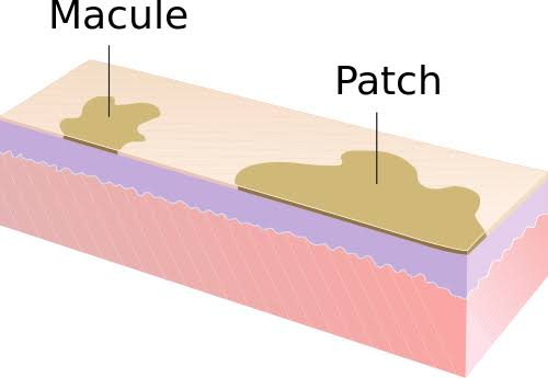
Macule:-
A macule is flat and non-palpable.
Different colors brown, white, purple and red are found in macule.
The size of the macule is less than 1 cm.
Example: follicles
2.Patch:- A patch is a macule-like structure (flat and non-palpable) but its size is more than 1 cm.
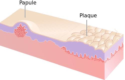
(3) Papule:-
A papule is an elevated palpable solid mass. whose size is less than 1 cm.Example: Verts
(4) Plaque:-
A plaque is a papule-like structure (elevated palpable) but larger than 1 cm in size.
Example: Psoriasis, keratosis
(5) Nodules:-
A nodule is an elevated palpable solid mass. which extends deep into the dermis layer. Nodules range in size from 0.5 to 2 cm. When the tumor size is more than 1-2 cm.
Example: Lipoma and Carcinoma
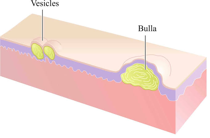
(6) Vesicle
A vesicle is an elevated palpable fluid-filled mass that is round or oval in shape. Its wall is thin and translucent.
Vesicle size is less than 0.5 cm.
Example: Herpes simplex, herpes zoster
(7) Bulla:-
A bulla is a vesicle-like structure but larger than 0.5 cm in size.
Example: Pemphigus
(7) Wheal:-
A wheel is a radius area with an elevated, irregular border.
Example : UTICARIA, insect bait
(8) Pustule:-
A pustule is a vesicle containing pus.
Example: Acne, Impetigo, Furuncle
(9) Cyst :-
A cyst is an elevated, fluid-filled semi-solid mass overlying the subcutaneous tissue and dermis layer.
Example: Sebaceous cyst
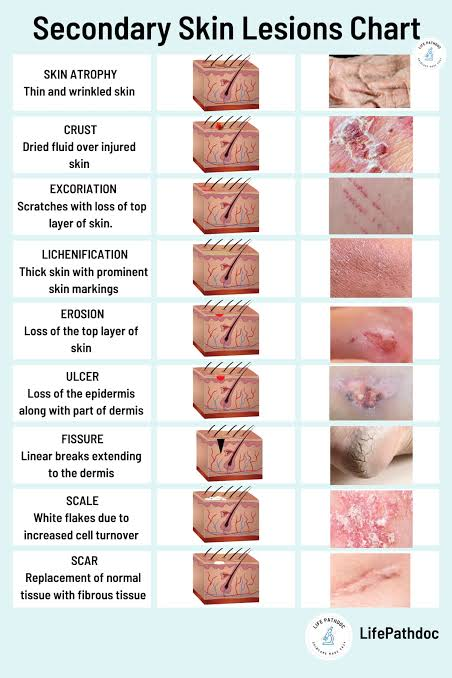
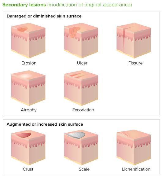
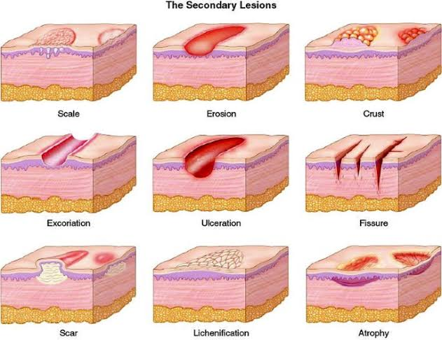
Secondary skin lesions
(Secondary skin lesions)
Secondary skin lesions develop from primary skin lesions. or occur as a result of scratching, infection, or trauma.
1) Erosion:
In erosion, the epidermis, the superficial layer of the skin, is lost or broken down. Also that area is found moist and depressed.
Example: Rupture vesicles and scratchmarks
2) Ulcer:
Epidermis and dermis layers are affected in ulcers. That is, loss of deep epidermis layer and necrotic tissue is seen
Example: Pressure ulcer
3) Fissure:
A linear break is seen on the skin in a fissure. That is, an incision is seen on the skin. which extends to the dermis layer. Fissures appear due to excessive drying of the skin and are painful swellings.
Example: Athlete’s foot
4) Scales:
Silver or white flakes appear on the skin due to the accumulation of dead epithelial cells under the skin.
Example: Dandruff, Psoriasis
5) Scar:
A mark seen on the skin after a wound or lesion has healed is known as a scar. Scars occur due to the replacement of dead tissue by connective tissue. Young scars are red or purple in color while mature scars are white in color.
Example: Surgical incision and healing wound
6) Keloid:
A keloid is an elevated, irregular, red hypertrophy scar that occurs due to excessive collagen formation during healing time.
Example: A keloid on the ear due to a surgical incision
7) Atrophy:
In atrophy, the skin becomes thin, dry and transparent, making the underlying vessels visible. Which is seen due to loss of collagen and elastin.
Example: Aged skin and arterial insufficiency
8) Lichenification:
The skin becomes thick and rough due to repeated, rubbing irritation and sketching.
Example: Contact dermatitis
9) Crust:
Crust is the dry exudate (crust) on the screen surface. Which serum is made up of blood and pus.
Example: Exudate left after vesicle rupture
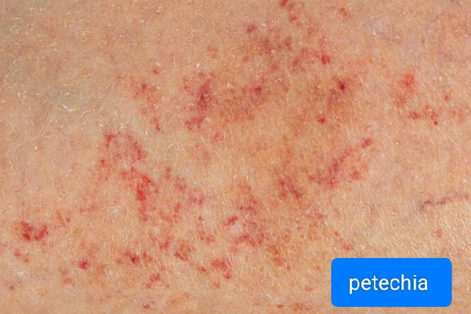
Vascular lesion:
(vascular lesion)
1) Petechia:
Petechiae are flat, round shaped, red or purple colored spots. Its size is found to be 1-3 mm. Which is seen due to blood leakage in the skin.
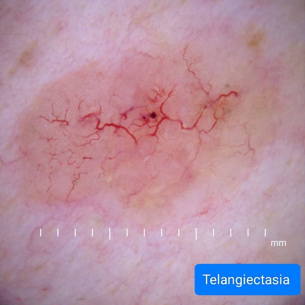
2) Telangiectasia :
Telangiectasia is also known as Venus star. Telangiectasia is a spider like bluish or red colored structure. Which is seen due to the dilatation of superficial vessels and capillaries in the skin.
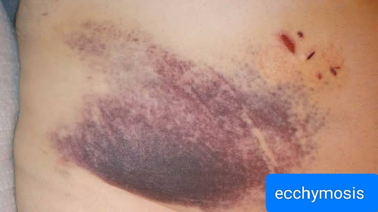
3) Ecchymosis:
Ecchymosis is a macular lesion of round or irregular shape. Which is larger in size than petechia. In ecchymosis, the skin is bruised in color due to the collection of blood under the skin.
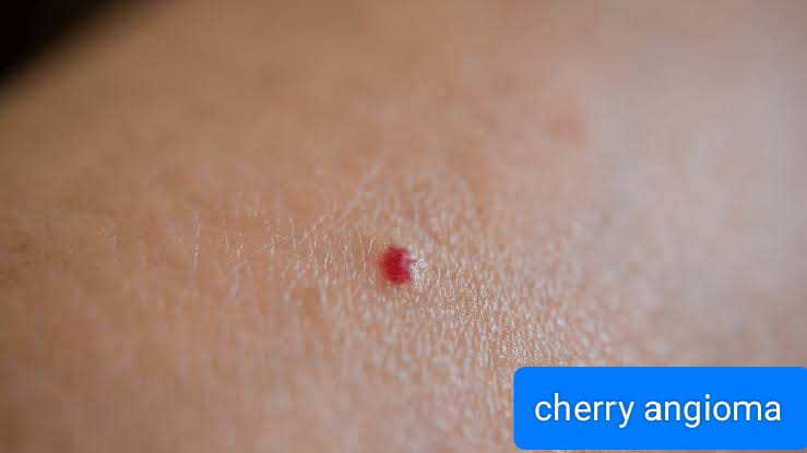
4) Cherry angioma:
Cheryengioma is a papule like structure of round shape, red or purple in color. Which is made up of small blood vessels. Which is seen due to age related skin changes, which are more common in the trunk and extremities.
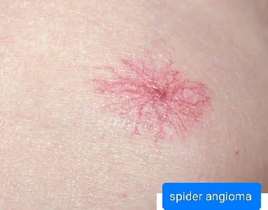
5) Spider angioma:
Spider angioma is a flat vascular lesion of bright red color. Which is seen due to dilation of the blood vessels under the skin. Spider angioma is seen during liver disease, vitamin B deficiency and during pregnancy.
Physical Examination :-
Palpation:
(palpation)
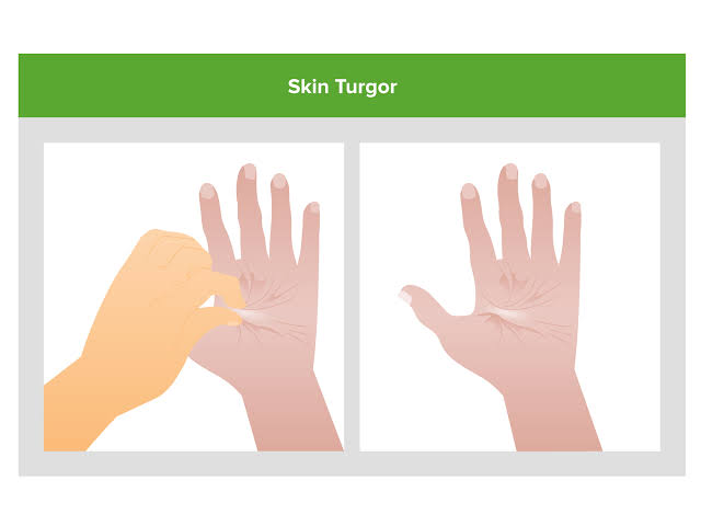
Skin temperature, turgor, mobility, moisture and texture are checked with the help of palpation.
Checking the patient’s body temperature and pulse.
Recesses and lesions in the skin can be identified by palpating the skin texture.
Use gloves when palpating lesions and lesions.
Before palpating the racemes, gently stretch the skin there to reduce redis color and observe the races more evenly.
Knowing the texture, shape and border of the lesion while palpating it.
Palpate lymph nodes in the skin.
Checking skin turgor and mobility.
Skin turgor and mobility refers to the elasticity of the skin.
Skin turgor decreases in old age.
Screen turgor flooding is seen in patients with dehydration.
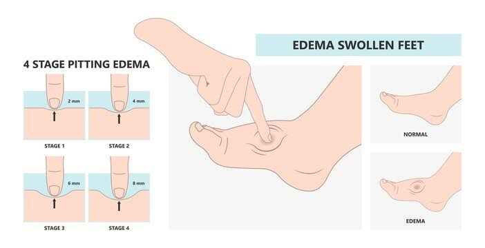
Finally checking whether edema is present in the leg or not.
Edema occurs due to excess fluid accumulation in the body.
When pressure is applied to this edema, if a pit or indentation is seen in it for a long time, it is called pitting edema.
Pitting edema is usually seen in the feet and ankles.
Pitting edema can be divided into grades as follows.
Grade : 1
Slight pitting is observed i.e. 2 mm deep pitting is observed and no distortion is observed.
Grade : 2
Deep pitting is observed i.e. 4 mm deep pitting is observed and no distortion is observed.
Grade : 3
Pitting is seen up to 6 mm deep and swelling is seen in the extremities.
Grade : 4
Pitting is observed up to 8 mm deep and accompanied by distortion.