ENGLISH SURGERY SPECIAL INSTRUMENTS VIVA-TABLE.GNM-SY
INSTRUMENTS USED FOR ENDOSCOPIC SURGERY
OESOPHAGOSCOPE
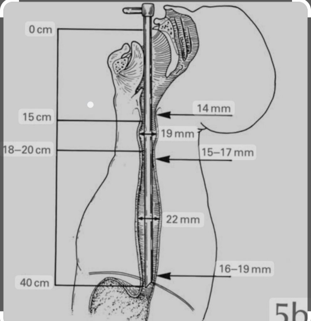
This instrument is used for examination of the inside of the esophagus.
1- Diagnostic use-Diagnostic use
Cancer in the esophagus
Cardiospasm
Benign stricture of the esophagus-benign stricture of the esophagus
Pharyngeal pouch-Pharyngeal pouch 2 – Therapeutic use-therapeutic use
To remove the foreign body-to remove the foreign body
To remove a benign tumor
(Esophagus) Taking a biopsy sample from the esophagus for histopathological examination.
How is esophagoscopy performed? -How is esophagoscopy performed?
Place the patient in supine (upright) position.
Administer general anesthesia and perform endotracheal intubation.
Elevating the patient’s neck and shoulders, lubricating the instrument, the instrument is then guided by the endoscopist’s left hand over the dorsum of the tongue and past the fairings.
The instrument is guided past the endotracheal tube, with the head extended slightly upward.
Proceeding from the larynx (vocal box), the crico-pharyngeal fold is now visible.
Extending the head and neck forward, this instrument exposes the middle thoracic esophagus.
This instrument can be seen up to 40 cm inside which is the cardiac end.
Anatomical sites of the esophagus
1 – Crico fairings from front teeth 15 cm
2 – Cross 25 cm from front teeth to arch of aorta – Cross 25 cm from front teeth to arch of aorta
3 – Cross left bronchus from front teeth 28 cm – Cross left bronchus from front teeth 28 cm
4 – 40 cm from front teeth to level of diaphragm – 40 cm from front teeth to level of diaphragm
Complications
Immediate
Esophagitis (infection in the esophagus)
Mediastinitis-Mediastinitis
Perforation
Injury to lips and teeth
Bronchitis in children-Late-Late
Esophageal stricture-esophageal stricture
Tracheoesophageal fistula
BRONCHOSCOPE-bronchoscope
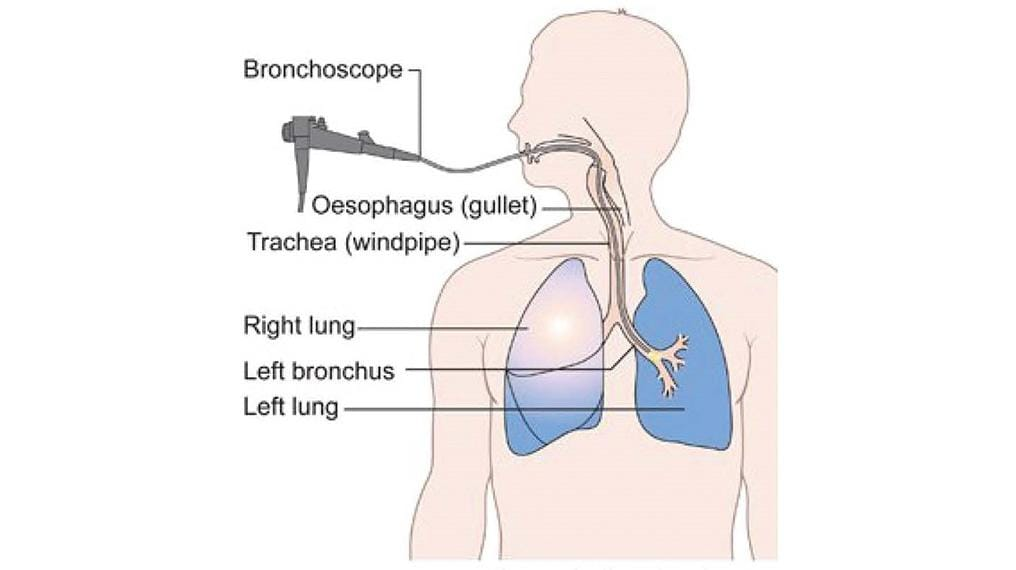
This is a hollow metallic tube. Its distal end opens into the side wall.
-Why are there holes in the side wall of the bronchoscope? Why are there holes in the side wall of the bronchoscope? To take respiration through a breathing tube that is not occupied by a bronchoscope.
uses It is used for both diagnostic and therapeutic purposes.
-Diagnostic use-Diagnostic use: in case of tracheobronchial pathology; In bronchogenic carcinoma, hemoptysis of undiagnosed pathology etc.
Therapeutic Uses:-Therapeutic Uses: For removal of foreign bodies, for biopsy from tracheobronchial tree and bronchial aspiration.
How is a bronchoscopy performed? The procedure is usually performed under local anesthesia in adults and general anesthesia in children. This instrument is inserted into the trachea through the rima glottis with the help of a laryngoscope. After proper adjustment of the patient’s head, the bronchoscope is slowly passed from the trachea to the serina. After that examination of different parts of the bronchial tree is done.
GASTROINTESTINAL FOBEROSCOPES (INCLUDING GASTROSCOPES, DUODENOSCOPES AND COLONOSCOPES)
There are three types of instruments based on their viewing angle:
1 – Forward or oblique (foreoblique) viewing for examination and biopsy. This is convenient for examining the esophagus, stomach and upper part of the duodenum.
2 – It is viewed from the side or lateral side for duodenal examination, biopsy and operative procedures. This is necessary when performing a thorough examination of the esophagus, stomach, duodenal, and biliary system. It includes the fundus of the stomach and the duodenal bulb.
3 – Forward viewing through two channels in complex operative procedures. It has a larger diameter than the other two and has channels of suitable dimensions for operating instruments. A lot of procedures are done in this.
UPPER GASTROINTESTINAL ENDOSCOPY-Upper gastrointestinal endoscopy
Objective – To perform examination and biopsy of esophagus, stomach and duodenum.
Position- Left lateral or seating
Anesthesia – Anesthesia- sedation with intravenous (IV) diazepam
Instruments and equipment required
A convenient fiberscope (e.g. forward or side view)
Fiberoptic light source-Fiberoptic light source
Mouth guard-Mouth guard
Biopsy forceps
Tongue depressor-tongue depressor
Pharyngeal spray-pharyngeal spray
Topical anesthetic, e.g. 1% lignocaine- -topical anesthetic ,e.g.1% lignocaine
Lubricant, e.g. lignocaine gel-lubricant, e.g. lignocaine gel
Esophageal bogies and tube sizes 16 and 18
Medical vacuum-Medical vacuum
Procedure
Sedate the patient with intravenous diazepam. -Sedate the patient with intravenous diazepam.
Plastic mouth guard-Plastic mouth guard
Lubricating the Fibroscope
Passing the endoscope from the mouth to the tongue
Ask the patient to swallow
Do not use force
Examination through the eye by inserting the instrument into the esophagus
Entering the instrument under observation into the stomach. – Entering the instrument into the stomach under observation.
LOWER GASTROINTESTINAL ENDOSCOPY – lower gastrointestinal endoscopy
Objective- To visualize, examine and biopsy the anterior part of the colon.
Position – Left Lateral
Anesthesia – Intravenous diazepam with sedation
Procedure
Sedate the patient with diazepam
Inserting a lubricated colonoscope
Viewing the rectum through the eyepiece
Advance the colonoscope under observation through the middle of the colon
Examination of the entire colon while withdrawing the instrument
It can also be used to obtain biopsy or cytology specimens.
LAPAROSCOPIC SURGERY – Laparoscopic surgery
Objective- Objective
Perform under observation operation in Abdominal Cavity.
Perform procedures using laparoscope and other related small instruments.
This surgical procedure is advantageous over open surgical procedure because;
Operative trauma is reduced.
Pain and discomfort after the operation is reduced
The process of patient recovery and return to activity is quick
A shorter hospital stay also reduces costs.
Today many procedures are performed with a laparoscope:
Gynecological surgery-gynecological surgery
General surgery-General surgery
Goal Bladder-Goal Bladder
Appendix-Appendix
Intra-articular surgery-Intra articular surgery
Anesthesia-Anesthesia
Local anesthesia or general anesthesia for intraluminal surgery
position – supine (upright)
procedure
Make a small 0.5 cm incision below the umbilicus
Removal of abdominal wall with thumb and finger and insertion of Vares needle
The rectus seat and peritoneum are entered through the needle and air is aspirated through it.
The needle can move freely and aspiration does not draw blood
Connect the inflator and the pressure on the gauge should be less than 4 mm
Start the gas flow at the rate of 1 liter per minute and stop it after 2.53 liters have been inflated.
Perform liver percussion (measure liver size) to confirm loss of liver tenderness.
Withdrawal of Veres needle and insertion of 10 mm trocore and cannula
Remove the trocar and insert the laparoscope, connecting the inspirator to the laparoscope
Inspecting the dominal KVT with an eyepiece
To make necessary openings related to procedures.
STERILIZATION OF ENDOSCOPE-Sterilization of endoscope
Endoscopes that are inserted through a trocar opening into the body CVT need to be sterilized using steam or ethylene oxide, parasitic acid, gas plasma if possible. Instruments that have come in contact with the mucosa can be disinfected using 0.2% glutaraldehyde for 45 minutes.
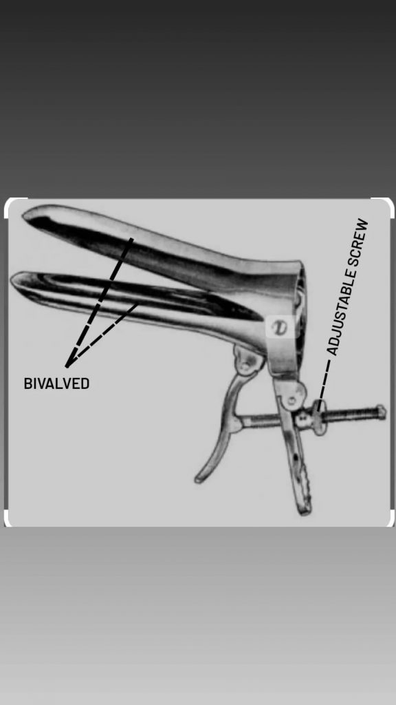
CUSCO’S SELF RETAINING ADJUSTABLE BIVALVED VAGINAL SPECULUM – CUSCO’S SELF RETAINING ADJUSTABLE BIVALVE
Identification point-identification point
This instrument has two hinged blades that can be opened and closed
It has two handles attached to each blade
It has an adjustable screw between the two handles, by adjusting the screw the handles and the blade can be opened or closed accordingly.
Use
1- To carry out regular examination of vagina and cervix
2- To examine the cervix in the following cases
cervical discharge or erosion-cervical discharge or erosion
cervical bleeding-cervical bleeding
Hypertrophied elongated cervix
genital prolapse
3 – to reach the cervix
To apply medication-to apply medication
Taking a swab for culture and sensitivity test – taking a swab for culture and sensitivity test
Obtain a biopsy specimen
To perform electrocautery in the cervix
Advantages
Since this is self retaining, no assistant is required
This allows both walls of the vagina to be grasped, thus eliminating the need for another instrument
This gives a clear view of the cervix
It can be adjusted based on the size of the vagina.
Disadvantages
The limited available space at the edges of this instrument introits is a drawback in many operative procedures.
How is it introduced?
Placing the patient in lithotomy position
Taking antiseptic and aseptic precautions – Taking antiseptic and aseptic precautions
Separating labia between left thumb and forefinger
Hold the blade of a sterile and lubricated instrument closed with the right hand
Slowly introduce the transverse diameter of this instrument towards the anteroposterior axis of the vagina.
Then rotate it to the right angle and adjust the screw between the two handles, so that the blade opens.
Sterilization
Boiling
(Autoclave) (121 degree Celsius temperature, 12 bar pressure, 30 minutes).
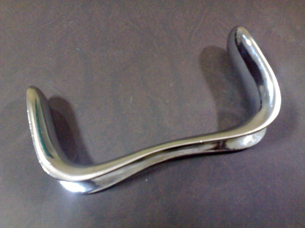
SIM’S SPECULUM
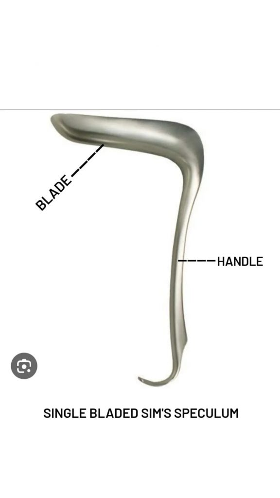
This instrument is designed to visualize the vagina and vulva simultaneously. It has two types:
1 – Single bladed
2 – Double bladed
1 – Single bladed
Identification points – Identification points
This instrument is very light and made of stainless steel
It has a handle and a blade. The blade is set at right angles to the handle
The blades are depressed in the middle which helps drainage of the secretion
2 – Double bladed sim’s speculum – double bladed sim’s speculum
Identification points-Identification points
It has two blades on its two poles
Its middle part is used for a grip or as a handle
Both the blades are of different sizes which is technically advantageous.
How to apply a speculum
Putting the patient in sim’s position or lithotomy position
Swab the vulva with an antiseptic solution
Performer should wear sterile gloves and be on the right side of the patient
Separating the labia with the thumb and finger of the left hand
Lubricate the instrument and hold it with the right hand, then slowly introduce its transfer diameter towards the anteroposterior axis of the vagina.
Now rotate the instrument through a 90 degree angle to bring it to the midline
position
This is an excessive left lateral position. Ask the patient to lie on the left side on the chest, with the right leg flexed and the left knee bent. Hold the patient’s left arm behind.
The patient can also be placed in the lithotomy position or left lateral position while applying the speculum.
But, sim’s position is more preferred. If any difficulty arises in this position, the patient is given lithotomy position.
uses
For examination of vulva, vagina and cervix
As an instrument in operations performed through the vaginal route
To collect material from cervix, for special investigation of vagina like cytology, histology and microscopic examination.
Advantages
The instrument is lightweight and comfortable for the patient
No complications are seen after its use
A double blade speculum is technically advantageous as it has two sizes of blades in a single instrument (smaller blade – naliparous women, larger blade – multiparous women).
Disadvantages
Since the instrument is very light, it requires a firm grip during examination
This is not a self-retaining device so assistance is required during the procedure
This instrument causes obstruction in the posterior vaginal wall, so it cannot be seen and operated.
What operative procedure does sim’s speculum not use? -What operative procedure in which sim’s speculum cannot be used
To remove the cyst from the posterior vaginal wall
Posterior colpoperineorrhaphy – Posterior colpoperineorrhaphy
To repair a recto-vaginal fistula
To repair a complete perineal tear – to repair a complete perineal tear
Before performing the speculum examination, the patient is instructed as follows
Ask the patient to empty the bowel beforehand
Ask the patient to empty the bladder before starting the examination
Do not ask the patient to go to the toilet if there is a history of urethral discharge or genital prolapse.
Basic preparation of an ideal speculum examination
The instrument should be sterilized and lubricated
Do not use antiseptic medicine when taking material for culture and sensitivity.
Good lighting should be available. -Good lighting should be available.
Sterilization
Sterilization
Autoclave
Plate and screw: Plate and scre
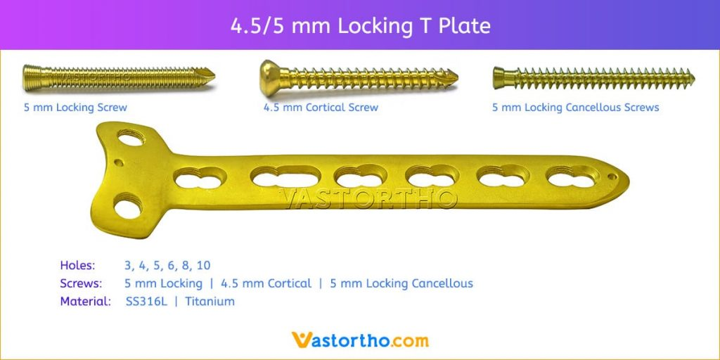
Late and screws are used to connect the internal part of the bone.
When there is a fracture in the bone, to connect the broken bone, the plate is attached to the bone and tightened by a screw, and the plate and screw are kept until the healing is complete, and after complete healing, the plate and screw is removed.
Cannula and screw: Cannula and screw:
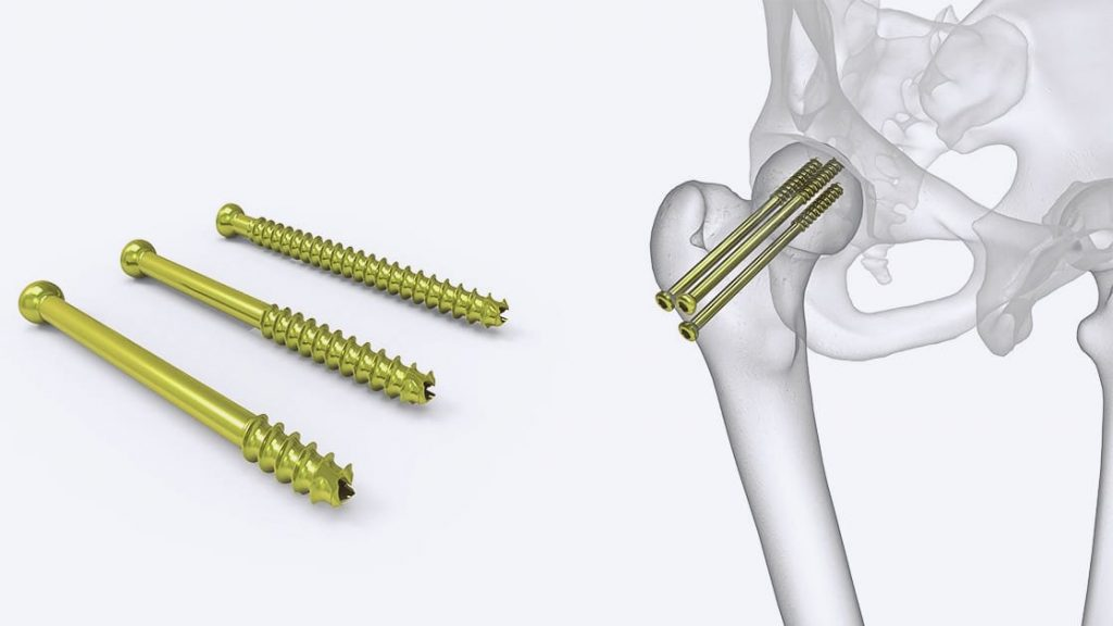
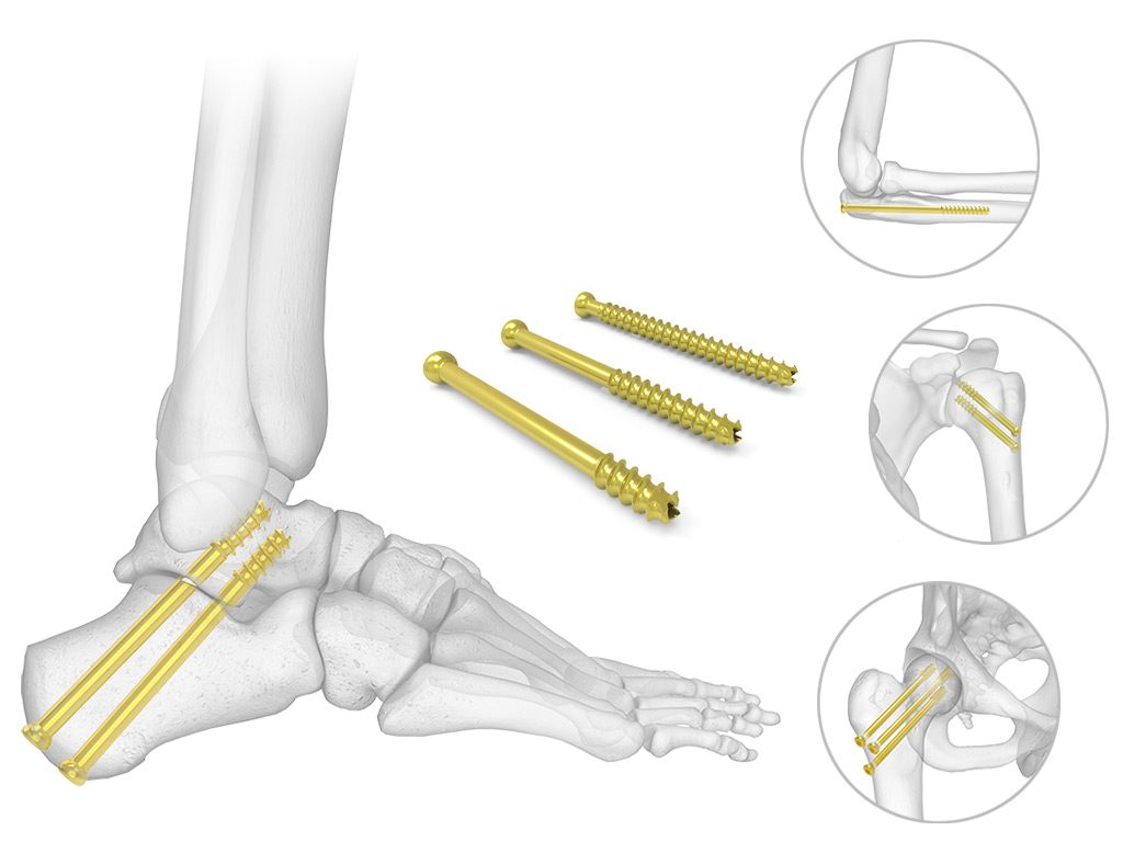
Cannulated screw is used to fix bone in orthopedic surgery when there is multiple fracture pattern.
A cannulated screw has a hollow pattern in between, due to which the guidewire is placed in the cannula.
Cannulated screw is used in bone and joint surgery to repair and secure artificial plant.
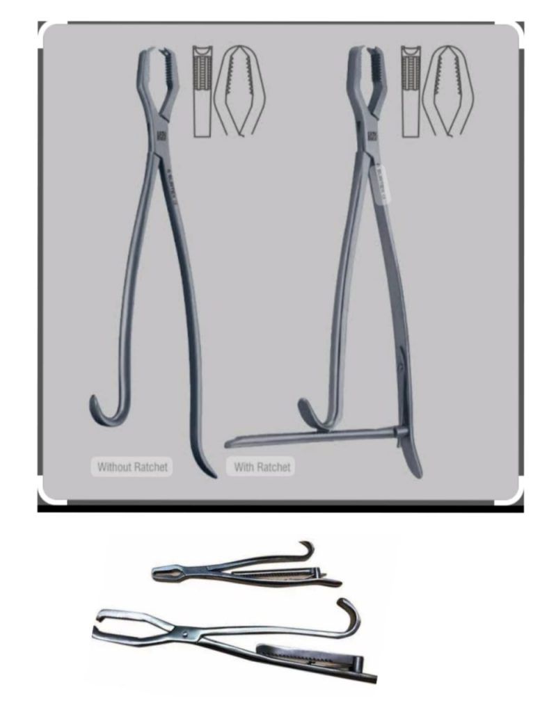
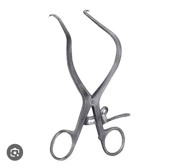
Lane bone clamp – Lane bone clamp
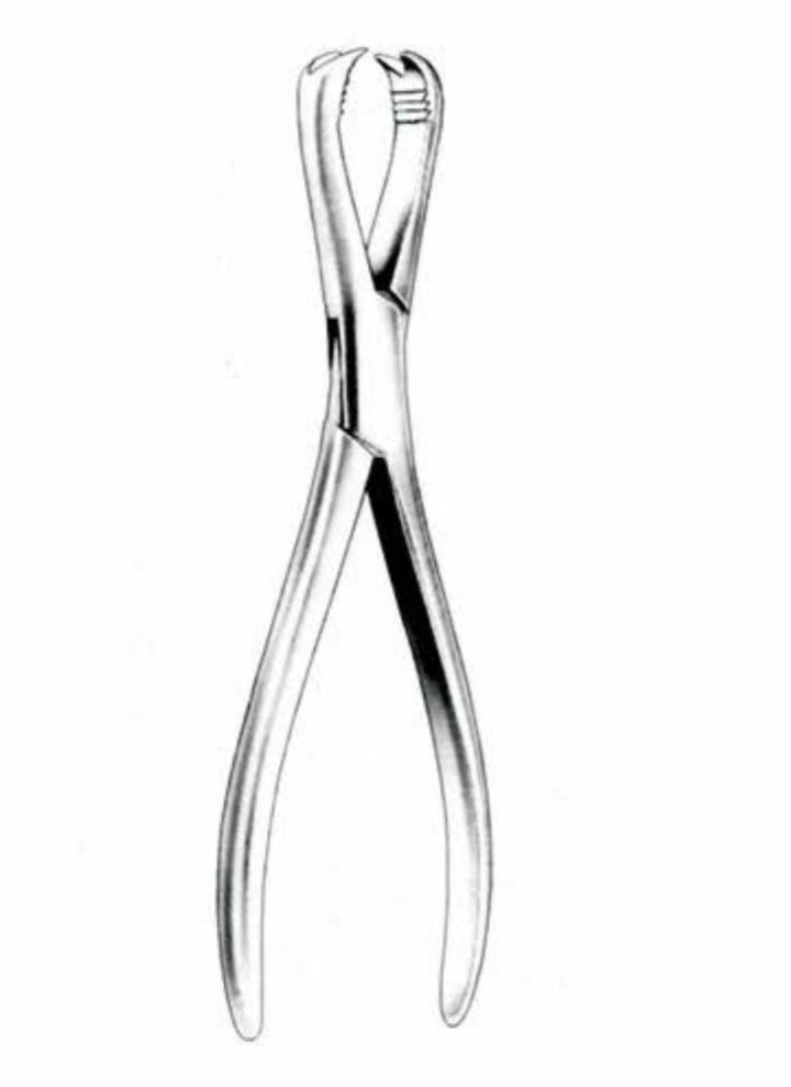
Introduction
The Lane Bone Clamp is named after its inventor, Dr. William W. Lane.
It consists of two parallel arms with serrated jaws at their ends and enables surgeons to perform complex procedures on bone with precision and control.
Uses
A lane bone clamp is a surgical instrument used in orthopedic procedures.
It is used to grasp and stabilize bone during surgery such as joint reconstruction and fixation of a fracture.
Its primary function is to hold the bone in place.
Prevents the surgeon from bone misalignment during the procedure. So that there is no change in the specific process.
Indications
1).Fracture fixation-.Fracture fixation :-
To hold the fractured bone in place during surgical repair.
2).Joint reconstruction-Joint reconstruction :-
such as ni (knee) or solder (pillar) procedures
Helps stabilize bone during reconstructive surgeries.
3). Corrective osteotomy:-
To help realign the bone by securing the bone in the desired position during corrective surgery.
4). Internal fixation:-
To support the position of screws or plates used for internal fixation of bone.
Orthopedic instruments
Rongeur-Rongeur

Introduction
A rongeur is a heavy duty surgical instrument.
It has a scoop-shaped tip with a sharp edge. It is used to access various spaces and to cut or trim specific bone and to extract bone.
Ronguere is a French word meaning rat or dog.
Uses
The rongeur is a versatile surgical instrument. -The rongeur is a versatile surgical instrument.
A medical procedure used to remove small pieces of bone or tissue.
They come in various shapes and sizes.
Commonly used in orthopedic, neurological and spinal surgery to precisely cut or grasp tissue during operations.
It is usually prescribed for various medical conditions or procedures where removal of specific bone or tissue is required.
Neurosurgery involves trimming the bone or tissue surrounding the spinal cord or nerve to remove bone fragments.
Indications- Indications
1).orthopedics-orthopedicus
Bone is cut or removed during joint reconstruction surgery, fracture repair, or corrective bone deformity.
2).Neurosurgery-Neurosurgery
Procedures such as spinal decompression or laminectomy remove fragile bone to relieve pressure on the nerve.
3).Oral and Maxillofacial Surgery – Oral and Maxillofacial Surgery
To remove small pieces of bone or to shape bone in teeth or facial processes.
4).Podiatry-Podiatry
Assisting in foot or ankle surgeries such as removing bone spurs or correcting deformities.
5). Plastic surgery-plastic surgery
To shape bone for reconstructive purposes such as facial reconstruction.
- Lawman bone clamp
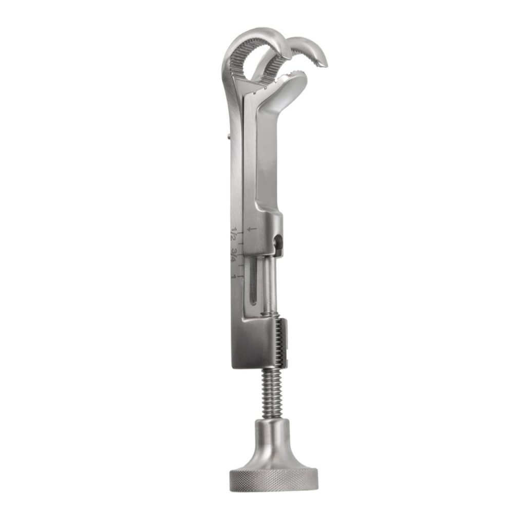
Introduction
The Loman Bone Clamp is a special surgical instrument.
It is used in orthopedic procedures to secure and stabilize the bone.
It is designed with a clamp mechanism that allows surgeons to hold bone fragments or bones together during surgery.
It comes in different sizes and configurations.
Uses
A Loman bone clamp is used in orthopedic surgery to hold a bone or bone fragments together during the healing process.
This clamp is an essential tool for maintaining bone stability and proper alignment during surgery or when applying fixation such as plates or screws.
indication
1). Fracture fixation
2). Joint reconstruction
3).Bone grafting-Bone grafting
4).osteotomy-osteotomy
5).Corrective surgery-corrective surgery
Orthopedic surgeries like etc. require this instrument.
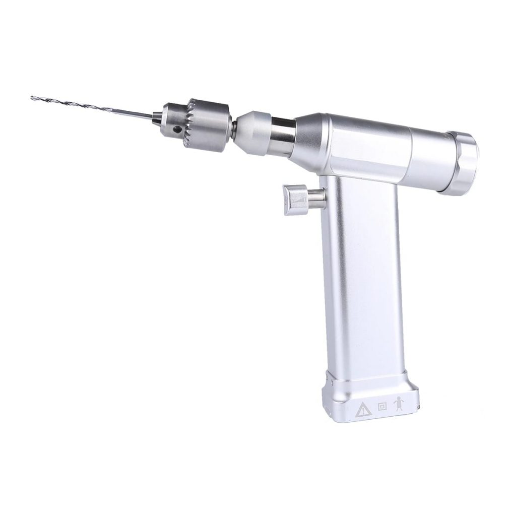
Bone drill
Or
Orthopedic twist drill-Orthopedic twist drill
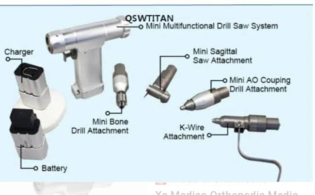
ndication-indication
Orthopedic surgery-orthopedic surgery
Craniomaxillofacial surgery.-Cranio maxillofacial surgery.
Ear, Nose, Throat Surgery.-Ear, Nose, Throat Surgery.
A bone drill is used to drill holes in the bone and insert nails and screws.
Sterilization Sterilization: Autoclave
Precautions
To check the part of bone drill before use.
Use with caution in surgery of elderly, osteoporosis and diabetic patients.
If the cutting speed is high, there is a risk of bone damage and cracking, so use it at a regular speed.
Complications
Osteonecrosis of tissue. -Osteonecrosis of tissue.
Micro crack.-Micro crack.
Break down the thrill-break down of thrill
This instrument is made of stainless steel. -This instrument is made of stainless steel.
bone saw – saw the bone
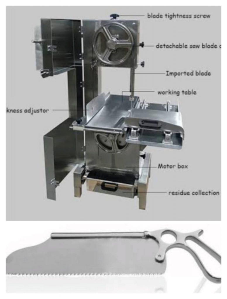
parts
handle
blade
Adjustment Mechanism: Adjustment mechanism: so that we can control the depth and angle according to the surgery.
Power Source:-Power Source: Power source is electric and pneumatic motor.
Use
This is a surgical instrument that is used to cut small and large bones. So as to get the best surgical outcome.
Like common orthopedic surgery
Joint replacement
Fracture repair-fracture repair
osteotomy.-Osteotomy.
The chosen subsequent is implanted by cutting the bone.
A bone saw cuts the bone with minimal damage to the bone’s surrounding tissue.
Sterilization-Sterilization
Autoclave (by removing the blade)
Sterilization is done by applying moist heat through saturated steam under pressure. This is the most common method.
Special features-Special features
Power source off-Power source off:
Some bone cutters require electric power. While some bone cutting requires battery power, the option is available, providing flexibility in the surgical setting.
Adjustable depth or angle– Adjustable depth or angle: In a surgical procedure, the surgeon adjusts the depth and angle depending on the surgery.
Disposable blades: Some cases have disposable blades to reduce the risk of cross contamination.
Safety feature: Modern bone saws have safety features. Like a blade guard to prevent accidental injuries.
Bone curette-Bone curette
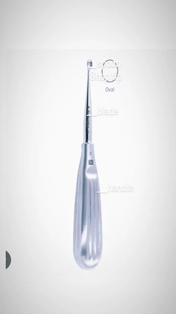
Parts name: Name of the parts
handle : By which it is held.
blade: The blade has a spun sap tip. Which is used for scraping (rubbing), and cleaning the tissue of the bone surface.
Use-Use This is a surgical instrument used for scraping and removing bone and tissue. Used in the following conditions.
=Bone grafting.-Bone grafting.
Treatment of certain bone conditions
= To remove infectious tissue from bone. (debrinement) To remove infective tissue from bone. (debrinement)
= Tumor excision-tumor excision
Remove cyst from bone
= Orthopedic surgery like irregularity or abnormality in bone. Orthopedic surgery like irregularity or abnormality in bone.
=Dental Procedures-Dental Procedure
Studylization:
Autoclave.-Autoclave.
Type of Bone Curate-Type of Bone Curate
Volk Man Curate-Volk Man Curate
Lucas Curate
Spoon curate
Diamond Curate-Diamond Curate
rongeur curate
Pituitary curettage-pituitary curettage
The choice of curettage depends on the surgery.
Complications
-infection.-infection.
-Damage Surrounding Structure.-Damage Surrounding Structure.
-Risk of building.-Risk of building.
Retractor (Orthopedic)
Hohmann retractor
Hohmann retractor is an orthopedic instrument. It minimizes tissue damage during surgery. It is overused in invasive hip and knee surgery.
Retracts the tissue around the margin of the joint.
Sterilization-Sterilization
=Steam autoclave only.-Steam autoclave only.
=270 °f for 4 min and dry time 30 min. -270 °f for 4 min and dry time 30 min.
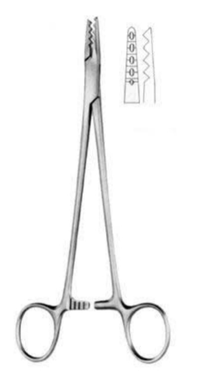
Lane bone holding forceps – Lane bone holding forceps
Use:
This is an orthopedic retractor. It is commonly used to grasp, hold and mobilize the fractured bone fragments.
To bring it back to its original anatomical position.
Sterilization
= Common Autoclaving Method – Common Autoclaving Method
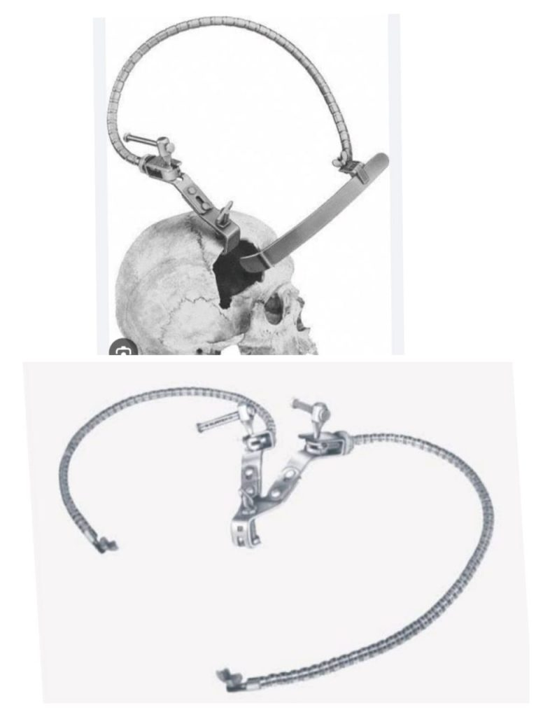
KIRSCHNER WIRE – Kirschner wire
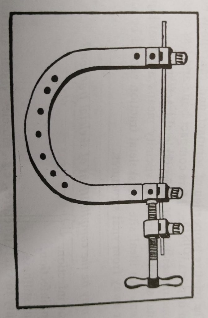
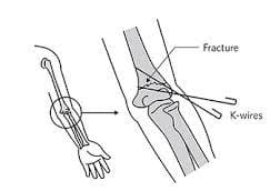
1909 by Martin Kirscher This wire is a thin, high tempered steel wire, less than 1 mm. It has a ‘U’ shaped striper and the ends of the striper are made very stiff and rigid by means of a ‘stretcher’ and a ‘handle’.
It is used to reposition small bone fractures, bone fragments.
Apart from this, it is used in orthopedic and other types of medical and veterinary surgeries.
This advantage can be obtained from any wire, even cut from bone. The wire can also become loose and infection can occur from friction with the strip.
Care- care
Do not let it soak in tub or other water, be careful while bathing.
Apply soap on the feet, pass water and dry the feet and clean the pin evenly with a towel.
Proper care of the pin, keeping it clean and protecting it with a clean dressing, keeping it safe with tape and wearing stiff shoes after the operation.
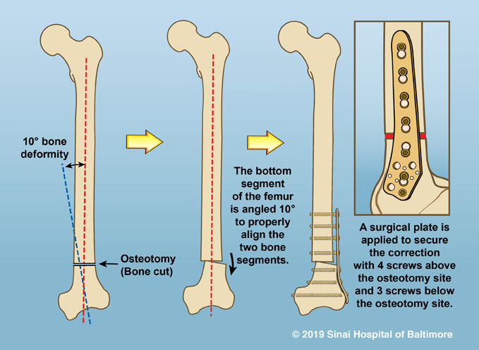
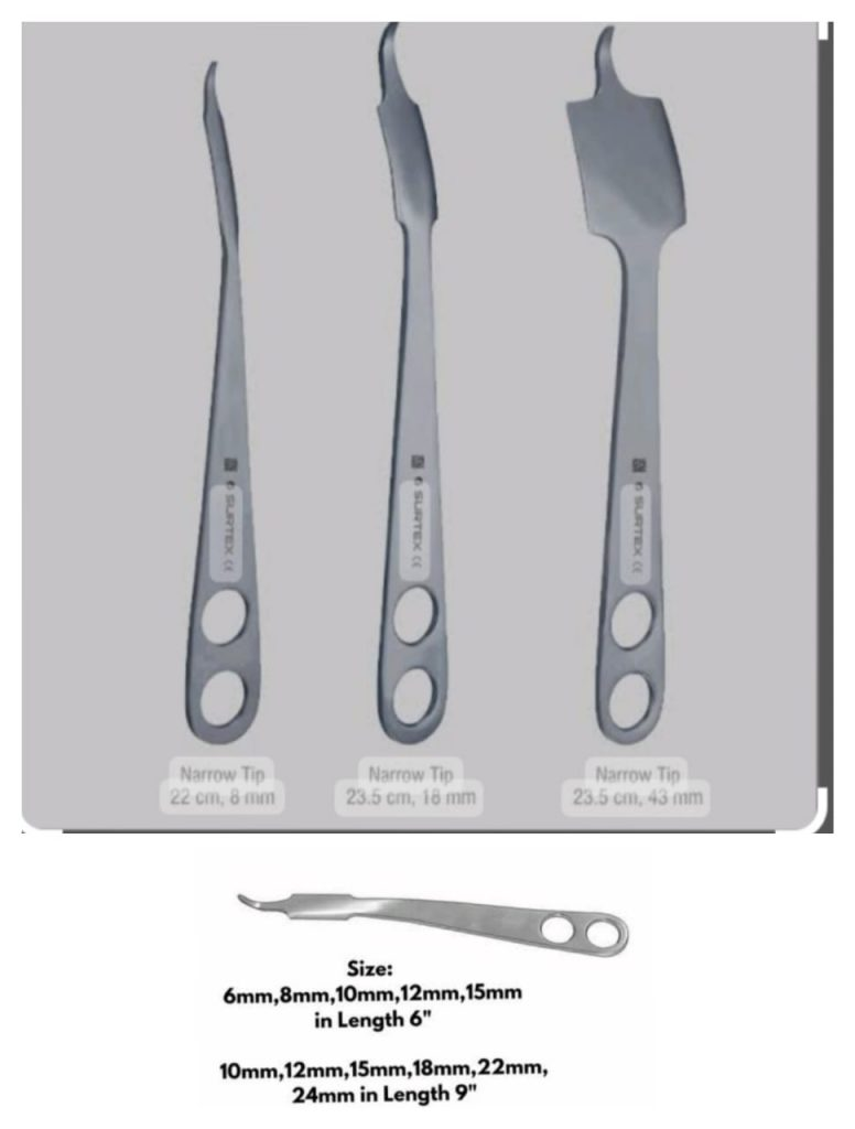
OSTEOTOME– Osteotome
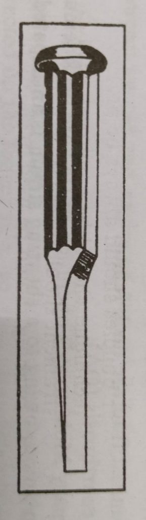
This instrument is similar to a chisel but its cutting edge has a gradual beveling on both sides.
It is used during osteotomy. Osteotomy means to correct the deformity caused by ankylosis by cutting the bone.
Type of osteotome
linear or transverse osteotomy
wedge or cuneiform osteotomy
TROCAR AND CANULA -type of osteotome
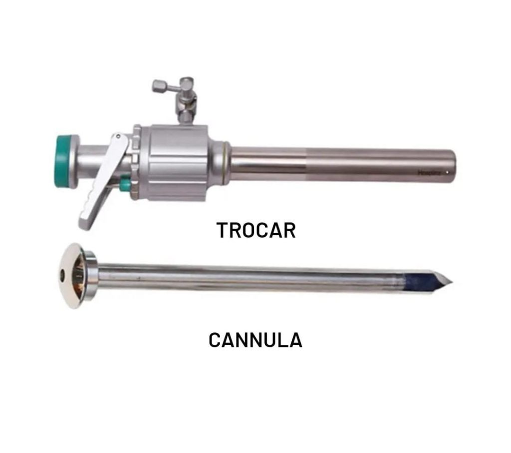
Fluid out of any KV or bag
Design instruments such as trocars and cannulas for extraction
have been done.
Spencer well’s ascitis trocar & cannula is mostly used in medical procedures like abdominal parasynthesis.
Identification point – identification point
A cannula is a hollow tube open at both ends.
Its base end is slightly larger.
The cannula is inserted through a needle, which is a metal rod with a solid, flat base and a sharp tapering tip.
After puncture, the trocar is withdrawn by its handle and a hollow cannula is left at the site to drain the fluid.
uses
To relieve shortness of breath in cases of ascites where there is a large accumulation of fluid.
Due to the improvement in renal function after parasynthesis, diuretics can work better; Drain a small amount of fluid to relieve tension within the abdominal cavity.
Abdominal parasynthesis is the removal of fluid from the abdomen.
There are two methods of parasynthesis:
Slow or Continuous Drainage Method – Slow or Continuous Drainage Method
A rapid drainage method that requires a trocar and cannula. -Rapid drainage method that requires trocar and cannula.
Precautions to be taken during paracentesis
(Precautions to be taken during Parasynthesis)
Taking proper aseptic and antiseptic precautions. Taking proper aseptic and antiseptic precautions.
Urinary bladder emptying (by the patient manually or with a catheter)
Apply abdominal binder (belt) to prevent sudden drop of intra-abdominal pressure and reflex shock.
Do not drain too quickly.
Initiate clinical assessment of the patient. Initiate clinical assessment of the patient.
Sealing the wound carefully and checking for infection. Sealing the wound carefully and checking for infection.
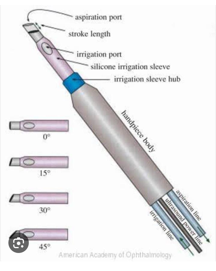
Phacoemulsification probe – Phacoemulsification probe
It is a surgical instrument used in cataract surgery.
Cloudy lenses are broken down by ultra violet vibration.
Irrigate, emulsify the lens by placing the incisors through the phacoemulsification probe.
Complications
Damage Surrounding Structure.- Damage Surrounding Structure.
= Post operative infection.-Post operative infection.
Sterilization: Autoclave.
Special precautions
Perform proper sterilization before use. -Proper sterilization before use.
Do operative check up so that separated part can be identified early. Do operative check up so that separated part can be identified early.
The correct mechanism involves inserting the probe into the eye through incisors. The correct mechanism involves inserting the probe into the eye through incisors.
Maintaining proper fluid balance. Maintaining proper fluid balance.
Minimizing Torsion Stress. Minimizing Torsion Stress.
Giving proper position to the patient during the procedure. Giving proper position to the patient during the procedure.
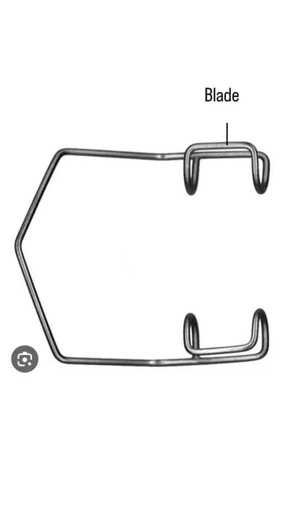
Eye speculum – eye speculum
Monitoring the temperature of the probe to prevent overheating.
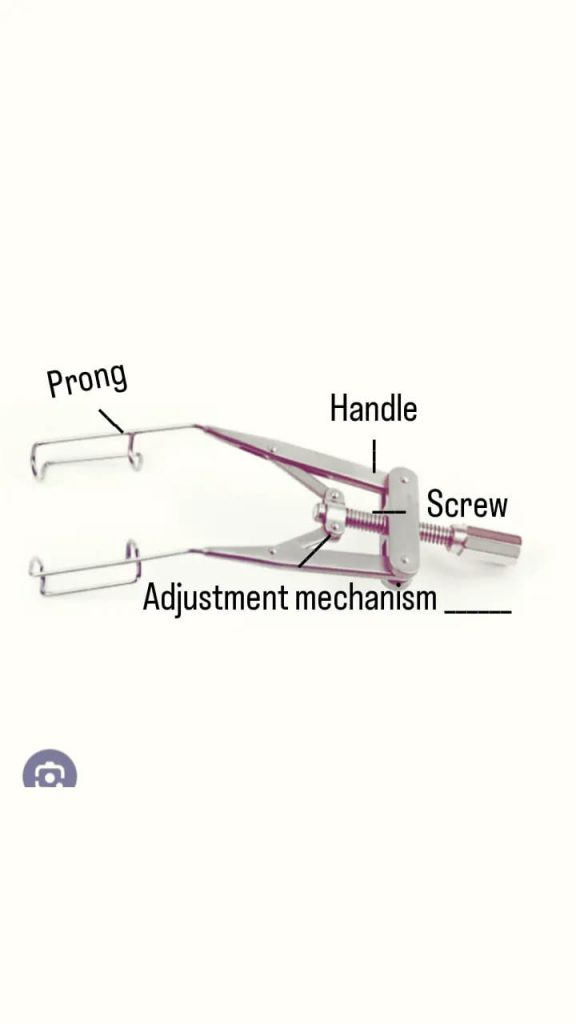
Eye (eye) speculum is such an instrument. which prevents the eye lead from closing so as to expose the ocular surface.
parts
-prong (blade)-prong (blade): hold the eye lead so that the eye is clearly visualized.
-Adjustment mechanism.-Adjustment mechanism.
–Handle.
Locking mechanism:
Some speculums have a locking feature. Which keeps the position of the blade secure.
Indications
Intra ocular operations like -Intra ocular operations like
Cataract surgery-cataract surgery
Glaucoma surgery-Glaucoma surgery
Keratoplasty-keratoplasty
during examination of i. -During examination of I.
Remove foreign body. -Remove foreign body.
Intra ocular lens implantation. -Intra ocular lens implantation.
Extra ocular surgery: –Extra ocular surgery:
Squint surgery.
Pterygium Surgery.
Also called kratz barraguer wire speculam – kratz barraguer wire speculam is also called
Studylization: Autoclave
Special precautions
Doing proper cleaning and sterilization can prevent the risk of infection.
Apply and remove the speculum in a manner that causes minimal discomfort and injury to the patient.
Selecting the size of the speculum according to the eye of the patient.
Micro surgical forceps – Micro surgical forceps
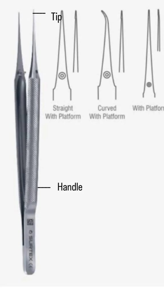
These forceps are made of stainless steel.
parts
-handle: Creates a handle to that portion.
-Tip: A specially designed part that grasps and manipulates tissue or objects.
Use
Grasp and handle tissue.-Grasp and handle tissue.
Dilates micro vessels.-Dilates micro vessels.
Remove large tissue from the surgical site for better examination. Remove large tissue from the surgical site for better examination.
Use in Ophthalmic Surgery, ENT. Surgery, Plastic Surgery, Cardiovascular Surgery, Neurosurgery.
Sterilization. Sterilization.
Autoclave
Boiling
Barraguer eye lid speculum – Barraguer eye lid speculum
This instrument is used in ophthalmic surgery.
parts
-Handle
-Blade: which retracts the eye lead.
Use
Eye lead surgery-Eye lead surgery
Blepharoplasty – Blepharoplasty
Reconstruction Procedure.-Reconstruction Procedure.
Ophthalmic Examination.-Ophthalmic Examination.
Corneal Procedures.
Prevents eye lead from closing in ophthalmic surgery. Prevents eye lead from closing in ophthalmic surgery.
A pair of blades is placed inside the eye lead conjunctiva. A pair of blades are placed inside the eye lead conjunctiva.
Sterilization-Sterilization
Autoclave.-Autoclave.
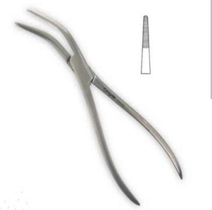
Van Buren forceps:
It is made of stainless steel.
use:
Van Buren forceps are used to grasp and mobilize bone during surgical procedures.
The tip of Van Buren forceps is useful for manipulating bone.
The serration (teeth) in the tip of this forceps prevents the bone from slipping, so that discomfort during surgery can be prevented.
Size: Size
23 cm
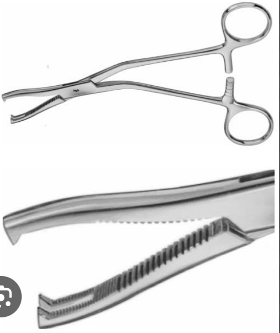
Bircher ganske forceps
It is made of stainless steel.
This forceps is a cartilage seizing forceps.
This is useful for seizing cartilage in orthopedic surgery.
Using these forceps, the orthopedic surgeon is useful for manipulating the articular surface of the cartilage and manipulating the cartilage, which is used during the joint repairing procedure.
Size: Size
20 cm
Dingman forceps: Dingman forceps
It is made of stainless steel.
Use:
This forceps is useful for holding the bone tightly during the surgical procedure and also for holding the hard tissue.
Size: Size
19 cm
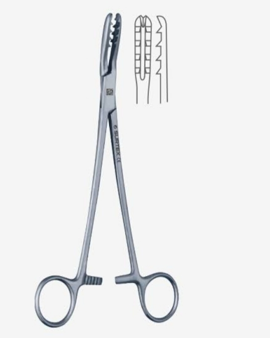
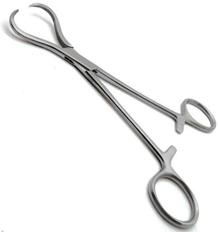
Martin bone holding forceps: Martin bone holding forceps
It is made of stainless steel.
Use:
This is a cartilage seizing forceps used in orthopedic surgery.
These forceps are used to clamp, retract and hold the tissue and cartilage of the bone surface.
The serration in the tip of the forceps strongly grasps the bone.
Size: Size
19 cm
Lewin bone holding forceps: Lewin bone holding forceps
It is made of stainless steel.
Use:
It has been specially designed to hold the bone in orthopedic surgery.
Size: Size
17.5 cm
Myringotomy knife-myringotomy knife
(Myringotomy knife)
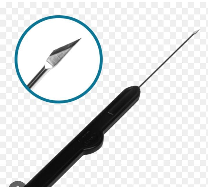
This is a sharp instrument.
Use
Myringotomy Life is used for grommet insertion to place an incision in the tympanic membrane.
Helps relieve fluid build up and pressure in the middle ear.
Indication
Middle ear infection, chronic fluid build up.
Hearing loss due to fluid build up, etc.
Size-size
Its size is commonly 2 to 5 inches.
And its specific size is used according to the age, anatomy of the ear.
Toric marker – Toric marker
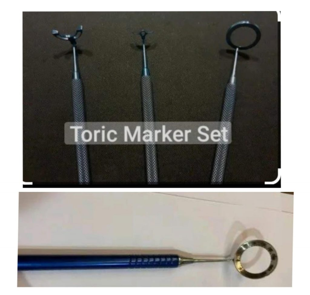
This is an ophthalmic instrument. It is used in cataract surgery.
Use
_Aligns the axis of astigmatism during surgery.
_Used in exchange of refractive lenses.
Sinuscope Sinuscope
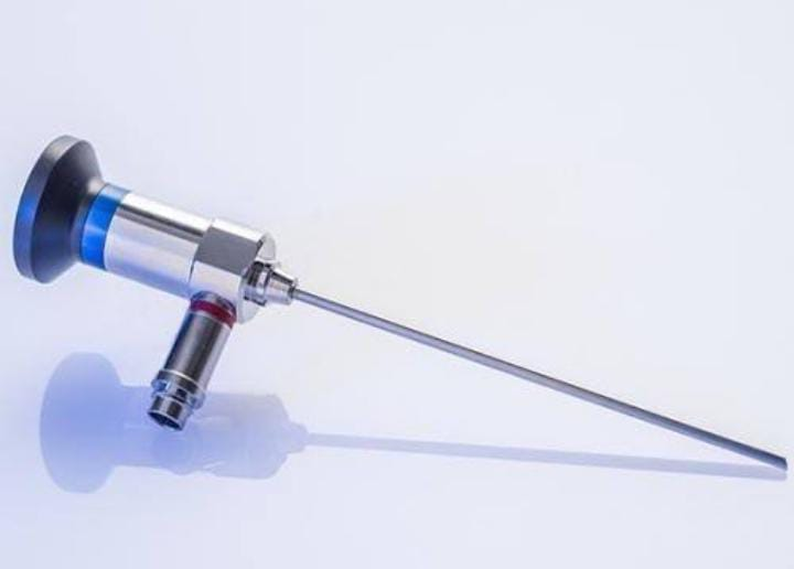
This is a fiber optic device. It is used to examine the nasal passages and sinuses.
Use
Frontal, maxillary and paranasal sinuses are used for endoscopic examination.
Used to diagnose sinus infections, polyps etc.
Types – types
Rigid (rigid) sinuscope
Flexible sinuscope
Contraindication – Severe nasal bleeding, anatomical abnormality, acute upper respiratory tract infection, unstable cardio vascular condition.
Size
Two diameters
2.7 mm and 4.0 mm
Sterilization-Sterilization autoclave or chemical disinfection.
Finochietto retractor
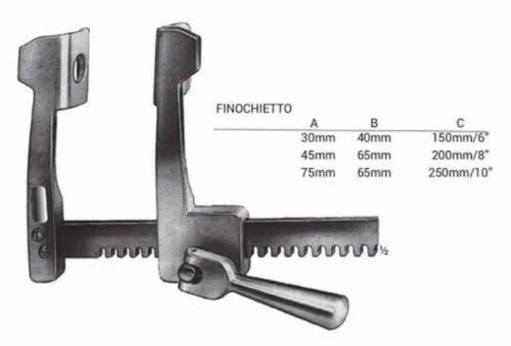
This is a rib retractor. It is used in surgical procedures.
Mainly used in chest, thoracic and cardiovascular surgery.
It holds the rib. When assessing internal organs including examining the heart and other structures.
Size- Size 65mm, 50mm, 200mm.
Sterilization =Steam only autoclave sterilization.- Only steam autoclave sterilization.
=270°f steam and 4 min. for. Dry time 30min.
Sternal saw & retractor system-Sternal saw and retractor system
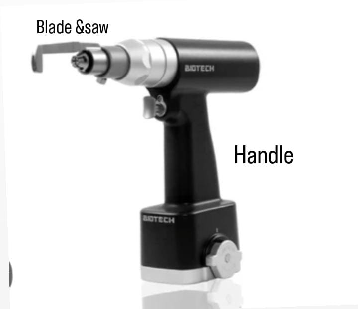
This is a bone cutter. Which is used to perform sternotomy, open the patient’s chest, breast bone or sternum spitting.
This instrument was designed by Dr. Edward p. It was introduced.
Type
- Re sternotomy handpiece (for cardiac surgery.)
- ASCO Steel Battery Operated Sternum Saw. Stainless steel sternum saw.
- biotech steel sternum
- Saw system.
Usage:
It shows a clear view of the surgical site.
Sterilization
Common method steam or autoclave.
Greenberg retractor-Greenberg retractor
This is a neurological instrument. It is used in neurosurgery.
Use
It is a surgical instrument used in neurosurgery to hold tissue and assess brain and spine procedures.
Helps the surgeon maintain a clear view.
Type
1. Self retaining retractor
_ Which does not require continuous manual assistance.
Hand held retractor
An assistant is required during the procedure.
Parts name: Blades, handle, locking mechanism
Sterilization- Common methods of sterilization are autoclaving, ethyl oxide gas and chemical disinfection.
Yasargil retractor – Yasargil retractor
This is a neurological surgical instrument.
It is named after Gazi Yasargil neurosurgeon.
It is commonly used in neurosurgery to gently hold brain tissue.
Provides a better view to the surgeon during the procedure.
Used in brain surgery, tumor removal procedures, vascular surgery and other complex neurosurgical interventions.
Use
Translabyrinthine removal, acoustic neuroma and middle fossa surgery.
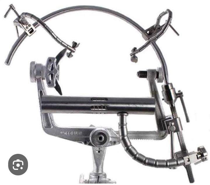
Surgical light and retractor -Surgical light and retractor
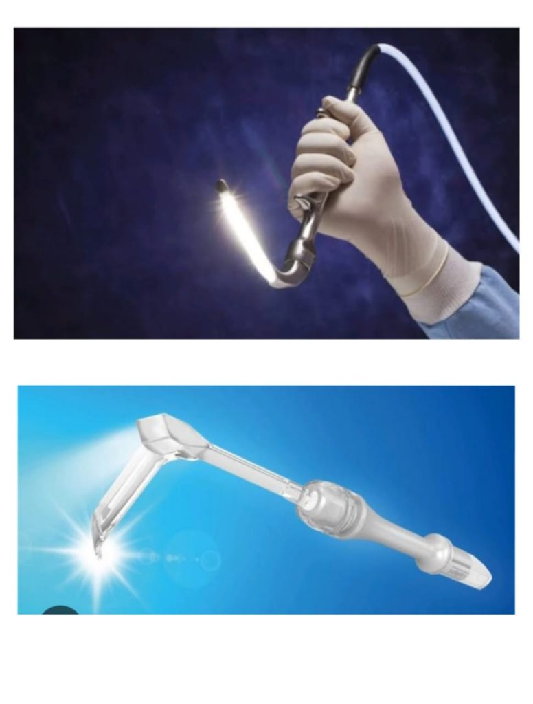
A surgical light provides visualization on the patient of the operative site in surgery during the procedure.
A surgical light provides bright light for hours. Provides light without excessive heat on patient or staff.
A surgical retractor helps to spread or separate a surgical incision or wound. Holds organs and tissues.
Retinal detachment instrument -Retina detachment instrument
Use
Detach retinal tissue to manipulate and hold.
Helps in elevating the extraocular muscles during eye surgery.
Sterilization-Sterilization Autoclave – Autoclave
Dish Advantage-Dish Advantage
infection
Bleeding-bleeding
Damage Surrounding Structure-Damage Surrounding Structure
SELF RETAINING RETRACTOR- Self retaining retractor
Weitlaner self retaining retractor -Weitlaner self retaining retractor
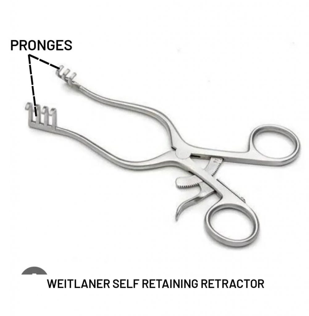
function
To open a narrow tunnel
Apply pressure around the wound wall by yourself
To leave the assistant’s hands free
Identification points Identification points
Four prongs
Its structure consists of four prongs which apply pressure on the wall of the shallow (superficial) wound. So that it can stay in proper position.
Use
Exposing the cerebellum in neurosurgery
In nerve or tendon surgery
-In Herniotomy, Plastic Surgery, Orthopedic Procedures, Mastoid Surgery etc.
Gelpi retractor- Gelpi retractor
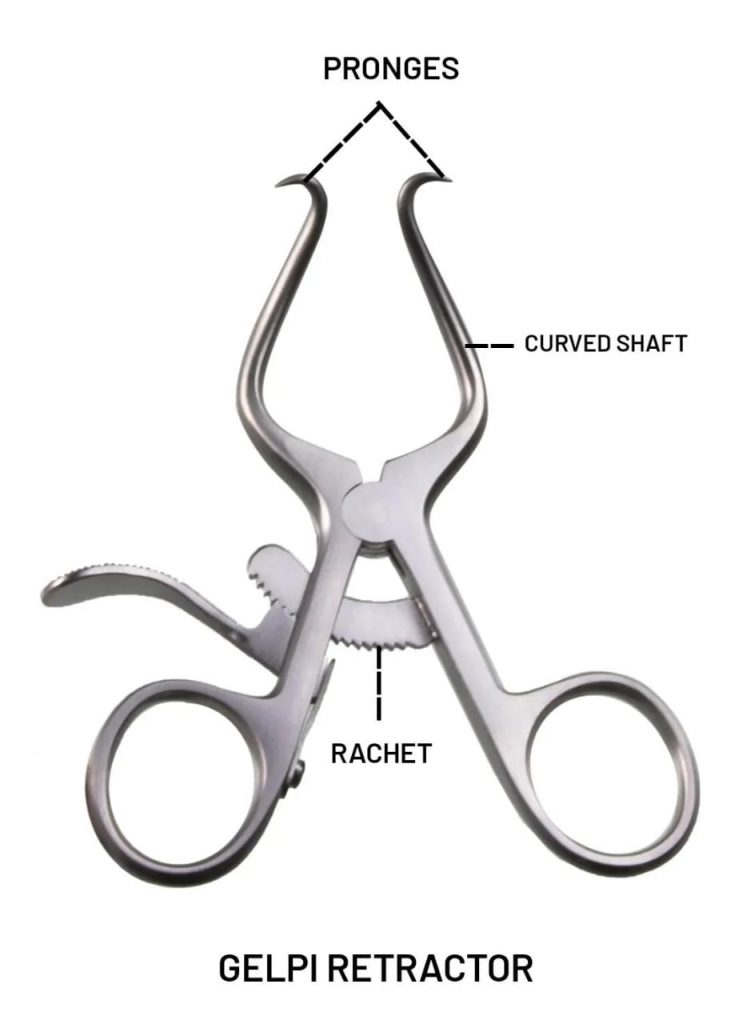
This is also a self retaining retractor
which does not require an assistant.
Identification points – Identification points
Two prongs
Both have prongs at the end of the curved shaft. The desired position can be given with the help of Rechet. Rechet works like a lever.
It is necessary to obtain the surgeon’s preference for its use.
Balfour retractor – Balfour retractor
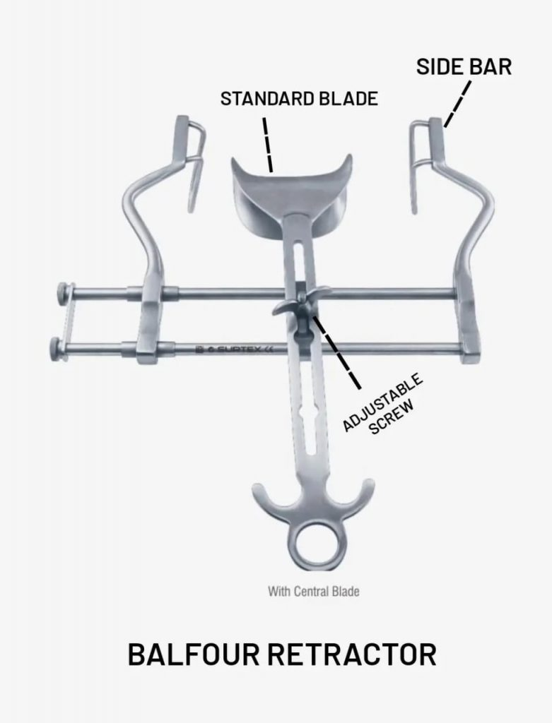
Identification points– Identification points
It has two side bars on the curved end which prevents it from slipping.
Both the side bars slide on the transverse bars
Pressure from the edges of the wound keeps the wound open
A central bar is attached to a horizontal bar to retract the corner of the wound.
Use
Used in long procedures
Saves the assistant from getting tired.
Sterilization of self retaining retractor – Sterilization of self retaining retractor
Autoclave-Autoclave
Sterilization method for all instruments:
Any article or instrument after use is taken care of by following steps.
In case of articles with blood content, all these articles are kept in sodium hypo chloride solution for 10 to 20 minutes so that the dish can be infected.
After this article is removed from sodium hypochloride, every part of it is carefully washed with running water and soapy water.
After that all these articles are dried and the article is checked for its proper working condition and if required lubricant is applied.
All these articles are then properly arranged in the drum and sent to the autoclave for reuse. Autoclave for 25-30 minutes at 121°C and 15 bar pressure, so that the article can be sterilized and safely reused.
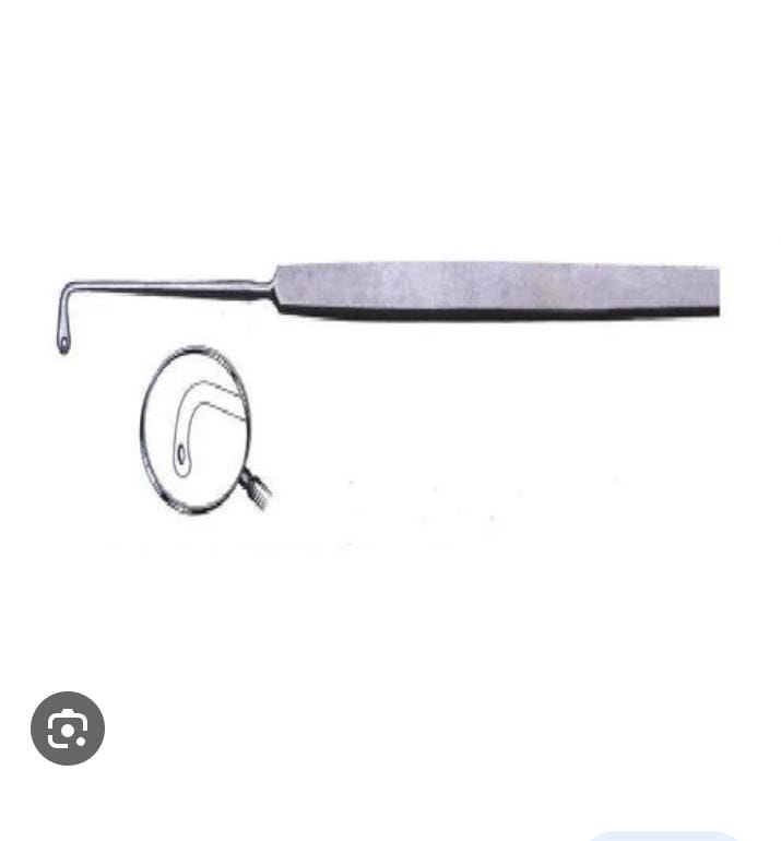
EAR CURRATTE – EAR CURRATTE
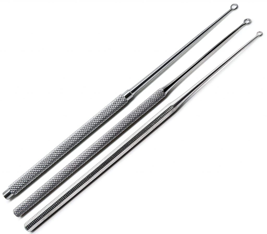
This is a surgical instrument used
It is done to clean the ear canal;
Called ear lavage or ear irrigation
is known as
Excess cerumen (ear wax) in the ear is removed using the loop of the blunt end of the curette. An ear curette is ideal for atraumatic clean (without tissue damage) of the ear canal.
It is used in ethmoidectomies, adenoidectomies and other ENT surgical procedures.
It is the smallest in size such as ; Billeau loops and Buck curette, from the largest such as; Barhnil (to remove adenoid tissue) is there.
A curette made of stainless steel can cause pain and discomfort to the patient during the procedure and can easily damage the ear canal and tympanic membrane.
ADENOID CURRATTE -Adenoid curette
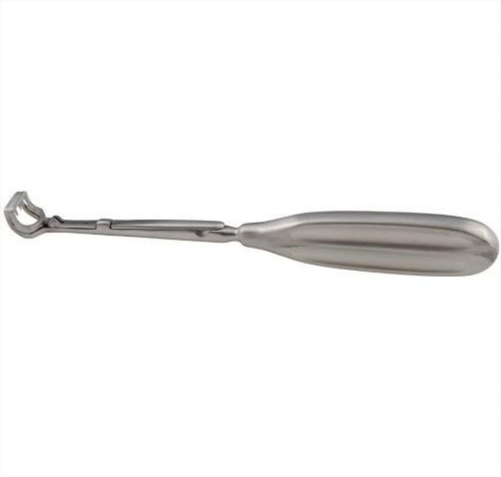
This is a surgical instrument used
adenoidectomy and adenotonsillectomy
In the procedure.
It has a sharp inner blade which provides smooth shaving of the adenoid after infection or inflammation.
It is used to remove excess tissue remaining from ear and tonsil.
Its use can cause primary and secondary haemorrhage as a complication and bleeding can also occur during the procedure.
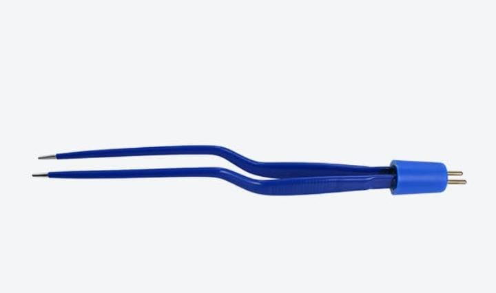
Bipolar forceps:
Bipolar forceps are made of stainless steel.
Bipolar forceps are used to grasp the tissue.
Apart from that, it is useful for dissection of small parts.
Used to ligate small blood vessels to large blood vessels.
Intraocular lens:
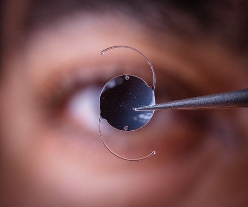
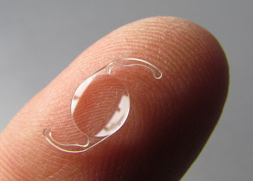
Intraocular lens is an artificial lens.
An intraocular lens replaces the natural eye in a patient with a diseased eye (cataract).
Intraocular lens is used after cataract surgery.
A CY intraocular lens is used for nearsightedness, farsightedness,
Astigmatism (the curvature of the cornea of the eye is more so that the vision becomes blurred),
Presbyopia (number due to age)
Lens is used in etc.
Abdominal retractor: Abdominal retractor
Richardson retractor– Richardson retractor
Bookwalter retractor- Bookwalter retractor
Richardson retractor: Richardson retractor

It is a hand-held instrument that has a single end and a right-angle blade.
Richardson retractor is used to expose, retract, push tissue, organ, muscle and bone during surgery.
Bookwalter retractor: Bookwalter retractor
This is a self-retaining retractor.
This retractor is a type of device which is used during chest and abdominal surgery to hold the organ after surgical incision.
Cardiovascular retractor: Cardiovascular retractor
Debaked vascular retractor:
The debaked vascular retractor is a ratcheted lock and finger ring retractor.
It is used to hold and manipulate delicate tissues during cardiovascular procedures.
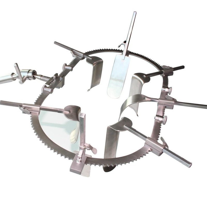
Sternal retractor: Sternal retractor
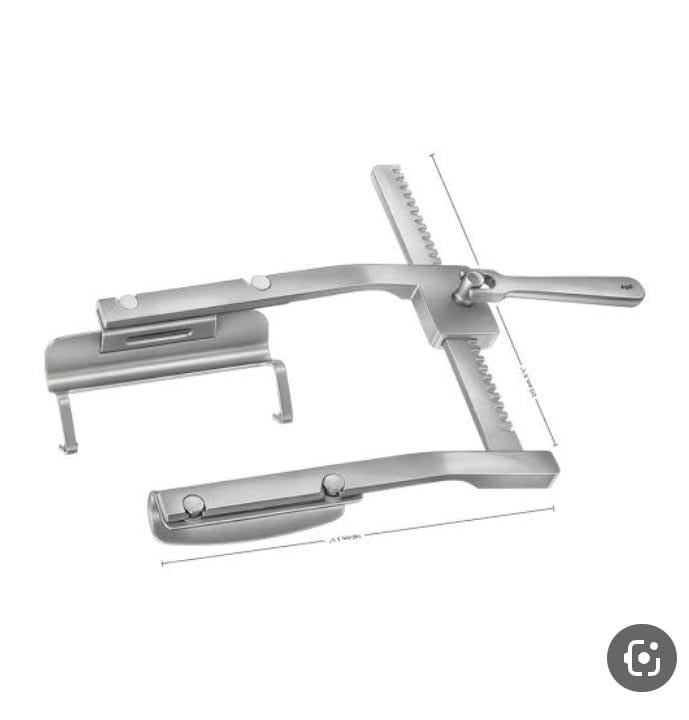
Sternal retractor is a specially surgical instrument.
It is used to reattach the thorax after mid-sternotomy and during surgery like respiratory and cardiac.
It is used to manipulate internal organs during CY surgery.