ENGLISH-NEW MIDWIFERY GNM TY UNIT 10 management of Complications of Purperium
Unit: 10 Management of Complications of Purperium (Management of Complications of Purperium):

Purperial Pyrexia:
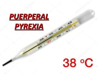
- Purple pyrexia is a condition in which the body temperature increases to more than 100.4°F (38 °C) within 14 days after delivery. It is called “Purple “Pyrexia” is called.
Or
- When the oral temperature reaches 100.4°F (38 °C) or higher on two separate occasions within 10 days after the first 24 hours of delivery, it is called “Purple pyrexia”.
Etiology:
- Unknown,
- Genital: puerperal sepsis.
- Extra genital: e.g.,
- Urinary tract infection (cystitis, pyelonephritis)
- Breast infection (mastitis, breast abscess),
- Intercurrent infections such as,
- Malaria, pulmonary tuberculosis, respiratory infection, and fever.
- Caesarean section wound abscess,
- Leg vein thrombosis
- Other causes:
- Mastitis,
- Pulmonary infection,
- Atelectasis,
- Pneumonia,
- Septic pelvic thrombophlebitis,
- Malaria,
- Pulmonary Tuberculosis.
Sign And Symptoms:
- Fever,
- Chills,
- Tachycardia,
- Uterine tenderness,
- Abdominal pain,
- Foul smelling lochia,
- Best Symptoms: Mastitis, localized pain, swelling and tenderness,
- Fatigue,
- Decreased appetite.
Diagnostic Evaluation:
- History Collection,
- Physical Examination,
- Vital Sign Monitoring,
- Complete Blood Count,
- Blood Culture,
- Urine Analysis,Urine Culture.
- Pelvic examination,
- Imaging studies,
- Breast examination
Management:
- Carefully treat the site of infection Assess.
- Collect a complete history of the patient such as headache, sneezing, coughing, burning urination, painful breasts.
- Perform a complete physical examination of the patient, including proper inspection and palpation of the uterus.
- Look for nokia and perineum and examine the legs.
- Take a throat swab, upper vaginal swab, and urine Send the midstream specimen to the laboratory and inform the doctor for the abnormality line in the report.
- Keep a record of the patient’s vital signs and provide proper nursing care for the fever condition.
- Provide the patient with proper antipyretic medication.
- Provide the patient with proper antibiotic medication.
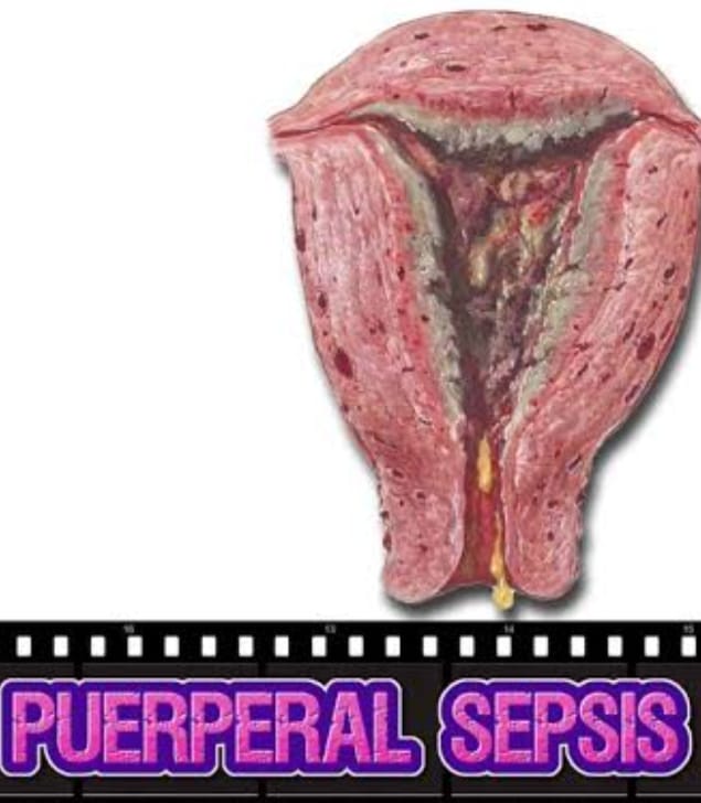
Purperial Sepsis:

- Pupillary sepsis is an infection of the genital tract that occurs as a complication of delivery.
Etiology:
- Endometritis,
- Endomyometritis,
- Endoparametritis,
- Or a combination of all three infections.
- Causative organism: Aerobic:
- –>
- Staphylococcus pyogenes,
- E. coli,
- Klebsiella,
- Pseudomonas,
- and non-hemolytic, Streptococcus,
- Staphylococcus aureus,
- Anaerobic: This involves:
- Anaerobic Streptococcus
- Bacteroides fragilis
- Clostridium welchi
- Clostridium tetani
Predisposing factors:
- Resistance General or local conditions,
- Conditions that increase the multiplication and virulence of the organism,
- Due to the introduction of the organism from outside,
- Increase in the prevalence of resistant organisms to antibiotics and chemotherapy.
- Malnutrition and Anemia,
- Pre-eclampsia (antipartem factor),
- Due to premature rupture of membranes,
- Sexual intercourse during late pregnancy,
- Due to chronic debilitating illness,
- Upper abdominal pain during internal examination after rupture of membranes or during manipulative delivery Introduction of sepsis into the genital tract.
- Dehydration and ketoacidosis during labor,
- Traumatic operative delivery,
- Hemorrhage – APH or PPH,
- Due to retained bits of placental tissue and membranes,
- Placenta previa.
Mode Of Infection :
Puerperal sepsis is specifically a wound infection. In this, the placental site, genital tract laceration or caesarean section wound can become infected in the following ways.
Endogenous:
- The organism is present in the genital tract before delivery and enters the genital organs through the bloodstream or the patient herself.
Autogenous:
- Here the organisms are present elsewhere in the body and are transmitted to the genital organs through the blood stream or droplet infection.
- Streptococcus beta-hemolyticus, E. coli, C. welchii and staphylococcus thus migrate from septic throat, pharyngeal and skin infections.
Exogenous:
- Exogenous infection is an infection transmitted from some source outside the patient. The organism is introduced by attendants, usually doctors or nurses. Infection may be dust-borne or in the form of droplets, during internal examinations, or through contaminated linen or blankets. Nowadays, Staphylococcus pyogenes is common.
Sign And Symptoms:
- Local Infection (Wound Infection):
- The primary sites of infection are the perineum, vagina, cervix, and uterus.
- Local wound infection can cause the area to become red, swollen and pus to form.
- Slight temperature, malaise and headache may be seen and in acute infection, high fever with rigors may be seen.
- Uterine Infection:
- Temperature and pallor increase in mild infection.
- Local discharge is excessive and foul smelling. There are.
- The uterus is tender and subinvoluted.
- In advanced infection, with high fever, rigors, lochia scanty and odorless, the uterus may be tender, soft and subinvoluted.
- Extrauterine:
- Pelvic tenderness (pelvic peritonitis),
- In Phoenix Tenderness (parametritis),
- Flesh (pelvic abscess) etc. are found in the pouch of Douglas.
- The patient has pelvic peritonitis (pyrexia, lower abdominal pain, tenderness, pus),
- General peritonitis,
- Thrombophlebitis,
- Septicemia etc. may occur.
Diagnostic Evaluation:
- History collection,
- Antenatal, intranatal and postnatal history
- Physical Examination,
- Imaging Studies,
- Pelvic CT Scan,
- MRI(Magnetic Resonance Imaging),
- Laboratory Tests:
- Urine Examination,
- WBCs( White Blood Cells count),
- Cervical canal swab,
- Intra uterine sampling of uterine cavity discharge,
- Blood culture.
Management:
Antenatal
Improve the nutritional status of the mother and eliminate any infection in the body.
Intranatal
Surgical asepsis during delivery, screening of group B streptococcus in high risk patients, antibiotics
Postpartum:
Initially one week of aseptic precautions, isolation and restriction of visitors.
Management (Management ):
- Maintain isolation in general care, provide adequate fluid, calories, antibiotics and oral iron.
- Properly maintain the patient’s intake output chart.
- Properly monitor the patient’s vital signs and maintain a local discharge chart.
- Surgical treatment: Remove pus and pain in the perineal wound.
- Heparin IV 7-10 days in septic pelvic thrombophlebitis.
- Debridement in pelvic abscess with colpotomy, laparotomy with unresponsive peritonitis.
- Provide treatment if patient is in septic shock.
Thrombo-embolic disorders/venous thrombo-embolic diseases. (Thrombo-embolic Disorder/Venous Thrombo-embolic Disease) :
Venous thromboembolic disease mainly involves 3 conditions.
1.Deep vein thrombosis.
2.Thrombophlebitis.
3.Embolism.
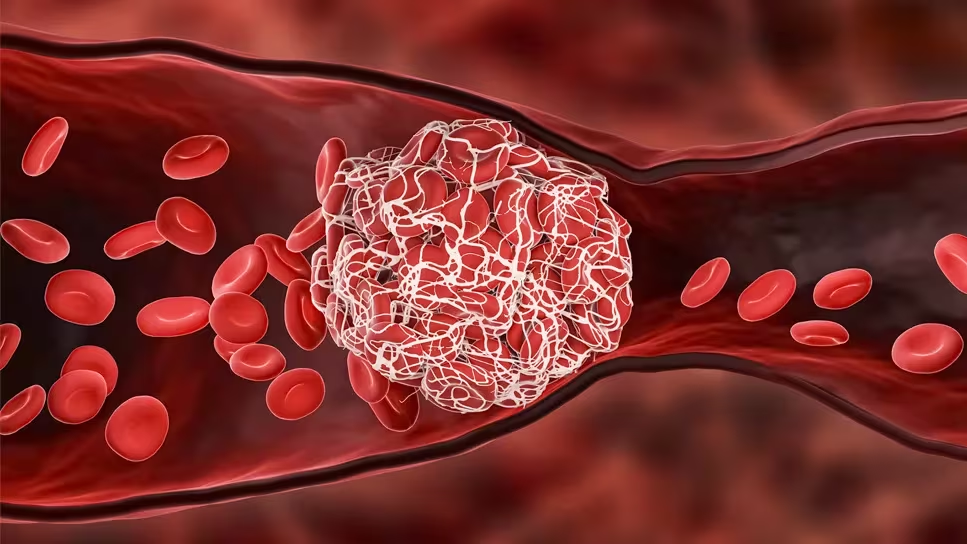
1.Deep Vein Thrombosis.
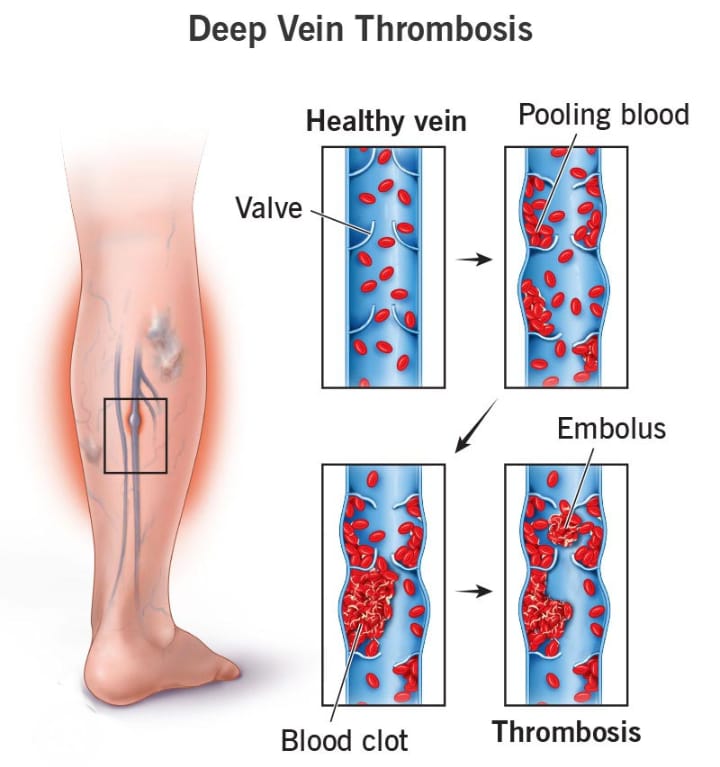
- Deep vein Thrombosis is a condition in which a blood clot forms in the venous system, mainly in the veins of the legs. This condition is called deep vein thrombosis. Thrombosis is significant in pregnant women because it increases the risk of pulmonary embolism, which is a major cause of maternal death.

Etiology:
- Increased concentration of coagulation factors in normal pregnancy,
- Increased concentration of progesterone activity and platelet count,
- Giving estrogen to suppress lactation.
- Increased venous stasis due to pressure of the gravid uterus on the inferior vena cava and iliac veins.
- Thrombophlebitis.
- Other risk factors:
- Increase in age,
- Multipara,
- Operative delivery,
- Obesity,
- Anemia,
- Heart Disease,
- Infection- Pelvic Cellulitis,
- Trauma,
- Immobility,
- Smoking,
- Previous Deep Vein,
- Or,
- Pulmonary embolism.
Sign And Symptoms:
- Pain in the calf muscles,
- Fever,
- Asymmetrical leg edema,
- Positive Homan sign (pain in the calf muscles when the foot is dorsiflexed), is seen.
Diagnosis :
- History collection,
Physical examination,
Doppler Ultrasound,
Venography,
Magnetic Resonance Imaging (MRI). - Management of Thromboembolic Diseases:
- Prevention of trauma, sepsis, dehydration and anemia in pregnancy and labor.
- Use of elastic compression stockings in surgery.
- Advice for leg exercises and early ambulation after operative delivery.
- Giving low molecular weight heparin to high-risk women.
Thrombophlebitis (thrombophlebitis):
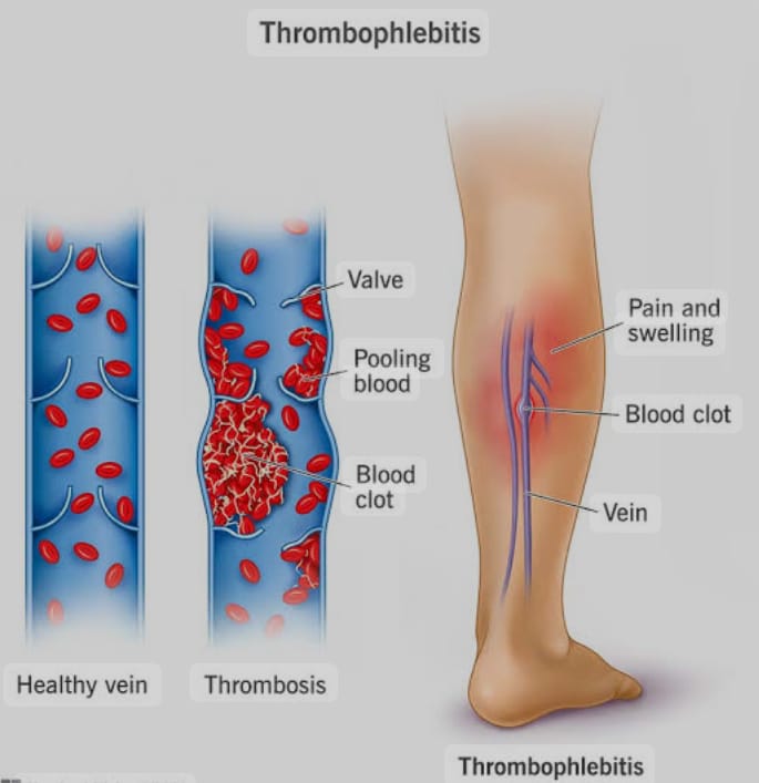
- Thrombophlebitis is a condition in which a blood clot forms in the wall of the vessels due to inflammation. This condition is called thrombophlebitis. Postpartum thrombophlebitis occurs from a thrombosed vein of the placental site. When it is localized in the pelvis, it is called pelvic thrombophlebitis. It travels from the right ovarian vein to the lungs through the inferior vena cava and from the left ovarian vein to the left kidney. Sometimes it extends retrogradely into the iliofemoral veins, causing “phlegmasia alba dolens” or white leg.
Etiology:
- Increased concentration of coagulation factors in normal pregnancy,
- Increased concentration of progesterone activity and platelet count,
- Giving estrogen to suppress lactation.
- Increased venous stasis due to pressure of the gravid uterus on the inferior vena cava and iliac veins.
- Thrombophlebitis.
- Other risk factors:
- Increase in age,
- Multipara,
- Operative delivery,
- Obesity,
- Anemia,
- Heart Disease,
- Infection- Pelvic Cellulitis,
- Trauma,
- Immobility,
- Smoking,
- Previous Deep Vein,
- Or,
- Pulmonary embolism.
Sign And Symptoms:
- Most often occurs in the second week of puerperium.
- Fever mild to high with rigors,
- Features of headache, malaise, tachycardia and toxemia should be observed.
- The affected leg should be swollen, painful, white and cold.
- In the calf muscles Pain,
- Fever,
- Asymmetrical leg edema.
Diagnostic Evaluation (Diagnostic Evaluation) :
- History Collection,
- Physical Examination,
- Doppler Ultrasound,
- Venography,
- Blood Investigations,
- Venous Ultrasound,
- Computed tomography (CT Scan),
- Magnetic resonance imaging (MRI).
Management:
- Prevention of trauma, sepsis, dehydration and anemia in pregnancy and labor.
- Use elastic compression stockings in surgery.
- Advise for leg exercises and early ambulation after operative delivery.
- Give low molecular weight heparin to high risk women.
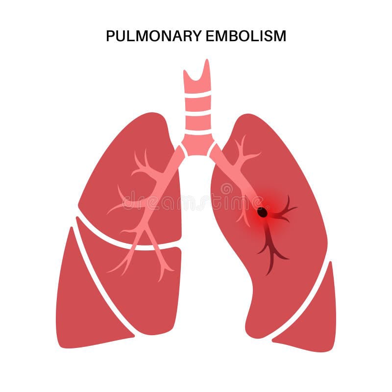
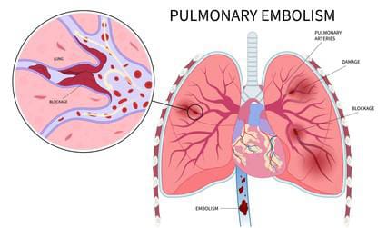
- Pulmonary embolism is caused by the formation of a thrombus (blood clot) in one or more pulmonary arteries. Obstruction and blockage are seen. This thrombus originates from the vascular system or right heart and travels to the pulmonary artery.
- Thus, a large clot breaks off from a thrombus vein and travels to the pulmonary circulation, causing pulmonary embolism. It is a leading cause of maternal death.
Pulmonary Embolism (Pulmonary Embolism):

Etiology :
- Deep vein thrombosis,
- Hypercoagulation,
- Venous stasis,
- Due to trauma,
- Due to inherited or acquired clotting disorders,
- Infection Due to.
- Prolonged immobilization
- Heart failure
- Heart disease
- Hormonal factors
- Advanced age
- Obesity
Sign And Symptoms:
- Rapid onset of dyspnea,
- Shortness of breath,
- Pleuritic chest pain,
- Tachycardia,
- Diaphoresis,
- Blood in sputum (hemoptysis),
- Cyanosis,
- Fever,
- Fainting,
- Calf & thigh pain.
Diagnostic Evaluation (Diagnostic Evaluation) :
- History Collection,
- Physical Examination,
- Chest X-ray,
- Ultrasound,
- Magnetic Resonance Imaging,
- Pulmonary Angiography,
- Ventilation Perfusion Scan,
- D-Dimer Test,
- Electrocardiogram,
- Doppler ultrasound,
- Lung scan.
Management:
Anticoagulant therapy
Provide anticoagulant drugs for the treatment of pulmonary embolism. Which prevents the formation of blood clots and helps in dissolving the clot. Give heparin, low molecular weight heparin medicine as anticoagulant drug.
Thrombolytic therapy
Provide thrombolytic therapy to rapidly dissolve the clot in life-threatening and emergency conditions. Which breaks down the clot. For example, tissue plasminogen activator (tPA)
Inferior vena cava filter
An inferior vena cava filter is used when anticoagulant therapy is contraindicated or ineffective. In which an inferior vena cava filter is placed, which prevents the clot in the lower area of the body from traveling to the lungs and getting deposited in the filter there.
Supplemental oxygen Supplemental oxygen should be provided to maintain adequate oxygen levels.
Emblemectomy In embolectomy, the clot (embolism) is surgically removed.
Nursing Management:
- The nurse plays an important role in assessing the woman for signs and symptoms of pulmonary embolism, which may include sudden chest pain, shortness of breath, rapid heartbeat, cough, and fainting.
- Provide the woman with a properly comfortable position.
- Check the woman’s vital signs, especially monitoring respiratory rate and breath sounds.
- Properly assess the woman for signs and symptoms of respiratory distress.
- Properly assess the mother for hypoxia.
- Properly administer intravenous fluids to the patient.
- Continuously monitor the woman’s oxygen level.
- The nurse should prioritize stabilizing the woman’s condition by ensuring adequate oxygenation, providing oxygen as needed, and initiating cardiac monitoring. Administer intravenous fluids to maintain hemodynamic stabilization, depending on the severity of symptoms.
- Collaborate appropriately with other health care personnel for the woman’s proper care
- Properly administer medications to the woman, which usually include anticoagulants (such as heparin or low molecular weight heparin) to prevent further clotting and provide thrombolytic medications.
- Provide complete education to the woman and her caregivers about the woman’s condition, its causes, its symptoms and signs, and its treatment.
- Properly monitor the effectiveness of the treatment provided to the woman.
- Provide adequate emotional and psychological support to the woman and her family members.
- Provide proper work and a comfortable environment for the woman.
- Provide education to the woman to take medication regularly.
Breast Engorgement :

- Breast engorgement is a complication of the postpartum period in which the breast tissue becomes swollen due to venous and lymphatic congestion. Breast engorgement condition is mostly seen in the puerperal period after the milk secretion starts i.e. on the 3rd or 4th day of postpartum. Breast engorgement is usually caused by the increase in secretion of breast milk after childbirth, causing the breasts to become overfilled and congested due to the accumulation of milk in the breasts.
- This condition is more common in breastfeeding mothers, especially in the early puerperal period when milk production and secretion increase. This condition causes the breasts to become full, firm and painful. And swelling and discomfort in the breast occurs, which is called breast engorgement.
Etiology:
- Milk does not come out of the lacteal system due to normal venous and lymphatic engorgement in the breast before lactation.
- Milk production increases during the postpartum period,
- Breast milk is removed in an inadequate amount,
- Milk accumulates in the breast due to not breastfeeding the baby properly.
- Due to not providing proper position to the child during breast feeding, the breast milk cannot be removed in an adequate amount.
Sign And Symptoms:
- 1) Swelling and Firmness
Due to accumulation of milk in the breast, the breast becomes tight, firm and Swollen. - 2) Tenderness and Pain The breast becomes tender and painful, and is especially painful around the areola and nipple.
- 3) Skin Changes
The skin over the breast becomes scaly and stretched. - 4) Difficulty in Breast Fitting
The child has difficulty in fitting the breast well. - 5) Fever and Malaise
Mother has fever and due to this she feels malaise and discomfort in the body in a generalized manner. - History collection,
- Symptoms assessment,
- Onset and Duration,
- Previous History,
- Medical History Collection.
- Physical Examination:
- This includes breast assessment,
- Skin Assessment,
- Nipple Examination,
- Ultrasound,
Diagnostic Evaluation:
Management:
1) Frequent and effective breastfeeding
Advise the mother to provide adequate best fitting on the demand of the child to prevent milk accumulation due to which milk is An adequate amount of milk can be removed from the breast and the condition of breast engorgement can be prevented.
Provide education to the mother about the best feeding technique so that the mother can provide the child with proper position during breastfeeding and remove milk in an adequate amount so that breast engorgement can be prevented.
2) Complete Ejection of Breast Milk
While feeding the child, only after complete breastfeeding on the first breast, the mother should feed the child on the other breast to ensure the best fitting. Provide education so that adequate amount of milk can be emptied from both breasts and the condition of engorgement can be prevented.
3) Breast Massage and Warm Compression
Provide education to the mother that it is best to massage the breast gently before feeding so that the engorged area can be softened.
Advise the mother to provide compresses with warm water on the breast before feeding so that discomfort can be relieved.
4) Manual Expression of Milk
If the child is not able to breastfeed properly, it is best to remove the excessive milk from the breast by using a breast pump or by expressing the milk by hand, which can prevent breast engorgement.
5) Comfort Measures Advise the mother to avoid wearing tight-fitting clothes. Provide the mother with proper work and a comfortable environment.
6) Pain Relief
If the mother is in pain, provide analgesic medication to relieve the pain.
Ex:
Acetaminophen,
Ibuprofen.
Breast Abscess :
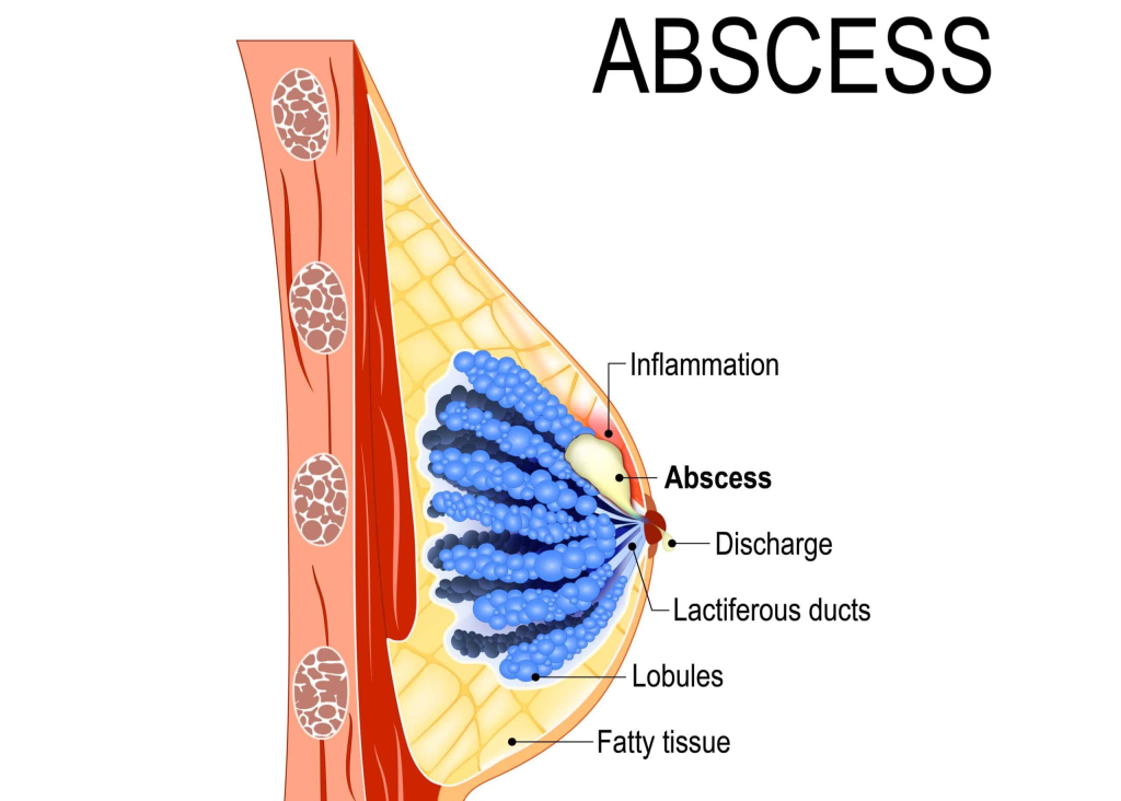
- Breast abscess is a condition in which the breast This condition is called a breast abscess when pus/abscess collects in the breast tissue due to infection and inflammation.
Etiology:
- Bacterial infections such as Staphylococcus aureus, including methicillin-resistant strains (MRSA),
- Lactation: Often seen in breastfeeding women due to blocked milk ducts or mastitis.
- Trauma: Injured breast tissue can introduce bacteria.
- Skin conditions: Skin conditions such as eczema, psoriasis, Can occur.
- Weak immune system.
- Diabetic patient.
- Due to surgery or invasive procedure.
- Chronic condition.
Sign And Symptoms (Sign And Symptoms) Symptoms):
- Localized pain,
- Swelling,
- Redness,
- Feeling warm,
- Fever.
Diagnostic Evaluation (Diagnostic Evaluation) :
- History Collection,
- Physical Examination,
- Ultrasound,
- Mammography,
- Fine Needle Aspiration,
- Culture and Sensitivity Test,
- Blood Test,
- Complete Blood Count (CBC) Test.
Management:
- Treatment involves incision and drainage, or serial percutaneous needle aspiration. Prepare the patient for the operative procedure.
- Start breastfeeding on the uninvolved side.
- Mechanically empty the infected breast every hour.
- Start breastfeeding on the involved side after the condition is treated.
- Provide the patient with instructions on antibiotics and analgesics.
- Midwives and doctors should wash their hands before handling the mother and baby.
- Take proper preventive measures to prevent breast abscesses.
Purperial Psychosis / Postpartum Psychosis:

- Pupillary psychosis or postpartum psychosis is a term that covers a group of mental illnesses in which a woman experiences a sudden onset of psychotic symptoms after childbirth. In which women may experience irritability, mood swings, hallucinations.
- Pupillary psychosis is a severe form of mental illness. It occurs in about 1-2 cases in 1000 women, which begins as early as the first 48-72 hours of delivery and most often within 2-3 weeks. It can be caused by hormonal changes (such as a sudden drop in estrogen levels after birth).
Etiology (Etiology):
- The causes of postpartum psychosis are not yet known, but it is common in women with a history of bipolar disorder or who have experienced postnatal psychosis after a previous birth.
Risk Factors:
- Having a family or personal history of psychiatric illness,
- Past history of postnatal psychosis,
- After the birth of the first baby.
Sign And Symptoms:
- Signs and symptoms appear suddenly 48 hours to 2 weeks after the birth of a child.
- Symptoms like mania, depression or schizophrenia illness are seen in patients with postnatal psychosis.
- Symptoms of mania such as,
Hyperactivity, euphoria, flights of ideas, insomnia, delusions, extreme excitement, restlessness, irritability, full of energy.
Severe depression with delusions, hallucinations (auditory), mutism, stupor or transient swings in hypomania.
Bizarre behavior,
Some mothers switch from depression to mania while others switch from mania to depression.
Typical features include confusion, extreme fear and ecstasy, perplexity, transient delusional ideas.
Management:
- Management The psychiatrist is consulted immediately and the patient is admitted to the hospital.
- Treat severe overactivity and delusions with antipsychotic drugs.
- Drugs include chlorpromazine and sublingual estradiol.
- In manic depressive psychosis, lithium is given.
- Provide electroconvulsive therapy in unresponsive or depressive psychosis.
- Do not breastfeed.
- Supervise the patient in cases of suicidal, infanticidal impulses.
- Psychosocial treatment includes counseling, psychotherapy, cognitive behavior therapy, family focused intervention, and social support provided by postnatal illness associations.