ENGLISH-MSN-II-CARDIAC DISORDER-PART-2
Common nursing diagnosis and care of cardiovascular disorder
Acute pain related to myocardial ischemia / reduced coronary blood flow as evidenced by facial expressions
Relieve Pain
- Assess the patient’s condition To do.
- Monitor vital signs.
- Assess pain level.
- Note location, duration, intensity, timing, etc. of pain.
- Assess factors triggering and relieving pain.
- Provide the patient with a Fowler or semi-Fowler position so that the lungs can expand properly. Can.
- Provide the patient with mind diversionary therapy. So that the patient’s attention is diverted from the pain.
- Provide relaxation therapy to the patient.
- Administer the medicine prescribed by the doctor.
- Assess the side effects and effectiveness of the medicine.
- Maintain records and reports.
Decrease cardiac output related to decrease cardiac contractility / reduced preload afterload contractility / decrease coronary blood flow as evidence by increase heart rate
Maintaining cardiac output / improving cardiac output
- Assess the patient’s condition.
- Monitor vital signs.
- Monitor cardiovascular status.
- Monitor ECG pattern.
- Auscultate heart sounds and rhythm.
- Assess peripheral pulse.
- Monitor hemodynamic parameters.
- Maintain intake output chart.
- Monitor urine output every hour.
- Provide the patient with a Fowler’s position or a high Fowler’s position.
- Provide IV fluids as prescribed by the doctor.
- Administer the medicine prescribed by the doctor.
- Assess the medicine for side effects and effectiveness.
- Maintain record report.
Ineffective cardiac tissue perfusion / impaired tissue perfusion related to reduce coronary blood flow as evidenced by decrease cardiac output, increase heart rate
Promoting adequate tissue perfusion / improving cardiac tissue perfusion
- Assess the patient’s condition.
- Monitor vital signs.
- Assess the patient’s skin for gross, moist cyanosis.
- Assess fluid volume status.
- Check skin temperature, peripheral pulse, capillary refill. Which can be used to determine whether tissue perfusion is present or not.
- Provide bed rest to the patient.
- Administer oxygen therapy.
- Provide mechanical ventilation if necessary.
- Provide fluid resuscitation.
- Administer medicine prescribed by the doctor To do.
- Monitor the side effects and effectiveness of the medicine.
- Maintain record reports.
Impaired gas exchange related to chest surgery / interruption in blood flow to the pulmonary alveoli as evidenced by dyspnea, cynosis
Improve gas exchange
- Assess the patient’s condition.
- Monitor vital signs.
- Monitor respiratory rate, rhythm, arterial blood gas, tidal volume, peak inspiratory pressure, extubation parameters.
- Assess skin, mucous membranes, nail beds for cyanosis.
- Breath sounds To auscultate.
- Provide the patient with a semi-fowler or fowler position.
- Provide the patient with bed rest.
- Restrict activity until the patient is hemodynamically stable.
- Provide the patient with knowledge about deep breathing exercises.
- Provide the patient with supplemental Provide oxygen.
- Administer medicine prescribed by the doctor.
- Maintain records and reports.
Ineffective thermoregulation related to infection as evidenced by increased body temperature
Maintain body temperature (body Maintain temperature)
- Assess the patient’s condition.
- Monitor vital signs.
- Monitor temperature every two to four hours.
- Maintain the patient’s room temperature.
- Provide cold applications to the patient To do.
- If the patient feels cold, avoid cold applications and provide a blanket.
- Maintain adequate hydration.
- Ask the patient to take oral fluids and administer IV fluids.
- Administer antipyretic and antibiotic medicines as prescribed by the doctor.
- Record and report To maintain.
Anxiety related to disease condition, uncertain prognosis, hospitilisation as evidence by verbalization, restlessness, agitation
Reduce anxiety
- Assess the patient’s condition.
- Assess signs of anxiety such as restlessness, sleeplessness.
- Pay attention to the patient’s psychological needs and listen carefully to the patient.
- Encourage the patient to express his/her feelings, discomfort and anxiety.
- Resolve all the patient’s doubts and queries.
- Help the patient to understand his/her Provide knowledge about the condition and treatment so that their anxiety is relieved and the patient becomes confident.
- Provide psychological support to the patient.
- Provide mind diversionary therapy and recreational therapy to the patient.
- Administer anti-anxiety agents. Activity intolerance related to fatigue,
dyspnea as evidenced by shortness of breath, abnormal heart rate
Increase activity level
- Assess the patient’s condition.
- Check the patient’s activity level.
- Provide bed rest to the patient initially.
- Then gradually encourage the patient to do range of motion exercises.
- Assist the patient with his/her activities.
- Provide the patient with rest between 2 activities.
- Check whether the patient experiences any breathing difficulty or palpitations during the activity.
- If present, stop the patient’s activity and provide rest.
- Ask the patient to avoid lifting heavy objects.
- Avoid doing work that puts strain on the heart Say.
Knowledge deficit related to disease condition and it’s prognosis as evidenced by communication with patient, patient questioning about him/her condition
Improve knowledge level (Improve knowledge level)
- Assess patient’s condition To do.
- Assess the patient’s knowledge about the disease condition and its treatment.
- Provide the patient with knowledge about the disease condition and its prognosis.
- Provide knowledge in a language that the patient can understand.
- Resolve the patient’s doubts and concerns.
Explain coronary artery disease
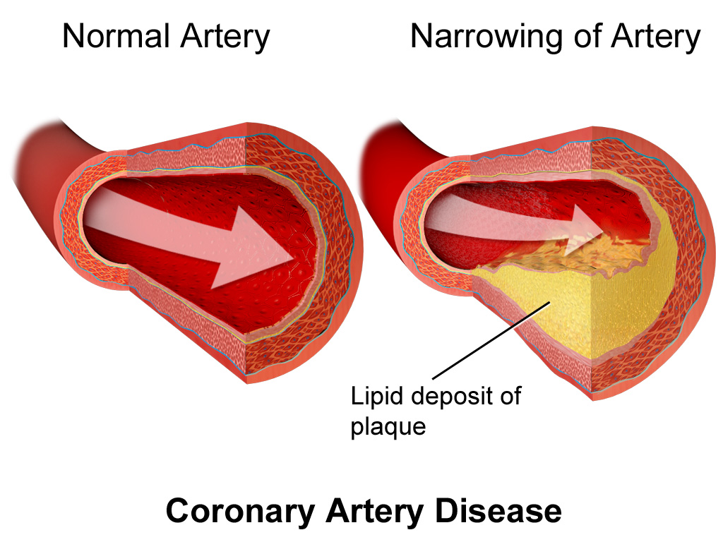
- Coronary artery disease is also known as ‘atherosclerotic heart disease’ .
- Coronary artery disease is a common type of heart disease. In which plaque (atheroma) develops in the innermost layer of the coronary artery that supplies blood to the heart, i.e. plaque is deposited.
- Due to which the coronary artery becomes narrowed and eventually blockage is seen in it.
- As a result, there is a decrease in coronary blood flow, so that the heart does not get sufficient blood supply and oxygen supply, i.e. myocardial ischemia is seen and finally a condition like heart attack is seen.
Define angina pectoris
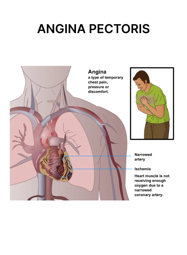
- Angina is also known as ‘ischemic chest pain’ .
- Angina pectoris is the medical term used for ‘chest pain’ and ‘discomfort’ caused by coronary heart disease.
- Angina is not a disease but a symptom of coronary artery disease.
- Plaque deposits in the coronary arteries cause them to narrow, which prevents adequate blood and oxygen from reaching the heart muscles, causing chest pain.
Write types of angina pectoris
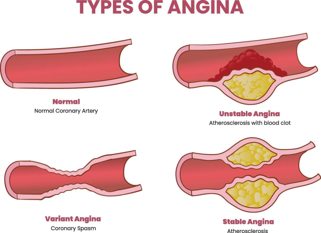
✓ Stable (classic) angina: In stable angina, chest pain occurs during exertion, exercise, or any stressful activity. This pain can be relieved by rest and medication.
✓ Unstable Angina: Unstable angina is the least common and most severe type. In which chest pain is seen during rest or minimal exertion. Hence it cannot be relieved by rest and medication. This is an impending sign of a heart attack.
✓ Variant Angina:Variant angina, also known as ‘Prinzmetal’ and ‘vasospastic angina’ , is characterized by chest pain due to spasm of the coronary arteries. This pain is seen during periods of rest and this pain is seen during midnight as well as early morning hours.
✓Refractory angina: Refractory angina is a severe and persistent form of angina in which chest pain is seen even after medication, lifestyle changes, angioplasty and bypass surgery. Therefore, enhanced external counter pulsation (EECP), spinal cord stimulation, heart transplantation are performed for treatment.
✓ Silent ischemia: In silent ischemia, the patient does not feel pain, i.e. there are subjective data absences. But it can be determined with the help of ECG, exercise stress test, Holter monitoring.
Write causes of angina pectoris
- Deposition of plaque in coronary artery
- Coronary artery spasm
- Arterial Embolism
- Narrowing of heart valve (aortic stenosis)
- Hypertrophic cardiomyopathy
- Severe anemia
Write sign and symptoms seen in angina pectoris
- Chest Pain
- During this pain, pressure, tightness, squeezing, heaviness, burning are felt.
- This pain is seen radiating to the neck, jaw, shoulder arm and back.
- Discomfort
- Shortness of breath
- Fatigue
- Weakness
- Sweating
- Nausea
- Vomiting
Write diagnostic evaluation of angina pectoris (Write diagnostic evaluation of Angina Pectoris)
- History Collection
- Physical Examination
- Electrocardiogram (ECG/EKG)
- Echocardiogram
- Coronary Angiography
- Exercise Stress Test
- Blood Tests – Cholesterol Level, Troponin, Creatine Kinase (CK-MB), Myoglobin
Write medical management of angina pectoris
- Oxygen therapy: Administer oxygen through a nasal cannula or mask. So that the heart muscles get oxygen.
- Vasodilator: Use nitroglycerin as a vasodilator drug. Which dilates blood vessels so that blood flow can improve. (Angina can be relieved by nitroglycerin, so nitroglycerin is the drug of choice)
- Antiplatelet: Antiplatelet drugs thin the blood and prevent blood clots. For example, aspirin.
- Beta blocker: Provide beta blockers to reduce the heart’s workload. They slow the heart rate and decrease blood pressure. For example, propranolol, atenolol
- Calcium channel blockers: Calcium channel blockers block the entry of calcium into the heart muscles, causing the muscles to relax and helping the arteries to widen and lower blood pressure. E.g. Amlodipine, Nifedipine
- ACE inhibitors: ACE inhibitors prevent the conversion of angiotensin I to angiotensin II and promote diuresis, vasodilation, and reduce the workload of the heart. For example, enalapril, captopril
- Statins: Statins decrease cholesterol levels and reduce the risk of plaque buildup in the arteries. For example, atorvastatin.
Write surgical management of angina pectoris
✓ Coronary artery bypass grafting (CABG)
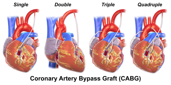
- CABG creates a new route for a narrow or blocked coronary artery or Bypass is done. In which healthy blood vessels are harvested from any part of the body such as chest, leg, arm and these harvested blood vessels are attached to the coronary artery and the blocked part is bypassed and normal blood flow is restored. CABG is mainly preferred in multiple coronary artery blockage conditions. Apart from this, CABG is done when medication and angioplasty fail.
✓ Percutaneous transluminal coronary angioplasty (PTCA)
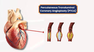
- PTCA is a minimally invasive surgical procedure used to open narrowed and blocked coronary arteries. In which a balloon attached to a catheter is inserted into the narrowing artery and then the balloon is inflated so that the artery widens and the plaque flattens against the artery wall, thereby improving coronary blood flow.
✓ Atherectomy
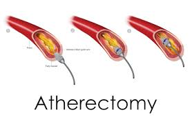
- Atherectomy is a surgical procedure used to remove plaque deposited in an artery. In this procedure, a small cutting device, blade, laser or drill attached to a catheter is inserted into the artery and with its help, the plaque is cut and the plaque is removed, thereby increasing coronary blood flow. Methods like directional, rotational, laser are used in atherectomy.
✓ Coronary stent (coronary stent)
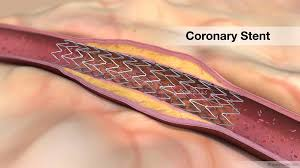
- A coronary stent is a small mesh-like tube or artificial support device made of metal or polymer. In which the stent is inserted into the narrow or blocked coronary artery with a balloon catheter and placed at the correct position, after which the balloon is inflated and the stent is expanded. After the stent is expanded, the balloon is deflated and removed, and the stent is permanently placed to keep the coronary artery open.
✓ Transmyocardial laser revascularization (TMR)
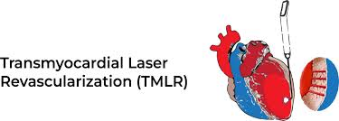
- Transmyocardial laser revascularization is a surgical procedure used to relieve severe angina caused by advanced coronary artery disease. This procedure uses a special CO2 laser to create small channels in the heart muscle, which increases blood flow to the heart. This method is not used at present.
Define coronary atherosclerosis
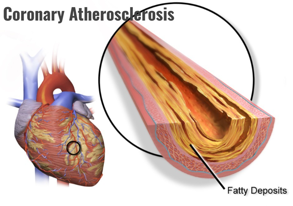
- Coronary atherosclerosis is a condition of coronary artery disease. In which plaque buildup occurs inside the coronary arteries.
- Coronary atherosclerosis is the buildup of abnormal lipid and fatty tissue inside the coronary arteries. Which is known as ‘atheroma’ and this condition is known as ‘atherosclerosis’.
- Which causes the artery to narrow and thicken and due to this, restriction in blood flow is seen.
Write causes and risk factor of Coronary atherosclerosis (Write causes and risk factor of coronary atherosclerosis)
Modifiable Risk Factors:
- High cholesterol level
- High blood pressure
- Smoking
- Diabetes
- Obesity
- Physical Inactivity
- Stress
- Unhealthy Diet
Non-Modifiable Risk Factors:
- Age
- Family history of coronary artery disease
- Gender
- Race
Write sign and symptoms of coronary atherosclerosis
- Chest Pain (Angina)
- Shortness of Breath
- Fatigue
- Arrhythmia
- Inadequate Cardiac Output
- Diaphaesis
Write diagnostic evaluation of coronary atherosclerosis
- History Collection
- Physical Examination
- Electrocardiogram
- Echocardiogram
- Exercise stress test
- Coronary angiography
- Coronary calcium scan
- Exercise thallium test
- PET scan
Write medical management of coronary atherosclerosis
- Oxygen therapy: Administer oxygen through a nasal cannula or mask. So that the heart muscles get oxygen.
- Vasodilator: Use nitroglycerin as a vasodilator drug. Which dilates blood vessels so that blood flow can improve.
- Antiplatelet: Antiplatelet drugs thin the blood and prevent blood clots. For example, aspirin.
- Beta blocker: Beta blocker may be given to reduce the heart’s workload. This slows the heart rate and decreases blood pressure. For example, propranolol, atenolol
- Calcium channel blockers:Calcium channel blockers block the entry of calcium into the heart muscles, causing the muscles to relax and helping the arteries to widen and lower blood pressure.
- ACE inhibitors: ACE inhibitors prevent the conversion of angiotensin I to angiotensin II and promote diuresis, vasodilation, and heart rate. Reduces workload. For example, enalapril, captopril
- Statins: Statins decrease cholesterol levels and reduce the risk of plaque buildup in the arteries. For example, atorvastatin.
Write surgical management of coronary atherosclerosis
✓ Coronary artery bypass grafting (CABG)
- CABG is a type of It is a surgical procedure used to treat coronary artery disease. In which a new route is created for a narrow or blocked coronary artery or it is bypassed. In which healthy blood vessels are harvested from any part of the body such as the chest, leg, arm and these harvested blood vessels are attached to the coronary artery and the blocked part is bypassed and normal blood flow is restored. CABG is mainly preferred in multiple coronary artery blockage conditions. In addition, CABG is performed when medication and angioplasty fail.
✓ Percutaneous transluminal coronary angioplasty (PTCA)
- PTCA is a minimally invasive surgical procedure used to open narrowed and blocked coronary arteries. In which a balloon attached to a catheter is inserted into the narrowing artery and then the balloon is inflated so that the artery widens and the plaque flattens against the artery wall, thereby improving coronary blood flow.
✓ Atherectomy
- Atherectomy is a surgical procedure used to remove plaque deposited in an artery. In this procedure, a small cutting device, blade, laser or drill attached to a catheter is inserted into the artery and with its help, the plaque is cut and the plaque is removed so that coronary blood flow increases. Methods like directional, rotational, laser are used in atherectomy.
✓ Coronary stent
- A coronary stent is a small mesh-like tube or artificial support device made of metal or polymer. The stent is inserted into a narrowed or blocked coronary artery with a balloon catheter and placed in the correct position. The balloon is then inflated and the stent is expanded. After the stent is expanded, the balloon is deflated and removed, and the stent is permanently placed to keep the coronary artery open. Transmyocardial laser revascularization (TMR) is a surgical procedure used to relieve severe angina caused by advanced coronary artery disease. This procedure uses a special CO2 laser to create small channels in the heart muscles, which increases blood flow to the heart. This method is not used today.
✓Endarterectomy:
- Endarterectomy is a surgical procedure in which plaque is removed from inside an artery. This procedure is mainly performed to remove blockages in the carotid artery.
Define myocardial infarction
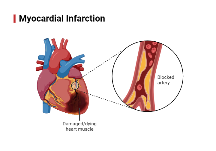
- Myocardial infarction is also known as ‘heart attack’ .
- Myocardial infarction is a serious medical emergency in which the blood flow to the heart muscle is blocked. Due to this, myocardial tissue is permanently damaged and destroyed and necrosis is observed.
Write causes and risk factors of myocardial infarction
Causes :
- Coronary atherosclerosis
- Plaque rupture
- Coronary artery spasm
- Coronary artery dissection
- Embolism
Risk factor :
- Risk factors include non-modifiable and modifiable risk factors.
✓Non-modifiable risk factors :
Non-modifiable risk factors cannot be controlled by ourselves, and these risk factors can also be controlled through exercise, lifestyle modification or medication. No.
- Age
- Gender (men are 3 times more prone to heart disease than women)
- Family history
- Race (African Americans have a higher incidence of heart disease)
✓ Modifiable Risk Factors:
Modifiable or controllable risk factors are those that we can control. Which are as follows :
- Smoking
- High blood pressure
- High cholesterol level
- Diabetes
- Obesity
- Physical Inactivity
- Unhealthy Diet
- Stress
- Type A Personality
- Lack of Estrogen in Women
- Elevated Serum Hemocysteine
Explain pathophysiology of myocardial infarction
Cholesterol is deposited in the walls of the main arteries.
|
This deposited cholesterol ultimately forms plaque on the walls of the arteries. This is known as atherosclerotic plaque. (This atherosclerotic plaque formation takes place over a long period of time, meaning it takes many years to establish.
|
Often this plaque ruptures, which activates the blood clotting process. Due to which platelets are activated and fibrin is deposited, and due to this, thrombus formation occurs in the coronary artery.
|
This thrombus completely blocks the coronary artery and restricts the blood flow to the myocardium.
|
Due to which myocardial cells and tissues are permanently damaged and elevation is seen in the ST segment and conditions like myocardial infarction are seen.
Degree of damage
Zone of Ischemia:In this zone or region, the heart muscles do not receive oxygenated blood supply and due to this, ischemia occurs. In which there is no injury or damage to the muscles.
Zone of Injury: In this zone, injury to the muscles occurs. The muscles become inflamed and damaged. This damage is reversible and can be restored by providing adequate oxygen.
Zone of necrosis: In this zone, due to the cut off of oxygen supply, the heart muscles are permanently and irreversibly damaged and necrosis is observed.
Write classification of myocardial infarction
✓ Clinical Classification of Different Types of Myocardial Infarction :
- Type – 1 : Spontaneous myocardial infarction occurs. Which occurs due to ischemia caused by plaque erosion, rupture, fissuring.
- Type – 2 : Myocardial infarction occurs due to secondary ischemia. Which occurs due to an imbalance between oxygen demand and supply. This imbalance is seen due to coronary artery spasm, coronary embolism, anemia.
- Type – 3 : Myocardial infarction is seen due to sudden unexpected cardiac death. Such as cardiac arrest.
- Type – 4 : Myocardial infarction is seen due to primary percutaneous intervention.
- Type – 5 : Myocardial infarction is seen due to cardiac surgery. Such as coronary artery bypass surgery
- In addition, myocardial infarction is classified based on anatomical structure, diagnostic method and location:
✓ According to the layers of the heart muscles involved:
- Transmural infarction:
In transmural infarction, a major coronary artery is affected and there is complete occlusion of the artery and in this, ST segment elevation is seen in the ECG (STEMI)
- Subendocardial infarction:
In subendocardial infarction, a small area of the subendocardial wall of the left ventricle, ventricular septum, or papillary muscles is affected and the ST segment is depressed. (NSTEMI)
✓ According to the presence or absence of ST segment elevation :
- ST – segment elevation myocardial infarction (STEMI) :
In this type, persistent ST-segment elevation is seen on the ECG. Which indicates complete occlusion of the coronary artery. Hence immediate reperfusion therapy is required. Such as thrombolytic drugs, percutaneous coronary intervention.
- Non ST – segment elevation myocardial infarction (NSTEMI) :
In this type, persistent ST-segment elevation is not seen on ECG but other signs of myocardial injury are present such as elevation in cardiac biomarkers. Invasive procedures are used for the management of NSTEMI. Such as coronary angioplasty, revascularization.
✓ According to the location of myocardial infarction:
- Left ventricular infarction:
Left ventricular infarction affects the left ventricle. That is, the blood flow to the left ventricle is blocked.
- Right Ventricular Infarction:
In right ventricular infarction, the right ventricle is affected in a heart attack.
Write sign and symptoms seen in myocardial infarction
- Chest pain or discomfort
- Pain may radiate to arm, jaw, neck, shoulder, back
- Pain is substernal, severe and diffuse
- Feeling of heaviness, squeezing
- Pain is not relieved by rest and medication
- Shortness of breath
- Diaphoresis (excessive sweating)
- Cool, clammy and moist skin
- Decrease Cardiac contractility
- Decrease cardiac output
- Nausea and vomiting
- Restlessness
- Dizziness
- Lightheadedness
- Fanting
- Confusion
- Disorientation
- Fever
Write diagnostic evaluation of myocardial infarction
- History Collection
- Physical Examination
- Electrocardiogram (ST segment elevation)
- Echocardiogram
- Coronary Angiography
- Blood Test (Cardiac Biomarker)
- Cardiac MRI and CT Scan
- Stress test
- Holter monitoring
- Coronary calcium scan
Write medical management of myocardial infarction
- Oxygen therapy: Administering oxygen through a nasal cannula or mask. So that the heart muscles get oxygen.
- Thrombolytic therapy: Using thrombolytic drugs to dissolve and break down blood clots. E.g. Streptokinase, Urokinase
- Anticoagulant drug: Use anticoagulant drugs to prevent blood clot formation and thin the blood. E.g. heparin, warfarin
- Vasodilator: Use nitroglycerin as a vasodilator drug. Which dilates blood vessels so that blood flow can improve.
- Antiplatelet: Antiplatelet drugs thin the blood and prevent blood clots. For example, aspirin.
- Beta blocker: Providing beta blockers to reduce the heart’s workload. Which slows the heart rate and decreases blood pressure. For example, propranolol, atenolol
- Calcium channel blockers : Calcium channel blockers block the entry of calcium into the heart muscles, causing the muscles to relax and helping the arteries to widen and lower blood pressure.
- ACE inhibitors: ACE inhibitors prevent the conversion of angiotensin I to angiotensin II and promote diuresis, vasodilation, and Reduces the workload of the heart. For example, enalapril, captopril
- Statins: Statins decrease cholesterol levels and reduce the risk of plaque buildup in the arteries. For example, atorvastatin.
- Pain relievers: Opioid drugs are used to relieve pain due to severe pain.
Write surgical management of angina pectoris
- Coronary Artery Bypass Grafting
- Percutaneous Transluminal Coronary Angioplasty
- Coronary Stent
- Atherectomy
- Trans Myocardial Laser Revascularization
Coronary artery bypass grafting (CABG)
- CABG is a type of surgical procedure used to treat coronary artery disease. In which a new route is created for a narrow or blocked coronary artery or it is bypassed. In which healthy blood vessels are harvested from any part of the body such as the chest, leg, arm and these harvested blood vessels are attached to the coronary artery and the blocked part is bypassed and normal blood flow is restored. CABG is mainly preferred in multiple coronary artery blockage conditions. Apart from this, CABG is done when medication and angioplasty fail.
Coronary stent (coronary stent)
- A coronary stent is a small mesh-like tube or artificial support device made of metal or polymer. The stent is inserted into a narrowed or blocked coronary artery with a balloon catheter and placed in the correct position. The balloon is then inflated and the stent is expanded. After the stent is expanded, the balloon is deflated and removed, and the stent is permanently placed to keep the coronary artery open.
Atherectomy
- Atherectomy is a surgical procedure used to remove plaque that has been deposited in an artery. In this procedure, a small cutting device, blade, laser, or drill attached to a catheter is inserted into the artery and used to cut and remove plaque, thereby increasing coronary blood flow. Atherectomy uses methods such as directional, rotational, and laser.
Transmyocardial laser revascularization (TMR)
- Transmyocardial laser revascularization is a surgical procedure used to relieve severe angina caused by advanced coronary artery disease. This procedure uses a special CO2 laser to create small channels in the heart muscles, which increases blood flow to the heart. This method is not used today.
Atherectomy
- Atherectomy is a surgical procedure used to remove plaque deposited in the arteries. In this procedure, a small cutting device, blade, laser, or drill attached to a catheter is inserted into the artery and used to cut and remove plaque, thereby increasing coronary blood flow. Atherectomy uses methods such as directional, rotational, and laser.
Write nursing management of myocardial infarction
Acute pain related to myocardial ischemia / reduce coronary blood flow as evidence by facial expressions
Relieve pain (Relieve pain to do)
- Assess the patient’s condition.
- Monitor vital signs.
- Assess pain level.
- Note the location, duration, intensity, timing, etc. of pain.
- Assess factors triggering and relieving pain.
- Provide the patient with a Fowler or semi-Fowler position so that the lungs can expand properly.
- Provide the patient with mind diversionary therapy. So that the patient’s attention is diverted from the pain.
- Provide relaxation therapy to the patient.
- Administer the medicine prescribed by the doctor.
- Assess the side effects and effectiveness of the medicine.
- Maintain the accord and report.
Decrease cardiac output related to decrease cardiac contractility / reduced preload afterload contractility / decrease coronary blood flow as evidence by increase heart rate
Maintaining cardiac output / Improving cardiac output
- Assess the patient’s condition.
- Monitor vital signs.
- Monitor cardiovascular status.
- Monitor ECG pattern.
- Auscultate heart sounds and rhythm.
- Assess peripheral pulses To do.
- Monitor hemodynamic parameters.
- Maintain intake output chart.
- Monitor urine output every hour.
- Provide the patient with Fowler position or high Fowler position.
- I.V. as prescribed by the doctor. Provide fluid.
- Administer the medicine prescribed by the doctor.
- Assess the medicine for side effects and effectiveness.
- Maintain record reports.
Ineffective cardiac tissue perfusion / impaired tissue perfusion related to reduced coronary blood flow as evidenced by decrease cardiac output, increase heart rate
Promoting adequate tissue perfusion / improving cardiac tissue perfusion
-
- Assess the patient’s condition.
-
- Monitor vital signs.
-
- Assess the patient’s skin for gross, moist cyanosis.
-
- Assess the patient’s skin for gross, moist cyanosis.
-
- Assess fluid volume status.
-
- Check skin temperature, peripheral pulse, capillary refill. Which can be used to determine whether tissue perfusion is present or not.
-
- Provide bed rest to the patient.
-
- Administer oxygen therapy.
-
- Provide mechanical ventilation if necessary.
-
- Provide fluid resuscitation.
- Administer medicine prescribed by the doctor To do.
- Monitor the side effects and effectiveness of the medicine.
- Maintain record reports.
Impaired gas exchange related to chest surgery / interruption in blood flow to the pulmonary alveoli as evidenced by dyspnea, cynosis
Improve gas exchange
-
- Assess the patient’s condition.
-
- Monitor vital signs.
-
- Monitor respiratory rate, rhythm, arterial blood gas, tidal volume, peak inspiratory pressure, extubation parameters.
-
- Assess skin, mucous membranes, nail beds for cyanosis.
-
- Auscultate breath sounds.
-
- Provide the patient with a semi-Fowler or Fowler position.
-
- Provide the patient with bed rest.
-
- Restrict activity until the patient is hemodynamically stable.
-
- Educate the patient about deep breathing exercises.
-
- Provide supplemental oxygen to the patient.
-
- Administer medicine prescribed by the doctor.
-
- Maintain records and reports.
Anxiety related to disease condition, uncertain prognosis, hospitilisation as evidence by verbalization, restlessness, agitation
Reduce anxiety
- Assess the patient’s condition.
- Assess signs of anxiety such as restlessness, sleeplessness.
- Pay attention to the patient’s psychological needs and listen carefully to the patient.
- Encourage the patient to express his feelings, discomfort and anxiety.
- Solve all the patient’s doubts and queries.
- Provide the patient with knowledge about his condition and treatment so that his anxiety is removed and the patient becomes confident.
- Provide psychological support to the patient.
- Provide mind diversionary therapy and recreational therapy to the patient.
- Administer antianxiety agents.
Activity intolerance related to fatigue, dyspnea as evidenced by shortness of breath, abnormal heart rate
Enhance activity level
- Assess the patient’s condition.
- Patient’s activity level Check.
- Provide bed rest to the patient initially.
- Then gradually encourage the patient to do range of motion exercises.
- Assist the patient with his/her activity.
- Provide rest to the patient between 2 activities.
- If the patient experiences any kind of breathing difficulty or palpitations during the activity Check for it.
- If present, stop the patient’s activity and provide rest.
- Ask the patient to avoid lifting heavy objects.
- Ask the patient to avoid doing work that puts strain on the heart.
Valvular heart disease (Valvular heart disease) Diseases)
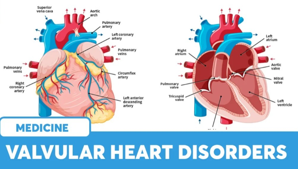
- Valvular heart disease is an abnormality or dysfunction in the valves in the heart.
- The heart only allows blood to flow in one direction. The valves in the heart are responsible for this. Because the valve prevents the back flow of blood.
- This valve keeps opening and closing, due to which the heart can flow blood in one direction.
- This valve has leaflets, cusps, through which the valve closes.
- There are mainly two types of problems in the valve: stenosis and regurgitation.
- In stenosis, the leaflets do not open properly wide, meaning the valve becomes narrowed. Due to which small amounts of blood flow through the valve.
- In regurgitation, the valve does not close properly, which causes small amounts of blood to leak backward. This is known as insufficiency or incompetence.
Define mitral stenosis
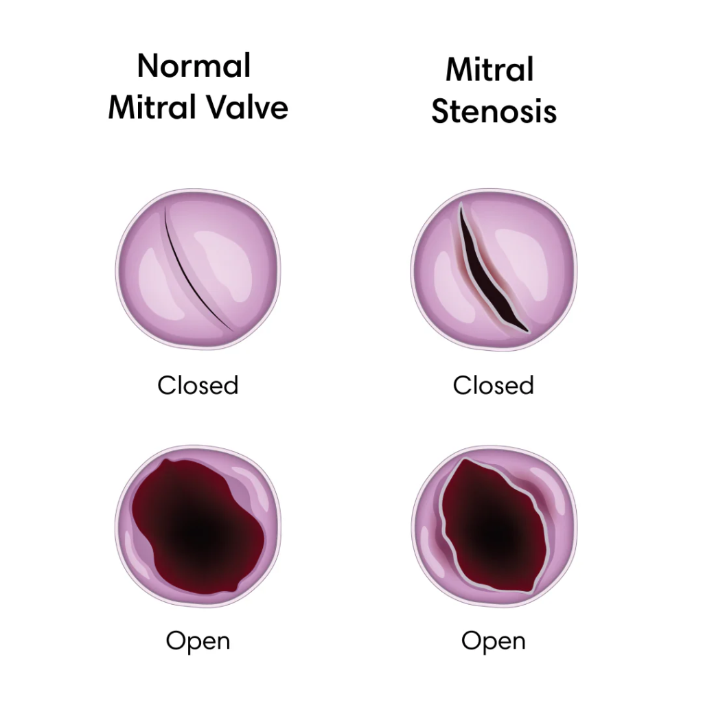
- Mitral stenosis is a form of valvular heart disease in which the opening or orifice of the mitral valve is narrowed, restricting blood flow from the left atrium to the left ventricle. Which causes blood to collect in the left atrium.
Write causes of mitral stenosis
- Rheumatic fever
- Congenital heart defect (which affects the mitral valve)
- Infective endocarditis
- Autoimmune disease (SLE)
- Age related degeneration
- Calcification of mitral valve
Write sign and symptoms seen in mitral stenosis (Write sign and symptoms seen in mitral stenosis) Stenosis)
- Shortness of Breath (After Exercise and at Night Time)
- Orthopnea
- Fatigue and Weakness
- Heart Palpitations
- Irregular Heart Beat
- Chest pain
- Swelling in ankles and feet
- Cough
- Frequent respiratory infections
- Loud, rumbling heart murmur
Write diagnostic evaluation of mitral stenosis
- History Collection
- Physical Examination
- Echocardiogram
- Electrocardiogram
- Cardiac Catheterization
- Chest X-ray
- MRI and CT scan
Write medical management of mitral stenosis
Diuretics: Pulmonary congestion and fluid overload Use diuretic drugs to reduce.
Beta blocker / calcium channel blocker:Use beta blockers and calcium channel blockers to control heart rate and relieve symptoms like dyspnea and palpitations.
Anticoagulant: Provide anticoagulant drugs to prevent blood clots and thromboembolism and to thin the blood.
Antiarrhythmic Medicine: Administering antiarrhythmic medicine to control the rhythm.
Write surgical management of mitral stenosis
Repair of valve
- Surgical repair of the valve is also known as ‘Commissurotomy’ . In which the fused leaflets are separated. The places where these leaflets meet are known as commissures. Commissurotomy is performed by two methods.
I) Open Commissurotomy: This method is considered as the traditional method. In which the leaflets fused through open heart surgery are directly separated.
II) Close Commissurotomy: The close commissurotomy method is not used at present. In which the surgeon makes a small incision on the heart and inserts a specially designed device through it and dilates the valve through it, i.e. the leaflets are separated.
Ballon valvuloplasty
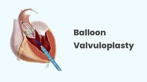
- Valvuloplasty is also known as ‘percutaneous balloon valvuloplasty’. Valvuloplasty is a minimally invasive procedure used for the management of heart valve disease. In this procedure, a deflated balloon attached to the tip of a catheter is inserted through blood vessels, mainly in the groin area, and guided to the heart valve, where the balloon is placed and inflated, thereby widening the narrow or stenotic valve and enlarging the valve opening. So that blood flow can be improved.
Valve replacement
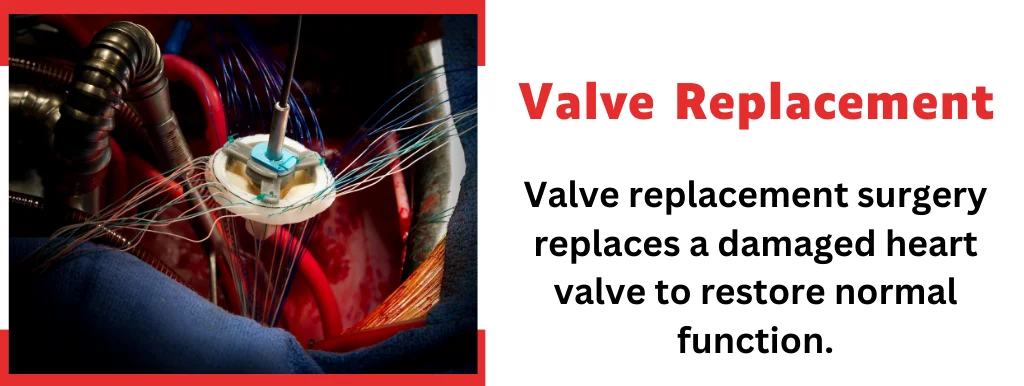
- Valve replacement is a surgical procedure used to manage severe valvular stenosis. In which the damaged heart valve is surgically removed and replaced with a mechanical or biological (bioprosthetic) valve. Mechanical valves are made of titanium, carbon, and other similar materials that do not interact with the body. In addition, lifelong blood thinners are given with it to prevent blood clots. While biological valves are made from animal tissue (porcine, bovine) such as valve tissue taken from pigs. Life-long blood thinners are not given in biological valve replacement.
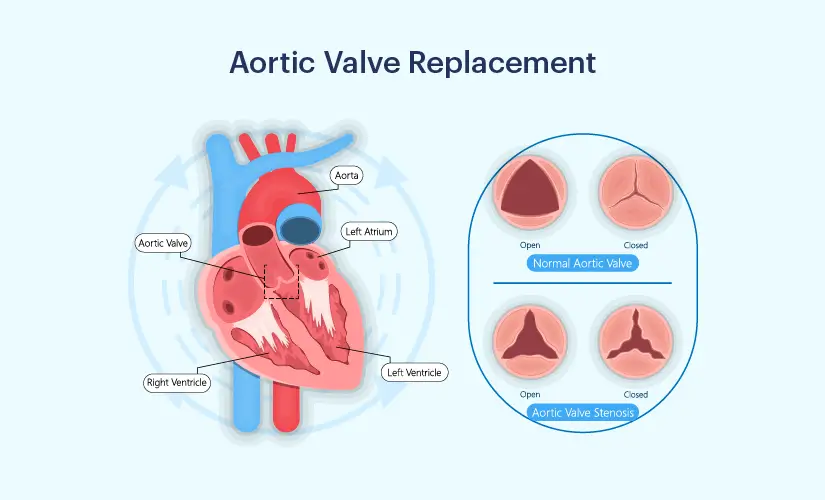
Define aortic stenosis
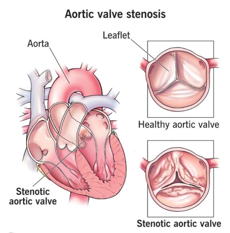
- Aortic stenosis is a form of valvular heart disease. In which the opening or orifice of the aortic valve is found to be narrowed, which restricts the blood flow from the heart to the aorta.
Write causes of aortic stenosis
- Calcification of aortic valve
- Congenital heart defect
- Rheumatic fever
- Endocarditis
- Degenerative changes
- Hypertension
- Hypercholesterolemia
- Marfan syndrome
- Ankylosing Spondylitis
Write sign and symptoms seen in aortic stenosis
- Chest pain
- Fancy, dyspnea
- Fatigue
- Shortness of breath (exertion)
- Heart palpitations
- Irregular heart beat
- Rapid pulse
Write diagnostic evaluation of aortic stenosis (right Diagnostic Evaluation of Aortic Stenosis)
- History Collection
- Physical Examination
- Echocardiogram
- Electrocardiogram
- Cardiac Catheterization
- Chest X-ray
- MRI and CT scan
Write medical management of aortic stenosis
Diuretics: Diuretics drugs are used to reduce pulmonary congestion and fluid overload To do.
Beta blocker / Calcium channel blocker: Use beta blockers and calcium channel blockers to control heart rate and relieve symptoms such as dyspnea and palpitations.
Anticoagulant: Provide anticoagulant drugs to prevent blood clots and thromboembolism and to thin the blood.
Antiarrhythmic Medicine : Administering antiarrhythmic medicine to control the rhythm.
Write surgical management of aortic stenosis
- Valvuloplasty
- Aortic valve replacement
- Transcatheter Aortic Valve Replacement
Ballon valvuloplasty
- Valvuloplasty is also known as ‘Percutaneous Balloon Valvuloplasty’ . Valvuloplasty is a minimally invasive procedure used for the management of heart valve disease. In this procedure, a deflated balloon attached to the tip of a catheter is inserted through blood vessels, mainly in the groin area, and guided to the heart valve, where the balloon is placed and inflated, thereby widening the narrow or stenotic valve and enlarging the valve opening. So that blood flow can be improved.
Aortic valve replacement
- Valve replacement is a surgical procedure used to manage severe valvular stenosis. In which the damaged heart valve is surgically removed through a large incision in the chest and replaced with a mechanical or biological (bioprosthetic) valve. That is, open heart surgery is performed. Mechanical valves are made of titanium, carbon, and other similar materials that do not interact with the body. In addition, lifelong blood thinners are given to prevent blood clots. While biological valves are made from animal tissue (porcine, bovine), such as valve tissue taken from pigs. Lifelong blood thinners are not given in biological valve replacement.
Transcatheter aortic valve replacement
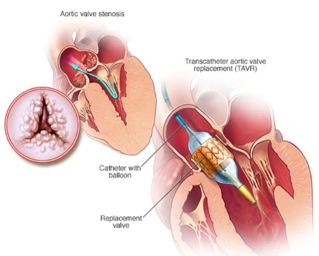
- This is a minimally invasive procedure. In which a catheter is inserted through the femoral artery in the groin area and placed up to the damaged valve. Then, with the help of a catheter, a new valve is placed in place of the damaged valve. The new valve is expanded and the leaflets of the old valve are pushed, and the function of the damaged valve is performed by this new valve.
Define Tricuspid stenosis
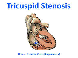
- Tricuspid stenosis is a valvular condition in which the tricuspid valve becomes narrowed, restricting the blood flow from the right atrium to the right ventricle.
Write causes of tricuspid stenosis
- Rheumatic heart disease
- Congenital heart defect
- Carcinoid syndrome
- Infective endocarditis
- Age Related Changes
- Systemic Lupus Erythematosus
- Calcification of Valve
Write sign and symptoms seen in tricuspid stenosis
- Abdominal swelling (ascites)
- Liver enlargement (hepatomegaly)
- Jugular vein distension
- Ankle edema
- Diastolic murmur
- Opening snap
- Cyanosis
- Fatigue
- Shortness of breath
- Palpitation
Write diagnostic evaluation of tricuspid stenosis
- History Collection
- Physical Examination
- Echocardiogram
- Electrocardiogram
- Cardiac Catheterization
- Chest X-ray
- MRI and CT scan
Write medical management of tricuspid stenosis
Diuretics: Diuretic drugs are used to reduce fluid overload and symptoms of right-sided heart failure.
Beta blocker / Calcium channel blocker: Use beta blockers and calcium channel blockers to control heart rate and relieve symptoms like dyspnea and palpitations.
Anticoagulant: Provide anticoagulant drugs to prevent blood clots and thromboembolism and to thin the blood.
Antiarrhythmic Medicine: Administer antiarrhythmic medicine to control the rhythm.
Antibiotics: Provide antibiotics to prevent bacterial infection.
Write surgical management of tricuspid stenosis
Repair of valve
- Surgical repair of the valve is called ‘Commissurotomy’. Also known as. In which the fused leaflets are separated. The places where these leaflets meet are known as commissures. Commissurotomy is done by two methods.
I) Open Commissurotomy: This method is considered as the traditional method. In which the fused leaflets are directly separated through open heart surgery.
II) Close Commissurotomy: The close commissurotomy method is not used at present. In which the surgeon makes a small incision on the heart and inserts a specially designed device through it and dilates the valve through it, i.e. the leaflets are separated.
De vega annuloplasty
- De vega annuloplasty is a surgical method used to manage tricuspid stenosis and regurgitation. In which the size of the tricuspid annulus is reduced and a ring-like structure is placed in it that supports the leaflets of the valve.
Tricuspid valve replacement
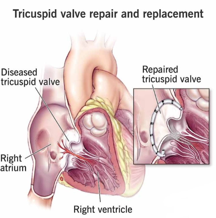
- Tricuspid valve replacement is a surgical procedure used for the management of severe valvular stenosis. In which the damaged heart valve is surgically removed through a large incision in the chest and replaced with a mechanical or biological (bioprosthetic) valve. That is, open heart surgery is performed. Mechanical valves are made of titanium, carbon, and other materials that do not interact with the body. In addition, lifelong blood thinners are given to prevent blood clots. While biological valves are made from animal tissue (porcine, bovine) such as valve tissue taken from pigs. Lifelong blood thinners are not given in biological valve replacement.
Define pulmonary valve stenosis (Define pulmonary valve stenosis)
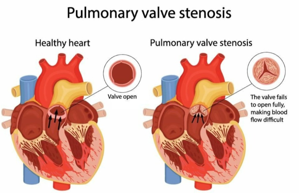
- Pulmonary valve stenosis is a narrowing of the pulmonary valve. Which blocks the blood flow from the right ventricle to the pulmonary artery.
Write causes of pulmonary valve stenosis (Write causes of pulmonary valve stenosis)
- Congenital heart disease / defect (Noonan syndrome, tetralogy of Fallot)
- Rheumatic fever
- Carcinoid syndrome
- Endocarditis
- Degenerative changes
Write sign and symptoms seen in pulmonary valve stenosis
- Heart Murmur (First Indication)
- Exercional Dyspnea (Shortness of Breath)
- Fatigue
- Chest Pain
- Palpitation
- Syncope
- Disease
- Cyanosis
- Right ventricular hypertrophy
- Rapid and irregular pulse
- Swelling in leg and abdomen
- Hepatomegaly
Write diagnostic evaluation of pulmonary valve stenosis
- History Collection
- Physical Examination
- Echocardiogram
- Electrocardiogram
- Cardiac Catheterization
- Chest X-ray
- MRI and CT Scan
Write medical management of pulmonary valve stenosis
Diuretics: Use diuretic drugs to reduce fluid overload.
Beta blocker/calcium channel blocker: Use beta blocker and calcium channel blocker to control heart rate and relieve symptoms like dyspnea and palpitations.
Anticoagulant:To prevent blood clots and thromboembolism and to thin the blood Provide anticoagulant drugs to thin.
Antiarrhythmic Medicine: Administer antiarrhythmic medicine to control the rhythm.
Prostaglandin E1 : If pulmonary valve stenosis is present in the neonate, then administer prostaglandin E1 to maintain ductal patency and adequate pulmonary blood flow.
Antibiotics :Provide antibiotics to prevent bacterial infection.
Write surgical management of pulmonary valve stenosis
Pulmonary valve repair
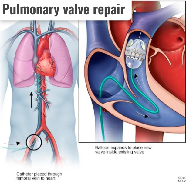
- Pulmonary valve repair involves making an incision in the chest and widening the pulmonary valve to improve blood flow.
Patch angioplasty (Patch Angioplasty)
- In patch angioplasty, a patch made of synthetic material or the patient’s own pericardium is used to widen a narrow section of the pulmonary artery or right ventricular outflow tract.
Balloon valvuloplasty (Valvuloplasty)
- Valvuloplasty is also known as ‘percutaneous balloon valvuloplasty’. Valvuloplasty is a minimally invasive procedure used for the management of heart valve disease. In this procedure, a deflated balloon attached to a catheter tip is inserted through the blood vessels, mainly in the groin area, and guided to the heart valve, where the balloon is placed and inflated. This widens the narrowed or stenotic valve and enlarges the valve opening. So that blood flow can be improved.
Pulmonary valve replacement
- Valve replacement is a surgical procedure used to manage severe valvular stenosis. In which the damaged heart valve is surgically removed through a large incision in the chest and replaced with a mechanical or biological (bioprosthetic) valve. That is, open heart surgery is performed. Mechanical valves are made of titanium, carbon, and other materials that do not interact with the body. In addition, lifelong blood thinners are given with them to prevent blood clots. While biological valves are made of animal tissue (porcine, bovine), such as valve tissue taken from pigs. Lifelong blood thinners are not given in biological valve replacement.
Inflammation and infections
Define pericarditis
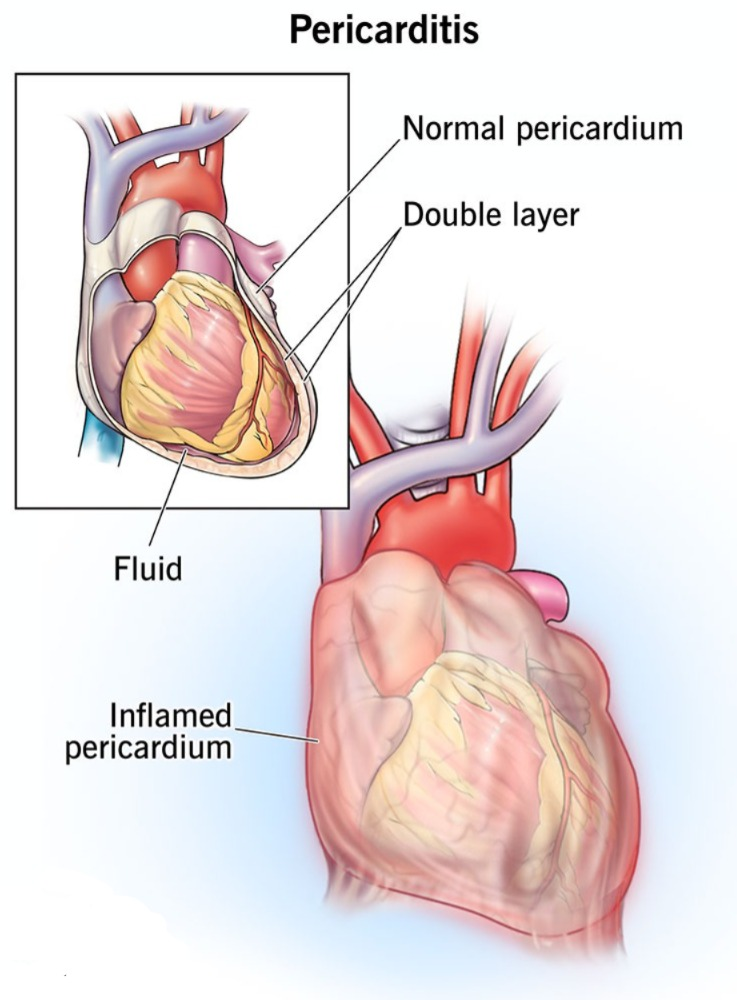
- Inflammation of the pericardium is known as pericarditis. The pericardium is the outermost layer of the heart which has a thin sac-like membrane-like structure.
Write classification of pericarditis
Pericarditis is classified based on the composition of the inflammatory exudate as follows:
Pericardial Effusion: In pericardial effusion, fluid accumulates in the pericardial sac, causing chest pain, dyspnea, and muffled heart sounds.
Constrictive Pericarditis: In constrictive pericarditis, chronic inflammation causes fibrous thickening and calcification of the pericardium, which restricts heart function and causes symptoms of heart failure.
Write causes of pericarditis
Idiopathic : The exact cause of pericarditis is unknown
Infection : Pericarditis can be caused by bacterial, viral or fungal infections. is.
Autoimmune disorders: Autoimmune conditions like rheumatoid arthritis, systemic lupus erythematosus, scleroderma can cause pericarditis.
Trauma: Blunt or penetrating trauma to the chest can cause pericarditis.
Cancer: Pericarditis can occur due to cancer metastasizing to the pericardium.
Medication:Drug-induced pericarditis is seen due to certain medications such as penicillin, phenytoin, procainamide.
Radiation therapy:Pericarditis can occur as a side effect of radiation therapy.
Write sign and symptoms seen in pericarditis
- Sharp and stabbing chest pain (radiating to the left shoulder and neck)
- Worsening when lying down and taking deep breaths (chest pain increases when lying down and taking deep breaths)
- Fever
- Cough
- Difficulty in breathing (dyspnea)
- Weakness and fatigue
- Swelling in legs, feet and abdomen
Write diagnostic evaluation of pericarditis (Right Diagnostic Evaluation of Pericarditis)
- History Collection
- Physical Examination
- Laboratory Tests (Blood Cell Count, C Reactive Protein, Erythrocyte Sedimentation Rate)
- Electrocardiogram
- Echocardiogram
- Chest X-ray
- Cardiac MRI and CT scan
- Pericardiocentesis
Write medical management of pericarditis
Medication and supportive care are used for the management of pericarditis.
Non-steroidal anti-inflammatory drugs: Use non-steroidal anti-inflammatory drugs such as ibuprofen, naproxen, aspirin to reduce inflammation and relieve chest pain
Colchicine: Colchicine is prescribed with NSAIDs to prevent recurrent episodes and reduce inflammation. (Colchicine is an anti-gout agent)
Corticosteroid: Use corticosteroid drugs when NSAIDs and colchicine medications are contraindicated or ineffective.
Analgesic: Provide analgesic drugs to relieve pain.
Antipyretic: Provide antipyretic drugs to reduce fever.
Pericardiocentesis: Pericardiocentesis is performed in cases of pericardial effusion and cardiac tamponade, in which The excess fluid in the pericardium is removed.
Pericardiectomy: A pericardiectomy is performed in conditions with chronic or recurrent pericarditis. In which part or the entire pericardium is surgically removed.
Write nursing management of pericarditis
Hyperthermia related to infection as evidenced by increased body temperature
Maintain body temperature
- Assess the patient’s condition.
- Monitor vital signs.
- Monitor temperature every two to four hours.
- Maintain the patient’s room temperature.
- Provide cold applications to the patient.
- If the patient feels cold, avoid cold applications and provide a blanket To do.
- Maintain adequate hydration.
- Instruct the patient to take oral fluids and administer IV fluids.
- Administer antipyretic and antibiotic medicines as prescribed by the doctor.
- Maintain records and reports.
Anxiety related to disease condition, uncertain prognosis, hospitilisation as evidence by verbalization, restlessness, agitation
Reduce anxiety
- Assess the patient’s condition.
- Signs of anxiety such as Assess for restlessness, insomnia.
- Pay attention to the patient’s psychological needs and listen carefully to the patient.
- Encourage the patient to express his feelings, discomfort and anxiety.
- Resolve all the patient’s doubts and queries.
- Provide the patient with knowledge about his condition and treatment so that his anxiety is removed and the patient feels confident. Can be done.
- Provide psychological support to the patient.
- Provide mind diversionary therapy and recreational therapy to the patient.
- Administer antianxiety agents.
Activity intolerance related to fatigue, dyspnea as evidenced by shortness of breath, abnormal heart rate
Increase activity level
- Assess the patient’s condition.
- Check the patient’s activity level.
- Provide bed rest to the patient initially.
- Then gradually encourage the patient to do range of motion exercises.
- Assist the patient with his/her activity.
- Provide rest to the patient between 2 activities.
- Check whether the patient experiences any breathing difficulty or palpitations during activity.
- If present, stop the patient’s activity and provide rest.
- Ask the patient to avoid lifting heavy objects.
- Ask the patient to avoid doing work that puts strain on the heart.
Define myocarditis (Define Myocarditis)
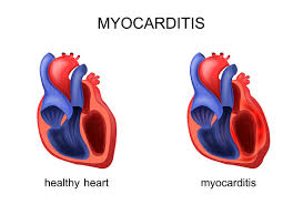
- Myocarditis is the infection and inflammation of the myocardium or heart muscle, which is the middle layer of the heart.
- Myocarditis causes the heart to become inflamed, which prevents it from pumping properly.
Write causes of myocarditis
Myocarditis is mainly caused by viral infections. It can also be caused by bacterial infections, fungal infections, parasitic infections, autoimmune disorders, and certain medications.
- Viral Infections: Common viruses such as adenovirus, coxsackievirus, influenza, enterovirus, herpes simplex virus can cause myocarditis.
- Bacterial Infections: Streptococcus, staphylococcus, tuberculosis, gonococcal infections can cause myocarditis.
- Fungal infections: Fungi like Aspergillus, Candida, Histoplasma can cause myocarditis.
- Parasite infections: Infections with parasites like Trypanosoma cruzi, Toxoplasma can cause myocarditis.
- Autoimmune disorders : Inflammation of the heart muscle is seen due to conditions such as autoimmune disorders such as lupus, rheumatoid arthritis, sarcoidosis and Wegener’s granulomatosis.
- Certain medications :Myocarditis can be caused by the use of certain medications such as drugs used for cancer treatment, immunosuppression medications.
- Toxic substances : Myocarditis can also be caused by exposure to toxic substances, such as heavy metals
Write sign & symptoms seen in myocarditis
- Various types of signs and symptoms are seen in myocarditis.
- Chest pain
- Shortness of breath
- Arrhythmia
- Rapid heart Beat
- Palpitation
- Fatigue
- Fever
- Headache
- Swelling in leg, ankle, foot
- Heart failure
Write diagnostic evaluation of myocarditis
- History collection
- Physical examination
- Electrocardiogram
- Echocardiogram
- Chest x-ray
- Cardiac MRI
- Endomyocardial biopsy
- Blood tests (cardiac enzymes, blood culture)
Write management of myocarditis (Right Management of Myocarditis)
✓ Heart Failure Management: If heart failure is present, the following medications should be used for its management.
- ACE Inhibitors : Use ACE inhibitor drugs to reduce afterload.
- Beta Blockers : Provide beta blocker group of medicines to control heart rate and reduce myocardial oxygen demand.
- Diuretics : Use diuretic drugs to manage fluid overload.
✓ Antiarrhythmic drug: Provide antiarrhythmic drug to control arrhythmia.
✓ Immunosuppressive therapy: Provide immunosuppressive drug to suppress immunity if myocarditis is caused by autoimmune disorder.
✓Antiviral therapy: If If myocarditis is caused by a viral infection, antiviral drugs should be administered to treat it.
✓ Antibiotic therapy: If myocarditis is caused by a bacterial infection, antibiotic drugs should be provided to treat it.
✓ Oxygen therapy: Provide supplemental oxygen if necessary. Write
nursing management of myocarditis
✓ Maintain body temperature
- Assess the patient’s condition.
- Monitor vital signs.
- Monitor temperature every two to four hours.
- Maintain patient’s room temperature.
- Provide cold applications to the patient.
- Avoid cold applications and provide blankets if the patient feels cold.
- Maintain adequate hydration.
- Asking the patient to take oral fluids and administering IV fluids.
- Administering antipyretic and antibiotic medicines as prescribed by the doctor.
- Maintaining records and reports.
✓ Maintaining cardiac output / Improving cardiac output
- Assess the patient’s condition.
- Monitor vital signs.
- Monitor cardiovascular status.
- Monitor ECG pattern.
- Monitor hemodynamic parameters.
- Maintain intake output chart To do.
- Provide the patient with a Fowler position or a high Fowler position.
- I.V. as prescribed by the doctor. Provide fluid.
- Administer the medicine prescribed by the doctor.
- Assess the side effects and effectiveness of the medicine.
- Maintain record reports.
✓ Increase activity level (increase activity level)
- Assess the patient’s condition.
- Check the patient’s activity level.
- Provide bed rest to the patient initially.
- Then gradually encourage the patient to do range of motion exercises.
- Assist the patient with his/her activities.
- Provide the patient with rest between 2 activities.
- Check whether the patient experiences any breathing difficulty or palpitations during the activity.
- If present, stop the patient’s activity and provide rest.
- Ask the patient to avoid lifting heavy objects.
- Avoid doing work that puts strain on the heart to say.
Define endocarditis
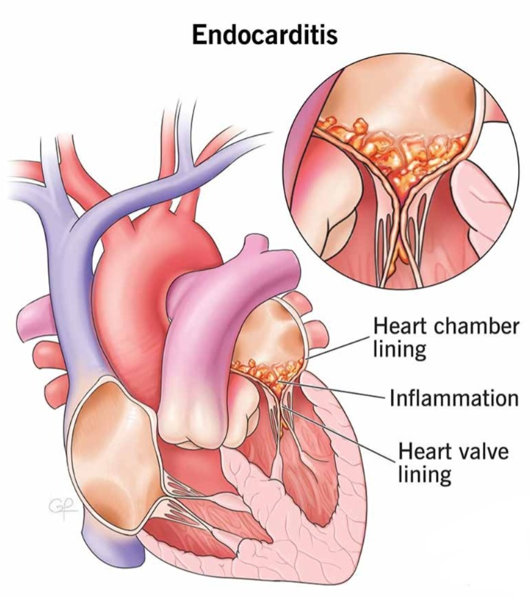
- Infection and inflammation of the endocardium is known as endocarditis. Which is the innermost layer of the heart.
- Infection of the inner lining of the heart chambers and heart valves is known as endocarditis.
- Which is mainly caused by bacterial, viral or fungal infections.
- In endocarditis, the heart valves are mainly damaged. (Mainly mitral valve)
Write causes of endocarditis (Write causes of endocarditis)
- Bacterial infection (Streptococcus, Staphylococcus, Enterococcus)
- Fungal infection (Candida, Aspergillus)
- Non-bacterial thrombotic endocarditis
- Autoimmune disease
- Trauma and injury
- Dental, urologic or gynecologic surgery, colonoscopy
- Immunosuppression
- Pre-existing heart condition
Write sign and symptoms of endocarditis
- Fever
- Chills
- Fatigue
- Shortness of breath Breath
- Chest Pain
- Night Sweats
- Heart Murmur
- Weight Loss
- Muscle and Joint Pain
- Swelling in Feet, Legs, Abdomen
Specific sign:
Petechiae (small red and purple spots on skin)
Osler’s node – osler’s node
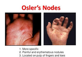
- (Painful raised red or purple subcutaneous lesion in finger and toes)
Janeway lesion :
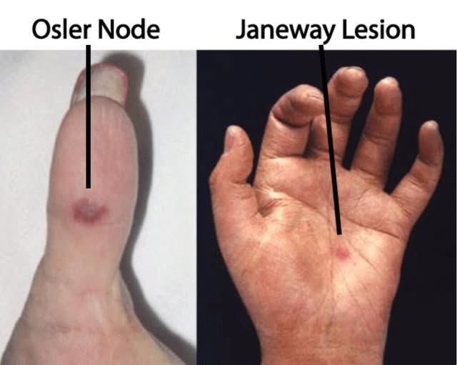
- Painless, flat red spot on the palm end Sol
splinter haemorrhage :
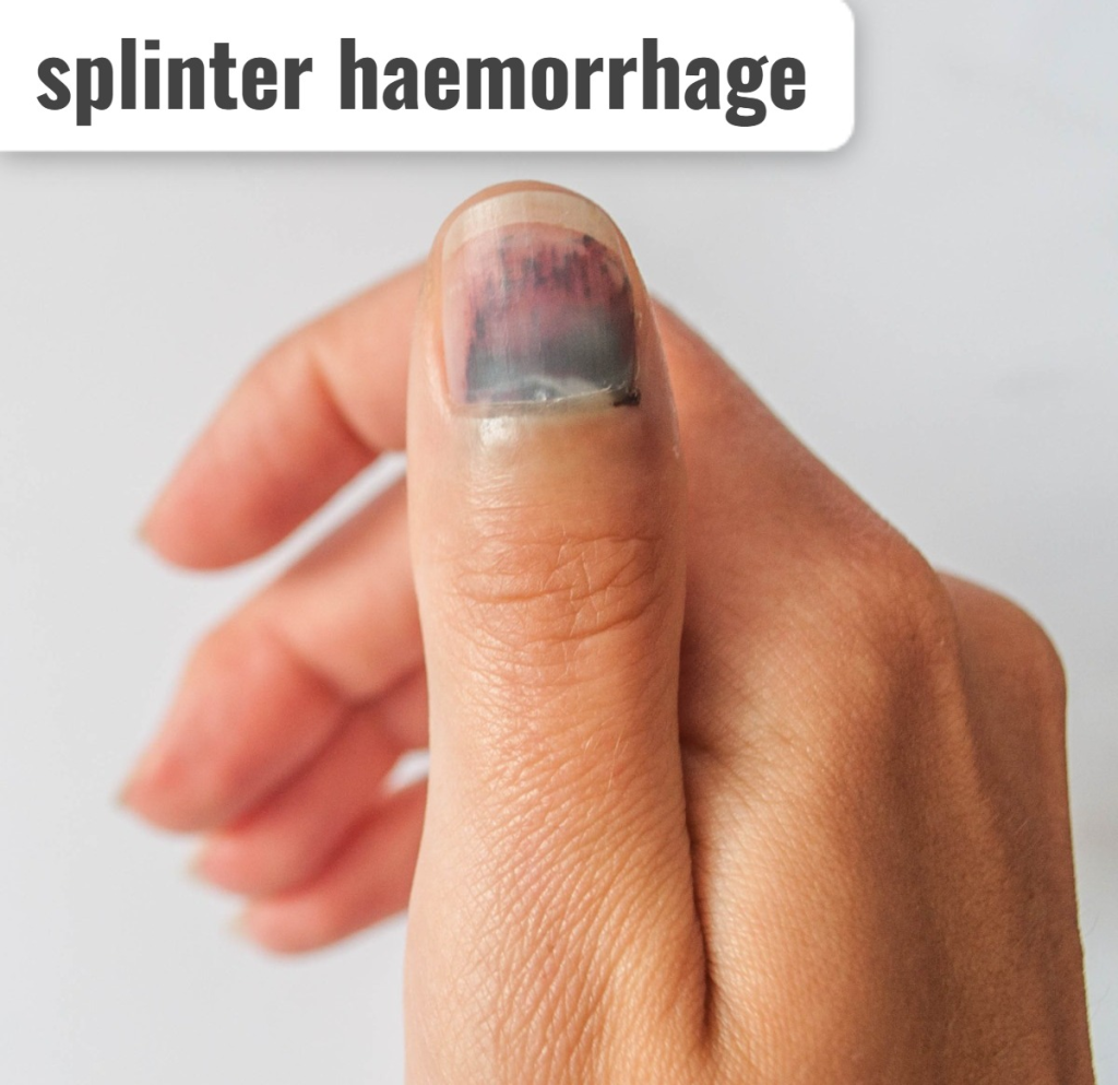
- Small, red and brown streak under the nail
Roth spot – roth spot :
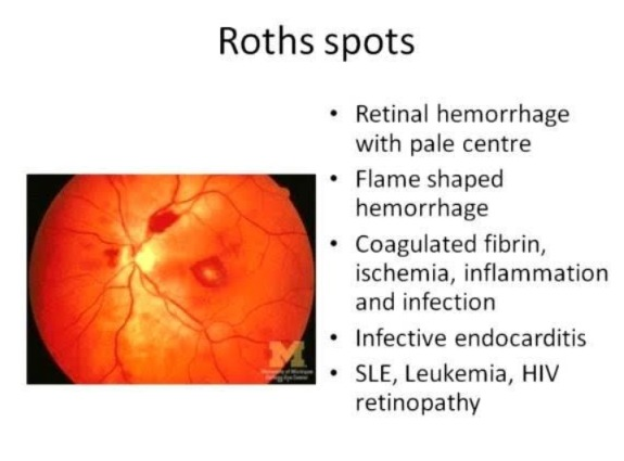
- (Retinal hemorrhage with pale center)
Write diagnostic evaluation of endocarditis
- History collection
- Physical examination
- Complete blood count
- Blood culture
- Inflammatory markers (CRP, ESR)
- Serological tests
- Echocardiography
- Electrocardiography
- Nuclear medicine tests
- Cardiac catheterization
Write management of endocarditis
✓ Antibiotics therapy :
- Empiric Antibiotics: Provide broad-spectrum antibiotics until blood culture results are available.
- Targeted Antibiotics: Administer targeted antibiotics after blood culture results. That is, administer antibiotics specific to the specific organism. This antibiotic course should be continued for 4 to 6 weeks.
- Staphylococcus aureus: In cases of Staphylococcus aureus bacterial infection, give nafcillin or oxacillin as well as vancomycin or daptomycin.
- Streptococci: In cases of Streptococci bacterial infection, give penicillin or ceftriaxone. Provide vancomycin in cases with penicillin resistance.
- Enterococci: In cases of enterococci bacterial infection, give ampicillin or vancomycin in combination with gentamicin.
✓Surgical intervention:
- Heart failure due to valve dysfunction In cases of uncontrolled infection, abscess formation, surgical intervention is performed.
- In which valve repair or valve transplantation is mainly performed.
Write nursing management of endocarditis
✓ Maintain body temperature (Maintain body temperature To do)
- Assess the patient’s condition.
- Monitor vital signs.
- Monitor temperature every two to four hours.
- Maintain the patient’s room temperature.
- Provide cold applications to the patient.
- If the patient feels cold, avoid cold applications and provide a blanket.
- Maintain adequate hydration.
- Instruct the patient to take oral fluids and administer IV fluids.
- Administer antipyretic and antibiotic medicines as prescribed by the doctor.
- Maintain records and reports.
✓ Maintaining Cardiac Output / Improving Cardiac Output
- Assessing the patient’s condition.
- Monitoring vital signs.
- Monitoring cardiovascular status.
- Monitor ECG pattern.
- Monitor hemodynamic parameters.
- Maintain intake output chart.
- Provide patient with Fowler position or high Fowler position.
- I.V. as prescribed by doctor. Provide fluid.
- Administer the medicine prescribed by the doctor.
- Assess the side effects and effectiveness of the medicine.
- Maintain record reports.
✓ Enhance activity level (increase activity level)