ENGLISH-MSN-II-unit 6 – CARDIAC DISORDER-PART-1
CARDIAC DISORDER:
TERMINOLOGY:
1) Acute coronary syndrome:
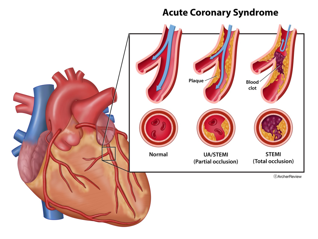
- Acute coronary syndrome A coronary It is caused by conditions that occur due to decreased blood flow due to arterial disease, including conditions like unstable angina, acute myocardial infarction.
2) Anemia:
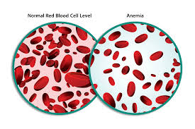
- Haemoglobin level being lower than normal. (Decreases the level of red blood cells or hemoglobin)
3) Aneurysm (aneurysm) :
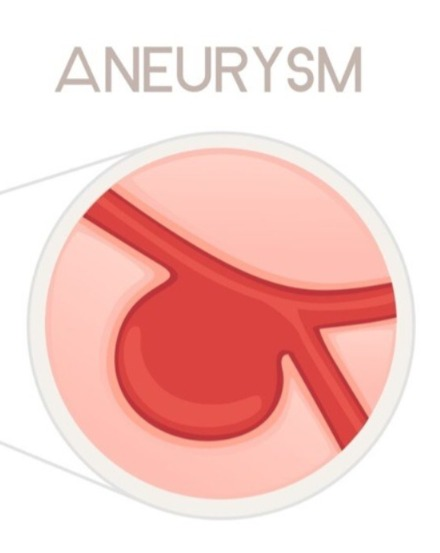
- Localized bulging or sac-like structure seen at weak points in the walls of blood vessels.
4) Angina pectoris (angina pectoris) :
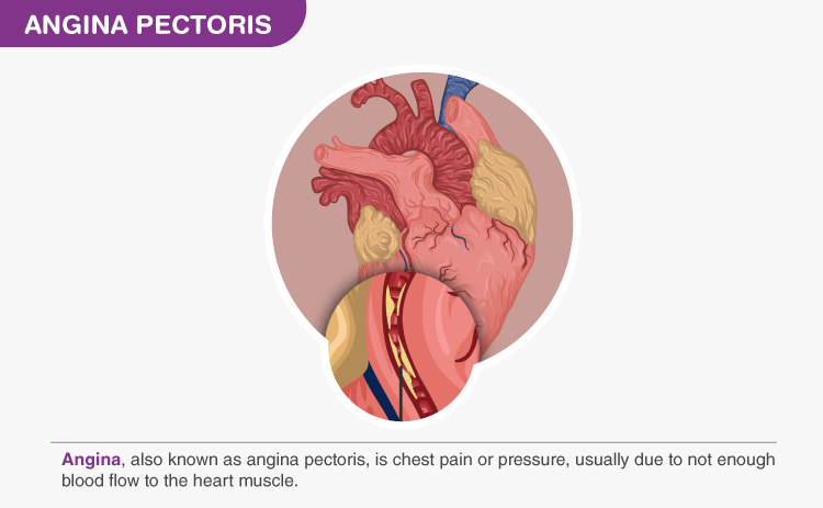
- Chest pain caused by reduced blood flow to the heart muscles is known as angina pectoris.
5) Angioplasty :
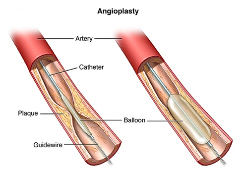
- Angioplasty is a surgical procedure used to open narrowed or blocked coronary arteries.
6) Aortic stenosis (Aortic stenosis):
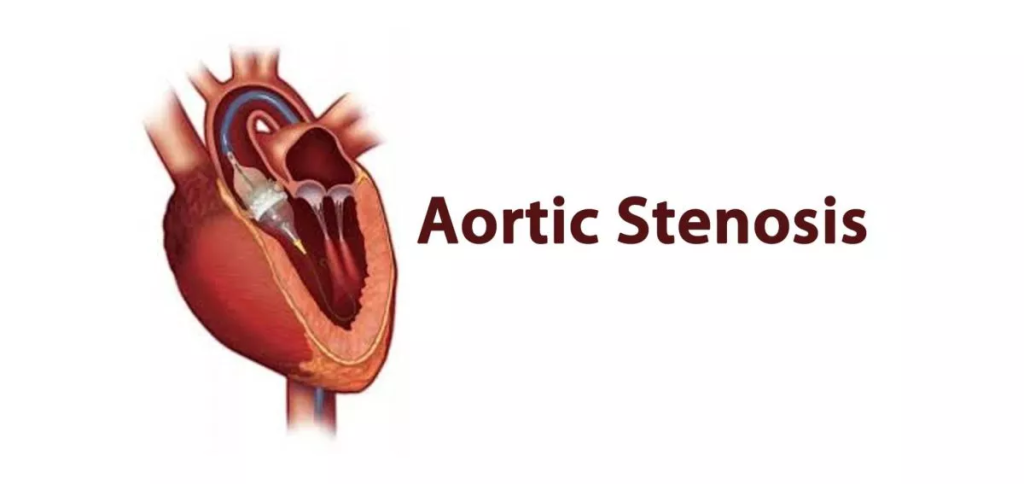
- Aortic valve narrow To happen.
7) Arterioscelrosis :

- Hardening and Thickening of the Artery.
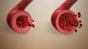
8) Atherosclerosis (atherosclerosis) :

- Atherosclerosis is a type of condition. In which the lumen becomes narrowed due to plaque buildup on the artery wall.
9) Athroma (Atheroma):
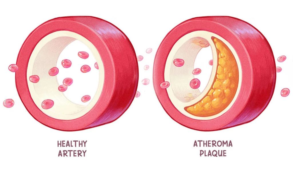
- Fatty deposits or plaque formations found on the walls of arteries are known as atheroma.
10) Buerger’s disease:
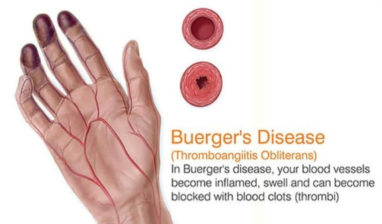
- In Buerger’s disease, inflammation and thrombosis are seen in the medium and small sized arteries in the arms and legs, due to which blood flow is reduced.
11) Cardiac arrest:
- Sudden and Unexpected Loss of Heart Function
12) Cardiac catheterization (Cardiac catheterization) :
- Cardiac catheterization is an invasive procedure used to diagnose heart conditions. Used to diagnose and treat.
13) Cardiac output (Cardiac output) :
- The amount of blood flow pumped by the heart in one minute is known as cardiac output.
14) Cardiomyopathy:
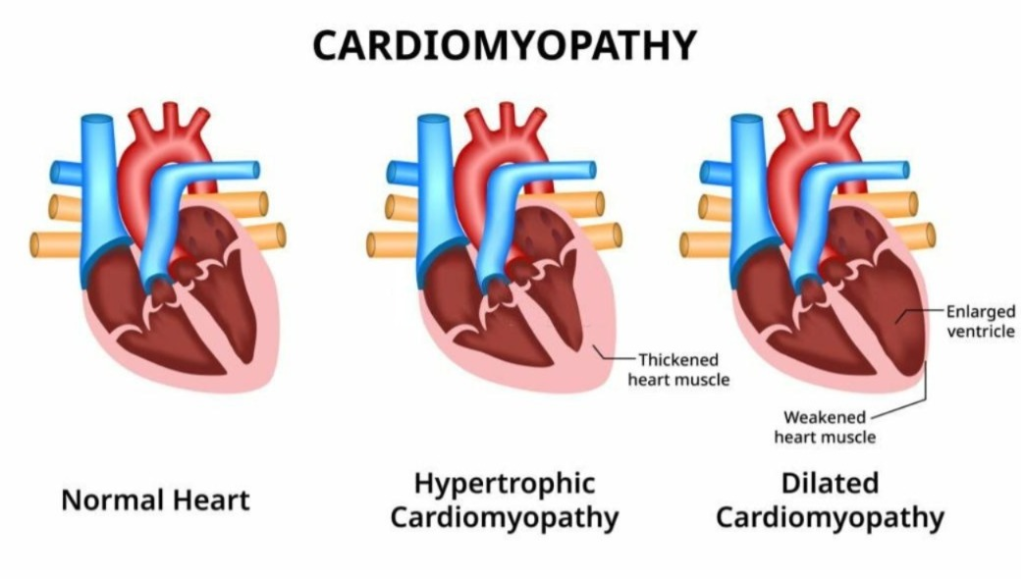
- Cardiomyopathy A heart muscle It is a disease in which the heart becomes enlarged, stiff and thick, due to which the heart cannot pump effectively.
15) Congestive heart failure (Congestive heart failure):
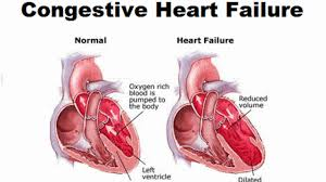
- Congestive heart failure is a chronic condition in which the heart is unable to pump blood effectively, causing fluid buildup in the lungs and liver.
16) Creatine kinase:

- Creatine kinase is a type of enzyme found in the heart, brain, and skeletal muscles.
17) Defibrillation:
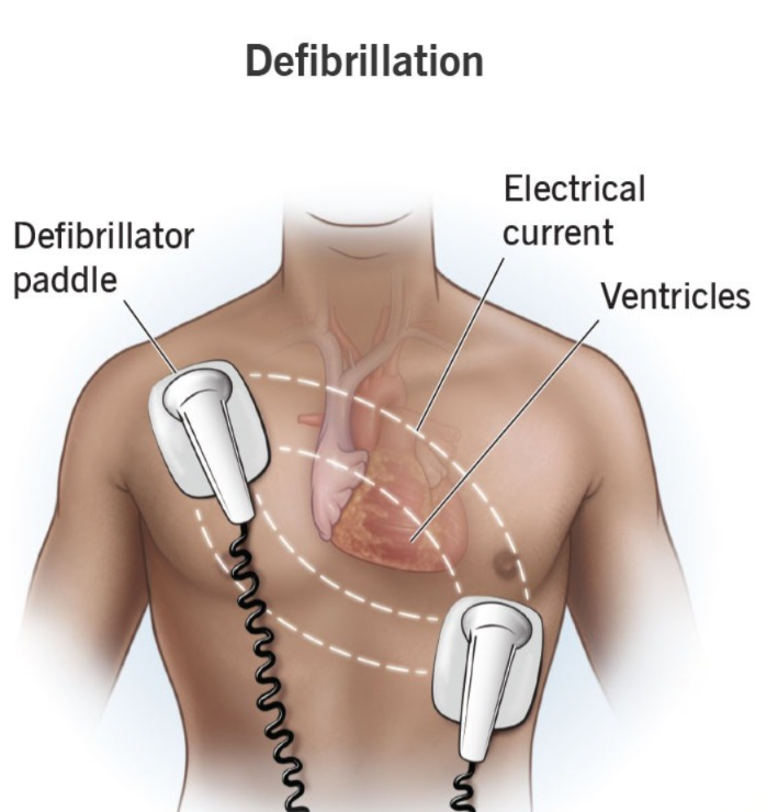
- Defibrillation is a medical procedure in which an electrical shock is delivered to the heart to restore a normal heart rhythm.
18) Dyslipidemia:
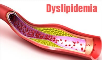
- Abnormal lipid level in blood
19) Dysrhythmia :

- Abnormal Heart Rhythm
20) Endocarditis (endocarditis) :
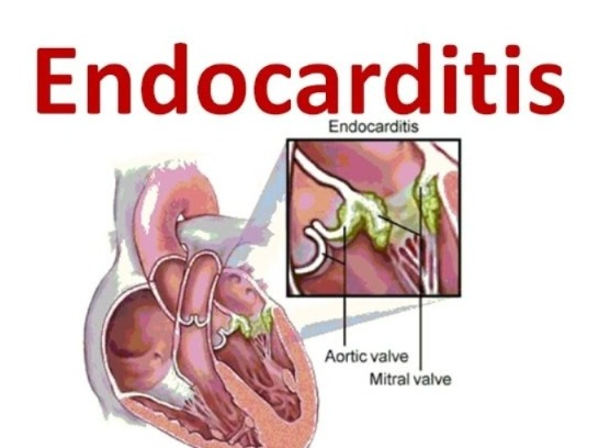
- Inflammation of inner layer of heart / Inflammation of Endocardium
21) Heart failure:
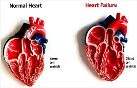
- In heart failure, the heart fails to pump enough blood.
22) High density lipoprotein (High density lipoprotein) :
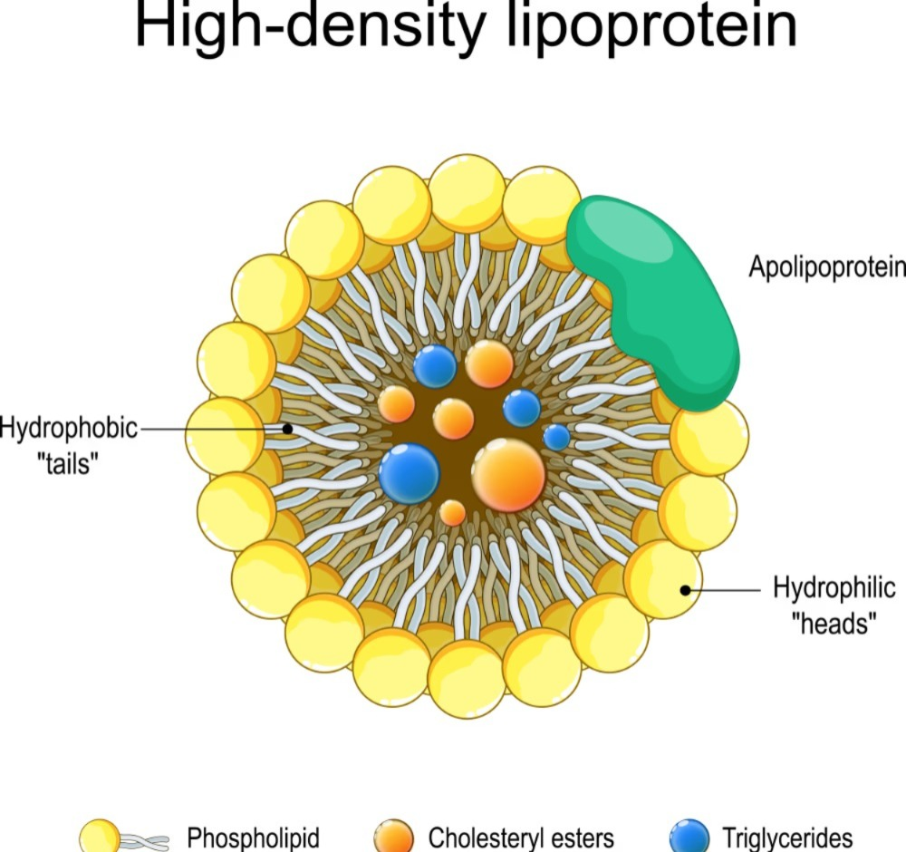
- High-density lipoprotein is a type of cholesterol known as good cholesterol. Also known as cholesterol because it helps remove cholesterol from the blood.
23) Hemophilia :
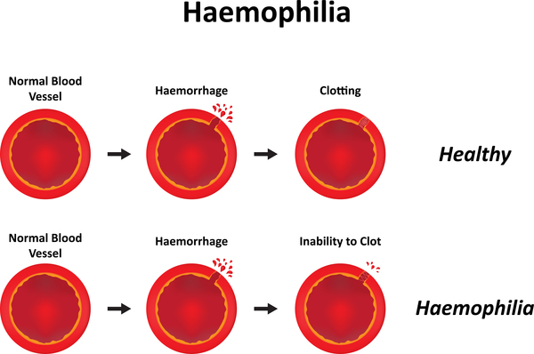
- Hemophilia is a genetic disorder in which the blood does not clot properly, which causes prolonged bleeding.
24) Hypertension :

- High level of blood pressure (below 140/90 mmHg)
25) Hypotension:
- Low level of blood pressure (below 90/60 mmHg)
26) Heterograft:

- Heterograft also known as xenograft is a type of tissue graft in which tissue graft from one donor species is transferred to a recipient of another species.
27) Homograft:

- Homograft is also known as allograft. Which is a type of tissue graft in which the donor and recipient of the tissue graft are of the same species.
28) Ischemia (Ischemia) :

- Reduces blood flow to the particular part of the body
29) Leukocytes (Leukocytes) :

- Leukocytes means white blood cells
30) Leukemia :
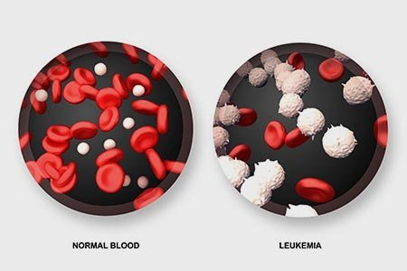
- Leukemia is a type of cancer that affects the blood and bone marrow.
31) Leukopenia :
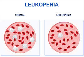
- Low level of white blood cells / Decrease number in white blood cells
32) Low density lipoprotein (Low density lipoprotein) :
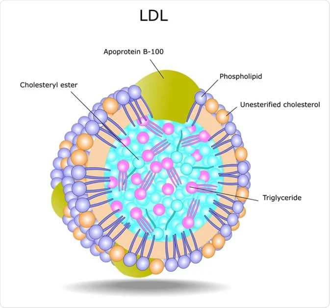
- Low density lipoprotein is a type of cholesterol also known as bad cholesterol. Due to the increase in its level, plaque formation is seen in the arteries.
33) Microcytosis:

- Red blood cells are smaller than their normal size.
34) Murmur :
- Abnormal heart Sound
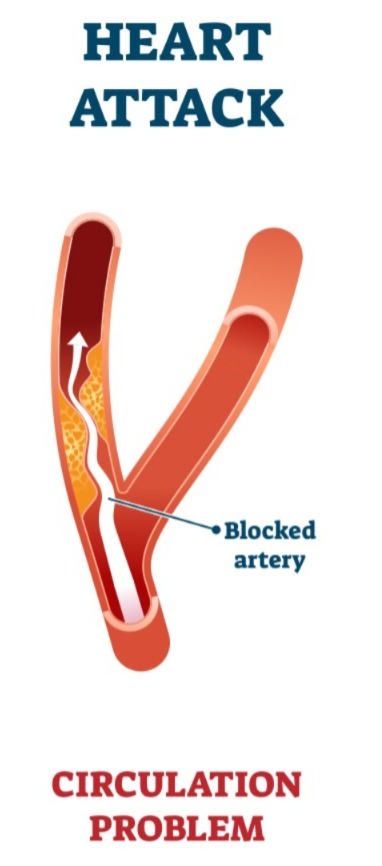
35) Myocardial infarction:
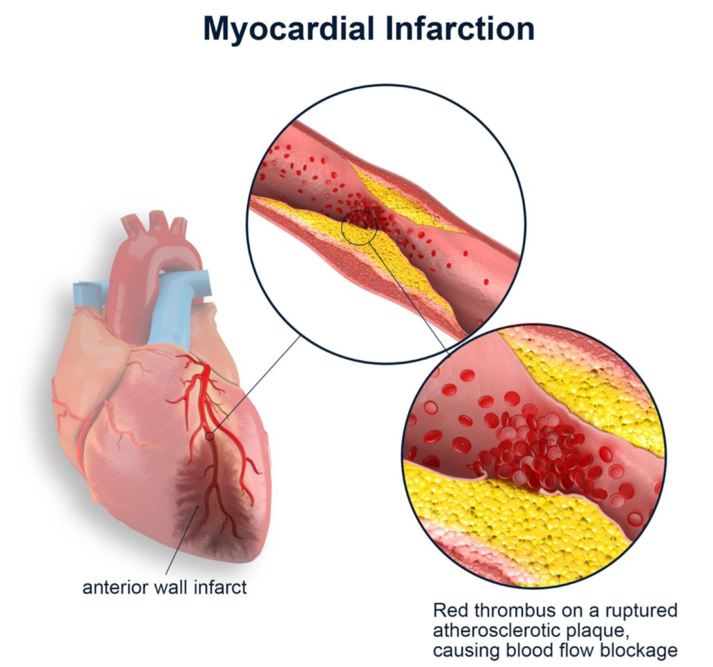
- This is a medical emergency in which the blood flow to the heart muscles is blocked.
36) Myocardial ischemia:
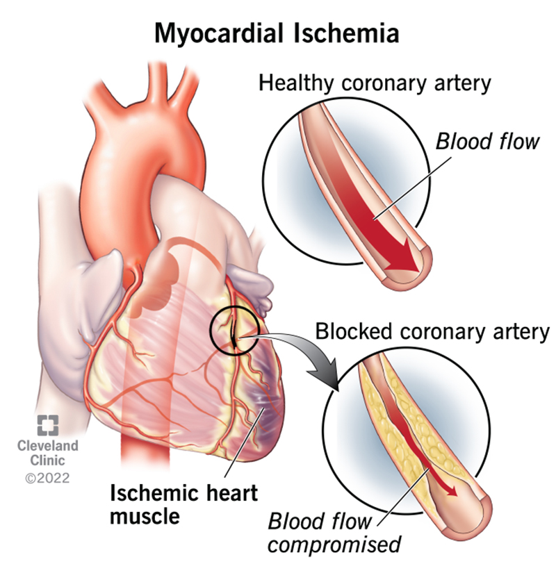
- This is a type of condition in which the heart muscles do not receive enough oxygen due to insufficient blood flow.
37) Myocarditis:
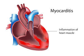
- Inflammation of myocardium.
38) Neutropenia:
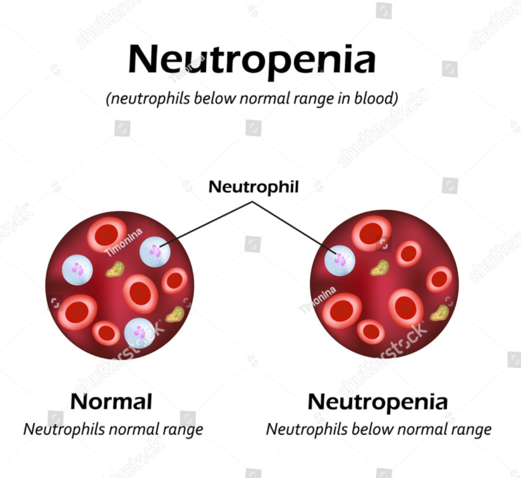
- Decrease number in neutrophils
39) Pancytopenia :
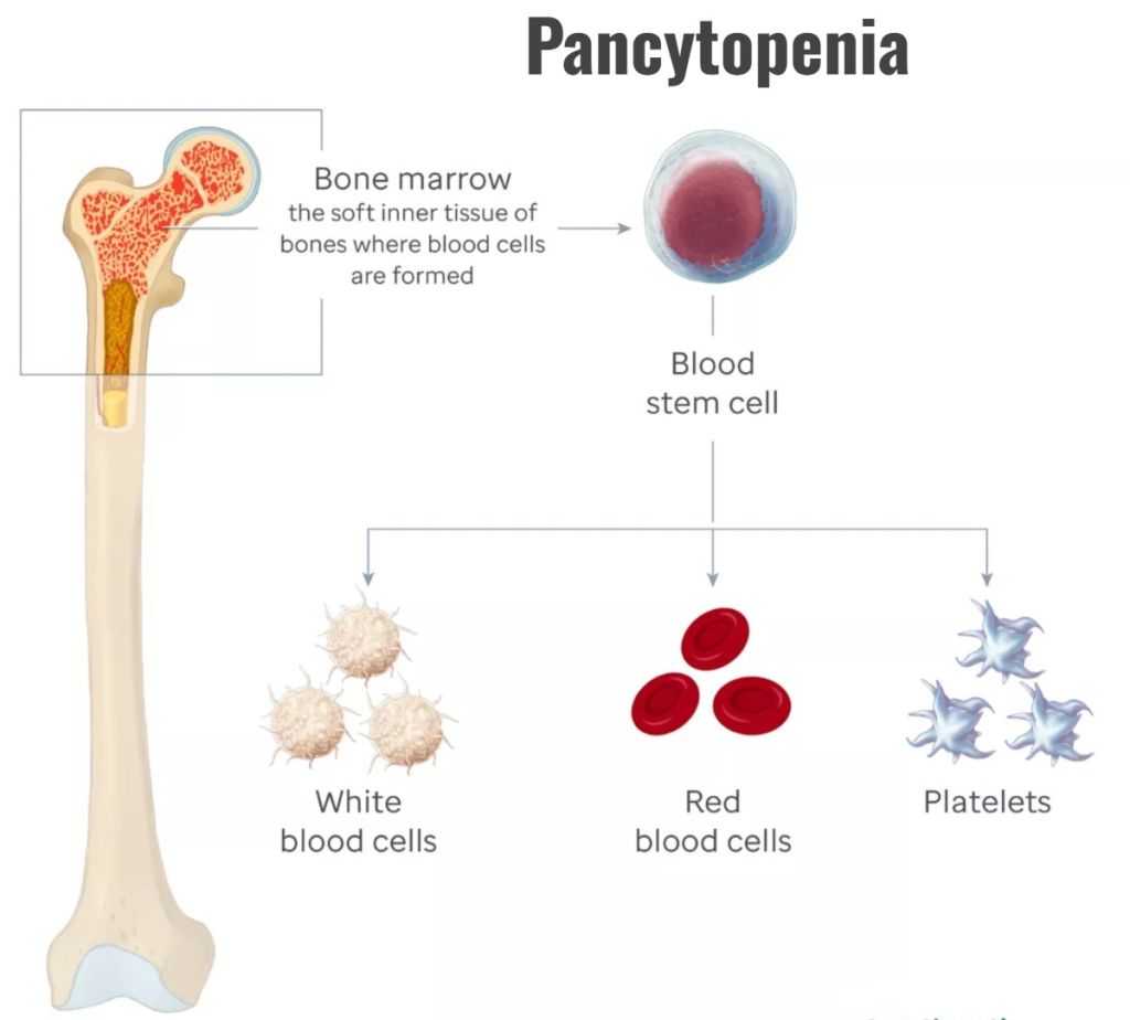
- Decrees in the number of three types of Blood cells (RBC, WBC, platelet)
40) Pericardiocentesis:
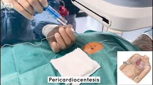
- Pericardiocentesis is a procedure in which a needle is inserted into the pericardial sac and the pericardial fluid is drained/removed.
41) Pericardiotomy :
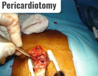
- Opening in Pericardium
42) Pericarditis :

- Inflammation of Pericardium
43) Poikilocytosis :

- Abnormal shape of red blood cell
44) Polycythemia:

- Increase the number of red blood cells
45) Postural hypotension:

- Postural hypotension as orthostatic hypotension Also known as. In which there is a sudden drop in blood pressure from a sitting or lying down position to a standing position.
46) Pulmonary edema (pulmonary edema) :
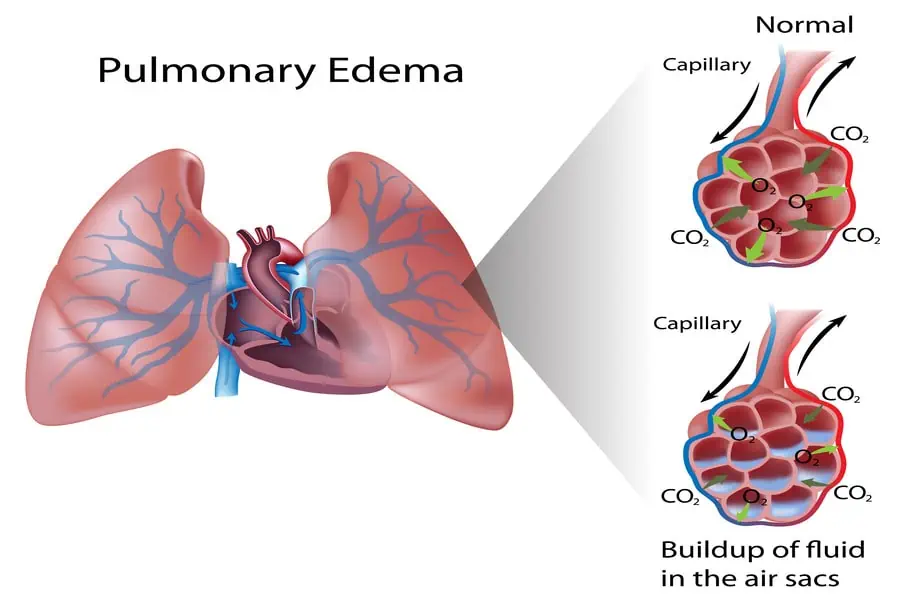
- Fluid accumulation in the lungs.
47) Regurgitation :
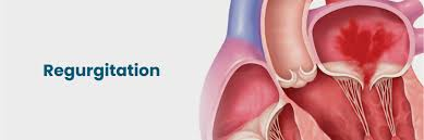
- Backward flow off Fluid. / Backflow of blood occurs due to the valves in the heart not closing properly, which is known as regurgitation.
48) Rubor :
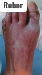
- Reddish blue discoloration of extremities indicating severe peripheral artery damage.
49) Stenosis :
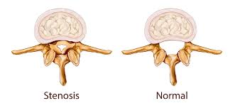
- Stenosis is the narrowing of an opening or passage in the body. To happen.
50) Stent :
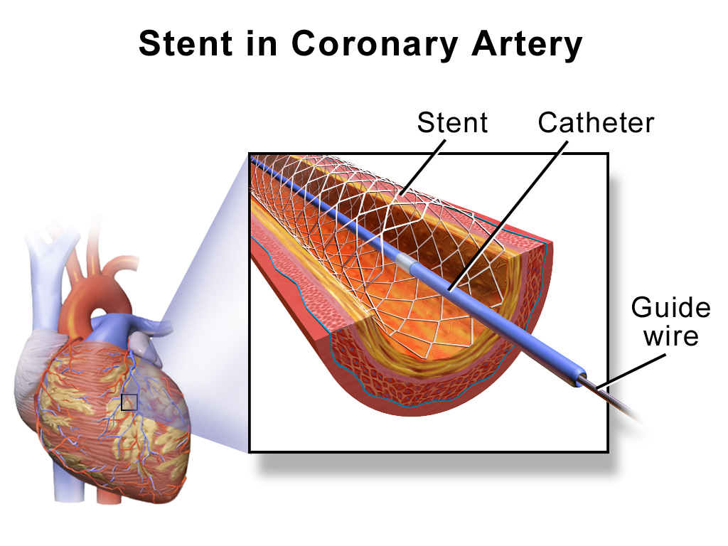
- A stent is a type of small mesh tube-like structure that is placed in an artery or duct to keep it open.
51) Streptokinase (Streptokinase) :

- Streptokinase is a type of medicine used to treat blood clots. happens to dissolve.
52) Thalassemia :
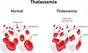
- Thalassemia is a hereditary hemolytic disease in which abnormal hemoglobin production is observed, due to which the condition of anemia is observed.
53) Thrombin :
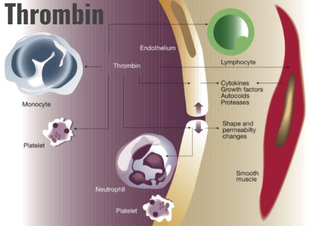
- Thrombin is present in blood plasma An enzyme that is needed to convert blood fibrinogen into fibrin so that blood can clot.
54) Thrombocyte :

- Thrombocyte is the term used for platelets.
55) Thrombocytopenia (Thrombocytopenia) :
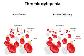
- Low platelet count means a decrease in platelet count.
56) Thrombosis:
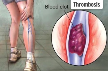
- Clot (thrombus) formation inside blood vessels.
57) Thrombocytosis:

- High platelet count means increase in platelet count To happen.
58) Thrombolytic:
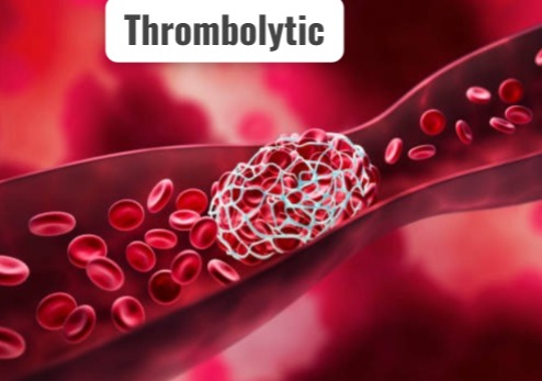
- Agents that break down or dissolve blood clots are known as thrombolytics.
59) Troponin:

- Troponin is a globular protein complex that is involved in muscle contraction. Associated with.
60) Valvuloplasty :
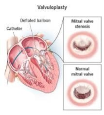
- Repairing of Narrowing and Stenosis Valve
61) Vasoconstrictor :

- Blood vessels constrict or Agent used for narrowing
62) Vasodilator :
- Agent used to dilate blood vessels
63) Ventricular tachycardia (Ventricular tachycardia) :
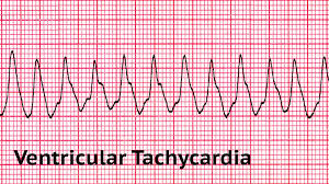
- This is a type of abnormal heart rhythm in which the heart beats fast and irregularly. Heartbeat is seen which originates from the lower chamber of the heart i.e. ventricle.
64) Vericose vein (Varicose vein) :
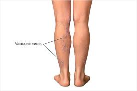
- A vein becomes enlarged, twisted, and swollen. which is mainly found in the leg.
Physical examination / objective data
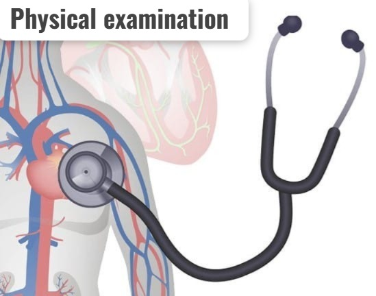
- Inspection, palpation, and auscultation methods are used in the examination of the cardiovascular system.
Inspection
✓ General Appearance :
Assess the patient’s general appearance. Such as assessing conscious level (alert, lethargic, comatose), mental status (oriented to time, place, person) and distress level.
✓ Inspection of skin:
- Check the color of the skin and mucous membranes.
- Check whether central cyanosis and peripheral cyanosis are present in the skin.
- Assess skin for temperature, texture, ecchymosis, xanthomas.
✓ Inspection of Extremities:
- Assess hands, arms, legs, and feet for skin and vascular changes.
- Swelling in the Extremities Inspect whether it is present or not.
- Check whether cyanosis and clubbing are present in the fingers and toes.
✓ Inspection of Chest:
- Check whether any kind of scar or surgical mark is present on the chest.
- Inspect the position of the trachea.
- Assess the shape of the chest.
- Check whether any dilated or engorged veins are visible on the chest wall.
- Assess the apex beat and other pulsations.
- In addition, examine the jugular vein.
Palpation

- The palmar surface of the finger is used in palpation.
- First check the body temperature.
- Palpat the pulse points in the body.
- Check blood pressure.
- Check whether swelling is present in the extremities with the help of palpation. Check whether pitting edema is present.
- Palpate rigidity of vessels.
- Palpate the chest for apical impulse or apex beat. Apical impulse is palpated in the mitral area.
- In addition, palpate the chest for thrill.
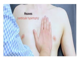
- Check whether there is a parasternal heave in the chest.
👉🏻For your knowledge Apical impulse
- The apical impulse is also known as the ‘point of maximum impulse (PMI)’. The apical impulse is a palpable beat in the apical area of the heart. It is felt at the 5th intercostal space and to the left of the midventricular line. It is felt during the contraction of the left ventricle during systole. Thrill
- A thrill is a palpable sensation caused by turbulent blood flow. Thrill is also known as a ‘palpable murmur’ and is associated with heart valve abnormalities and ventricular septal defects.
Parasternal heave (parasternal heave)
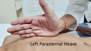
- Parasternal heave is a noticeable outward movement of the chest wall. It can be seen and felt on the left side of the sternum. It indicates increased activity of the enlarged right ventricle of the heart. (The heel of the hand is palpated parallel to the edge of the left sternum.
Auscultation
- Auscultation is used to listen to heart sounds, rhythm, and rate with the help of a stethoscope.
- Abnormal heart sounds, murmurs, and irregular heart rhythms can be detected with the help of auscultation.
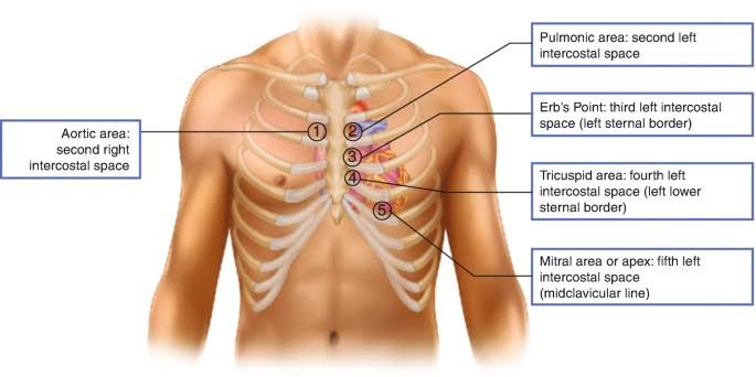
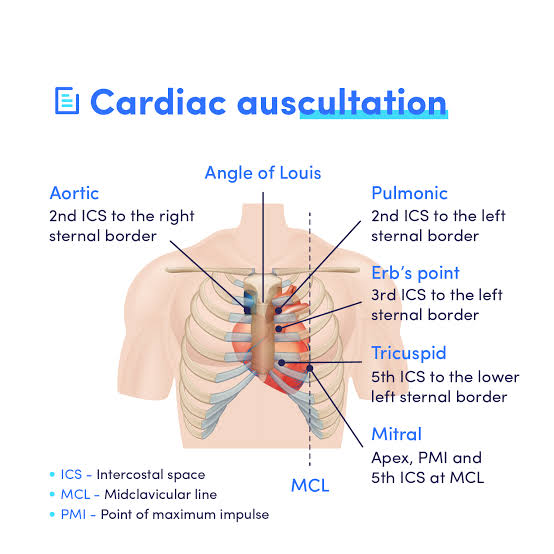
- Auscultate the valve area with the help of a stethoscope. The diaphragm of the stethoscope is used to auscultate it.
- In this, the mitral valve, tricuspid valve, aortic valve and pulmonary valve are auscultated. Which are auscultated in the given area as shown in the picture below.
For your knowledge
Normal heart sound
- S1 and S2 sounds are considered as normal heart sounds. S1 and S2 sounds are heard due to the closure of the atrioventricular valve and semilunar valve. Hence, S1 and S2 sounds are heard during the normal cardiac cycle.
S1 sound :
- Atrioventricular valve i.e. tricuspid valve and mitral valve are closed Due to
- S1 sound is heard. A lob sound is heard during S1 sound. S1 sound is heard during systole. S1 sound can be heard well in the apical area.
S2 sound :
- The S2 sound is heard due to the closure of the aortic and pulmonic valves. A double sound is heard during the S2 sound. The S2 sound is heard during diastole. The S2 sound is best heard in the aortic and pulmonic areas.
Abnormal heart sound
- Abnormal heart sounds are heard when there is a structural or functional problem or abnormality in the heart. Abnormal heart sounds include S3 gallop, S4 gallop, murmur, opening snap, systolic click, friction rub. S3 and S4 sounds are called gallops because they sound like a galloping horse.
S3 sound:
- The S3 sound is also known as the ‘ventricular gallop’ and ‘protodiastolic gallop’. This is a type of extra heart sound that is heard immediately after the S2 heart sound, i.e., a lub-dub-dub sound is heard. The S3 heart sound is heard during early diastole. The S3 sound is caused by rapid ventricular filling. The S3 sound is normally heard in children, youth, and athletic persons, while in the elderly it is considered abnormal and indicates left ventricular failure, mitral regurgitation. The S3 sound can be heard well on the apex side of the heart with the bell of a stethoscope.
S4 sound :
- The S4 sound is also known as the ‘arterial gallop’ and ‘presystolic gallop’. This is a type of extra heart sound. Which is heard just before the S1 heart sound, i.e. lub-lub-dub is heard. The S4 sound is heard during late diastole. S4 sound is seen due to atria contracting during non-ventricular contraction. S4 sound is considered totally abnormal. S4 sound indicates hypertensive heart disease, aortic stenosis, myocardial infarction. The S4 sound can be heard best on the apex side of the heart with the bell part of the stethoscope.
Opening snap :
- Normally, no sound is heard when the valve opens, but when valvular disease is present, a sound is created by the leaflets at the time of valve opening during systolic and diastolic, which is known as the opening snap. Opening snap is an abnormal high pitched diastolic sound that is heard during the opening of the AV valve and is heard during early diastole, i.e. the S2 sound bar is heard immediately. For example, mitral stenosis
Systolic click :
- Like opening snap, when stenosis occurs in the semilunar valve, a short high pitched sound is heard at the time of opening of the valve, which is called a systolic click. Systolic click is heard during early systole, i.e. immediately after the S1 sound.
Friction rub :
- A harsh and grating sound heard during systole and diastole is known as friction rub. It is mainly heard due to abrasion of the inflamed pericardial surface during pericarditis. Friction rub can be best heard using the diaphragm of a stethoscope.
Murmur:
A heart murmur is an abnormal heart sound that occurs due to turbulent blood flow in the heart and blood vessels. The intensity, timing, and duration of the murmur sound depend on its cause. Murmurs are of two types: systolic murmurs and diastolic murmurs
- Systolic Murmur:A systolic murmur is a type of heart murmur that is heard during contraction, or systole. Systolic murmurs are heard in conditions such as aortic stenosis and mitral regurgitation.
- Diastolic murmurs: Diastolic murmurs are a type of heart murmur that is heard during relaxation, or diastole. Diastolic murmurs are heard in conditions such as aortic regurgitation and mitral stenosis.
Diagnostic test for heart and cardiovascular system
Laboratory test:
- Complete blood in laboratory tests Count, cardiac markers, blood chemistry tests, lipid profile and coagulation studies are performed.
✓ Complete Blood Count :
- Red Blood Cell Count
- White Blood Cell Count
- Platelets
- Differential Count
✓Cardiac Biomarker :
- Creatine Kinase
- Cardiac Troponin
- Myoglobin
- Lactic Dehydrogenase
✓ Blood Chemistry Test :
- Electrolyte Levels
- Lipid Profile
- Blood Glucose Level
✓Coagulation Study :
- Prothrombin Time
- Activated Partial Thromboplastin Time
- Partial Thromboplastin Time
Cardiac enzymes
- When heart cells or muscles are damaged (such as a heart attack), proteins and enzymes are released from them and are known as cardiac enzymes. These enzymes dissolve in the blood and their levels are found in high levels in the blood. Therefore, the level of cardiac enzymes is measured through a blood test and cardiac conditions are identified. Cardiac enzymes include creatine kinase, lactate dehydrogenase, cardiac troponin, myoglobin.
Creatine kinase
- Creatine kinase is a type of enzyme that is found in the tissues of the heart, brain and skeletal muscles. The enzyme found in the heart is known as CK-MB, the enzyme found in the brain is known as CK-BB and the enzyme found in the tissues of skeletal muscles is known as CK-MM. Therefore, when cardiac cells are damaged, creatine kinase is released into the blood and the level of CK-MB in the blood increases. The level of CK-MB increases within four to six hours of cardiac cell damage and reaches its peak level within 12 to 24 hours and returns to normal levels within 48 to 72 hours. Therefore, an increase in CK-MB levels indicates a condition of heart attack.
Cardiac troponin (Cardiac troponin)
- Troponin is a type of protein that is found only in cardiac muscles. It plays an important role in the contraction of cardiac muscles. Troponin is found in two forms: troponin T and troponin I. Troponin T is a highly sensitive indicator of myocardial damage. When heart muscle is damaged, troponin is released from the muscles and enters the blood. Troponin T is a highly sensitive indicator of myocardial damage. The level of this troponin is elevated within 4 to 6 hours of heart muscle damage, reaches a peak level at 10 to 24 hours, and remains elevated for up to 7 days after muscle damage. Cardiac troponin is a crucial marker for diagnosing heart attacks and is also useful for assessing the severity of heart muscle damage.
Myoglobin
- Myoglobin is a monomeric protein. It is also known as an oxygen storage protein because it can bind to oxygen and release oxygen when needed. That is, it stores oxygen. Myoglobin is a protein found in both cardiac and skeletal muscles. Therefore, when cardiac or skeletal muscles are damaged, myoglobin is released into the blood and its levels in the blood increase. Myoglobin levels increase within 1 hour of acute myocardial infarction, peak within 4 to 12 hours, and return to normal levels within 18 hours.
Lactic dehydrogenase (LDH)
- Lactic dehydrogenase is an enzyme found in many tissues of the body, including the heart, liver, kidney, lung, and red blood cells. Cell. Lactic dehydrogenase plays an important role in converting lactate to pyruvate during anaerobic metabolism. There are five types of lactic dehydrogenase: LDH1, LDH2, LDH3, LDH4, LDH5. Of these, LDH1 and LDH2 are present in the heart, kidney and red blood cells, while LDH3 is present in the lungs and LDH4 and LDH5 are present in skeletal muscles. Normally, LDH2 is higher than LDH1. But when cardiac muscles are damaged, the level of LDH1 exceeds LDH2. LDH levels increase within 8 to 12 hours of MI, peak at 24 to 48 hours, and return to normal levels within 5 to 7 days.
Lipid profile
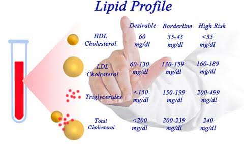
- Lipid profile is a blood test that measures various types of lipids in the blood, such as cholesterol, high-density lipoprotein, low-density lipoprotein, triglycerides, phospholipids. This test can assess whether there is a risk of cardiovascular disease.
X-ray
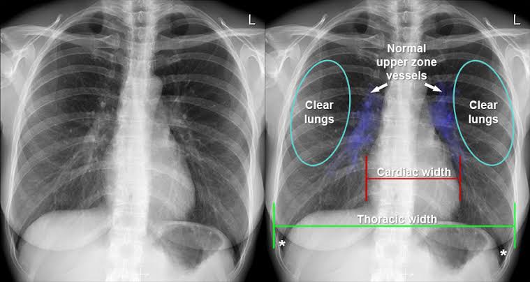
- A chest X-ray uses a small amount of X-rays to create a 2D image of the area. In this, mainly the anterior, posterior and lateral side views are taken. With the help of X-rays, the size, shape and position of the heart can be known. Chest X-ray is used to diagnose conditions such as pericardial calcification, pericardial effusion.
Computed tomography scan (CT scan)
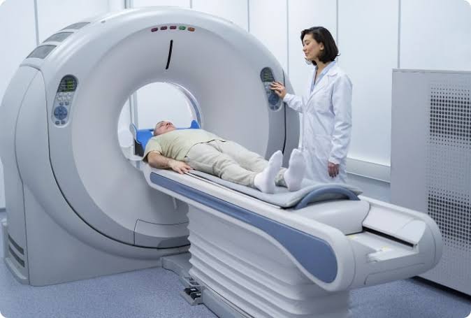
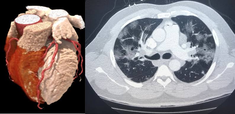
- A computed tomography scan (CT scan) is also known as computerized axial tomography (CAT). It is an imaging technique that uses X-rays and computer processing to create detailed cross-sectional images of the body. Which provides valuable information about the body’s internal organs, bones, soft tissues, and blood vessels. During a chest CT scan, it provides images of the heart and great vessels, which can be used to evaluate cardiac masses, as well as diseases of the aorta and pericardium.
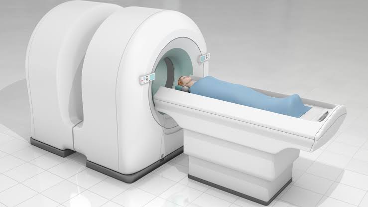
Magnetic resonance imaging (MRI)
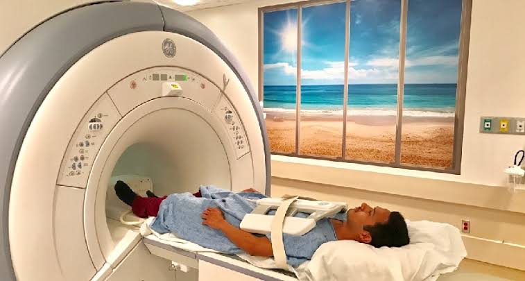
- Magnetic resonance imaging is a non-invasive medical imaging technique that provides a 3D image of the heart. This test uses powerful magnets and radio waves to create a cross-sectional image of a given area and provides detailed information about the heart, lungs, and blood vessels. MRI is used to detect abnormalities of the heart chambers and valves, as well as to identify tumors and coronary artery disease.
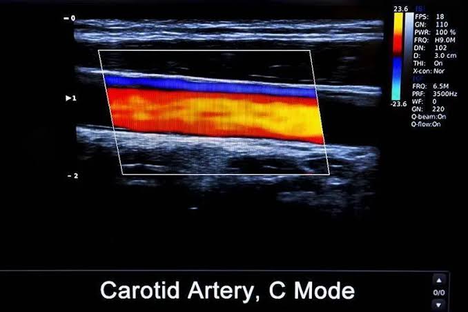
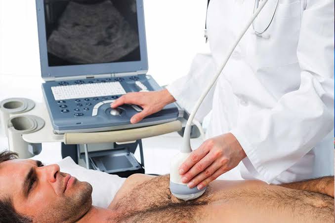
Doppler ultrasound
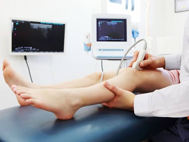
- Doppler ultrasound is a non-invasive imaging technique that uses sound waves to evaluate blood flow. This test measures blood flow by changing the frequency of the sound waves and reflecting them off blood cells. Doppler ultrasound is used to identify blood clots, narrowing arteries, and problems associated with heart valves.
Duplex ultrasound
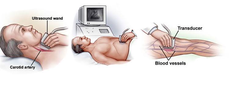
- This is a non-invasive imaging method that combines Doppler ultrasound and traditional ultrasound imaging. In this test, the speed and moving capacity of blood flow are measured using a transducer. It is useful for detecting conditions such as blockages in blood flow, such as arterial stenosis.
Gated blood pool scan
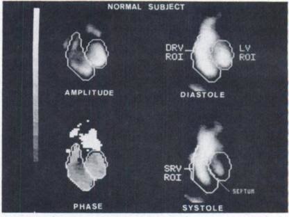
- Gated blood pool scan is also known as ‘radionuclide ventriculography’ and ‘multigated acquisition scan (MUGA)’. It is a nuclear medicine imaging technique used to assess the function of the heart chambers, especially the ventricles. In this test, a small amount of radiotracer is injected into the bloodstream. This tracer circulates through the heart, and a special camera captures an image of the area. This image provides information about ventricular function, ejection fraction, ventricular volume, and cardiac output. Gated blood pool scan is used to diagnose and monitor heart failure, cardiomyopathy, coronary artery disease.
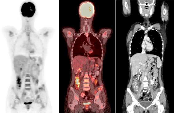
Positron emission tomography
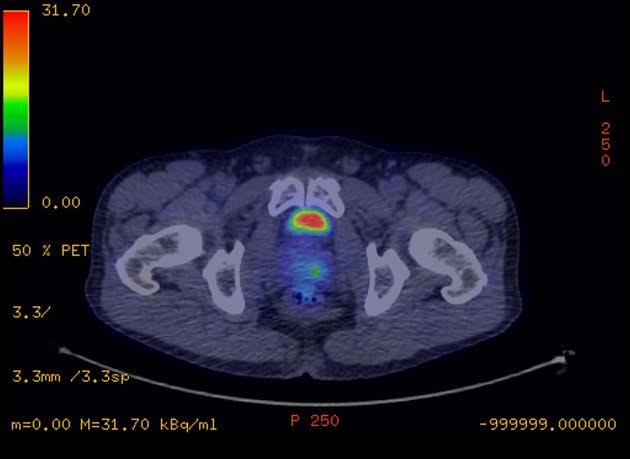
- Positron emission tomography is a nuclear medicine imaging technique that provides 3D images of organs and tissues of the body. In this test, a radioactive tracer, nitrogen 13 ammonia, is injected into the bloodstream. This radioactive tracer emits positrons. This positron colloid combines with electrons in the body and releases gamma rays. The area is then scanned to show the distribution of the radiotracer in the body. Therefore, PET scans are used in medical fields such as oncology, neurology, and cardiology. PET scans provide information about blood flow, metabolic function, and cardiac function. Therefore, PET scans are used to evaluate myocardial ischemia, myocardial metabolic function.
Angiography
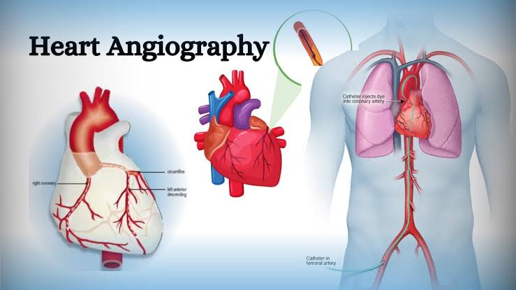
- Angiography is a type of imaging test used to visualize blood vessels in various parts of the body. Angiography includes arteriography and venography. In which a contrast agent is injected into the blood stream and an X-ray image of the given area is taken and blockages, obstructions, blood clot formation and other conditions in the blood vessels are identified. For example, aneurysm. Coronary angiography is performed for the heart.
Phlebograohy (Phlebography)
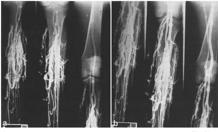
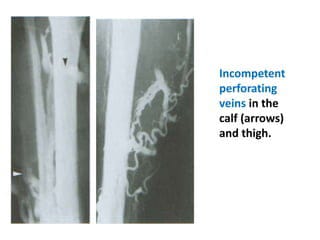
- Phlebography is also known as venography. It is a diagnostic imaging technique used to visualize veins in the body, especially those in the extremities. In this test, a contrast dye is injected into a vein in the foot or hand and an X-ray image of the given area is taken and the vein is visualized. Phlebography is used to identify conditions such as deep vein thrombosis, varicose veins, venous insufficiency, and venous malformations.
Radioisotope imaging
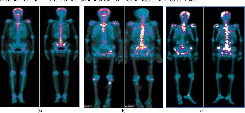
- Radioisotope imaging is also known as ‘nuclear medicine imaging’. It is a medical imaging technique in which a radioactive substance – a radioisotope – is injected intravenously and this substance emits gamma rays. Thus, images of organs, tissues and physiological processes are taken with the help of a special camera. This image is used to detect cancer, heart disease and bone disorders.
Technetium pyrophosphate scan (Tc PYP)
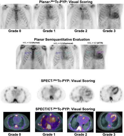
- A technetium pyrophosphate scan is a nuclear imaging test used to diagnose cardiac amyloidosis. This is a condition in which abnormal proteins accumulate in heart tissue. During this test, the radioactive tracer technetium pyrophosphate is injected into the bloodstream. This radioactive tracer binds to the amyloid protein deposited in the heart so that an image can be taken of that area with the help of a special camera and it can be identified through this image.
Fluroscopy
- This is an imaging technique in which the heart can be visualized on an X-ray screen. In which a real-time moving image of the body’s internal structure is created by continuously using an X-ray beam. With its help, cardiac and vascular pulsations can be seen, as well as the shape and size of the heart. Therefore, it is used to provide guidance during catheter insertion and positioning of intravenous electrodes during cardiac catheterization.
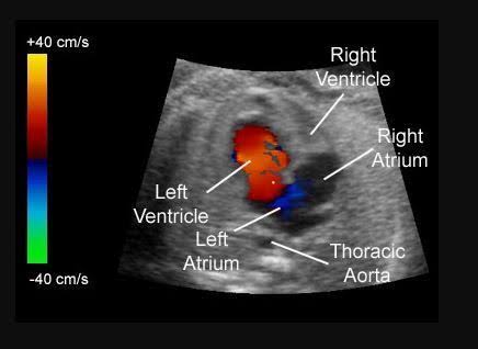
Echocardiograohy (Echocardiography)
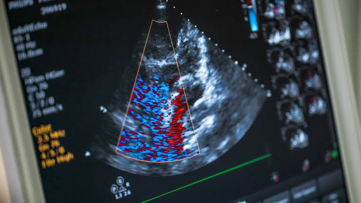
- Echocardiography is a non-invasive test that uses sound waves to create an image of the heart. It provides detailed information about the heart’s structure and function. It provides information about the shape and size of the heart chambers, pumping function, heart valve movement, and blood flow. Echocardiography is used to diagnose and monitor conditions such as heart valve disorders, congenital heart defects, and heart failure.
Electrocardiogram (ECG / EKG) (Electrocardiogram)
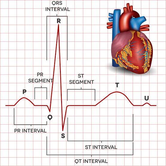
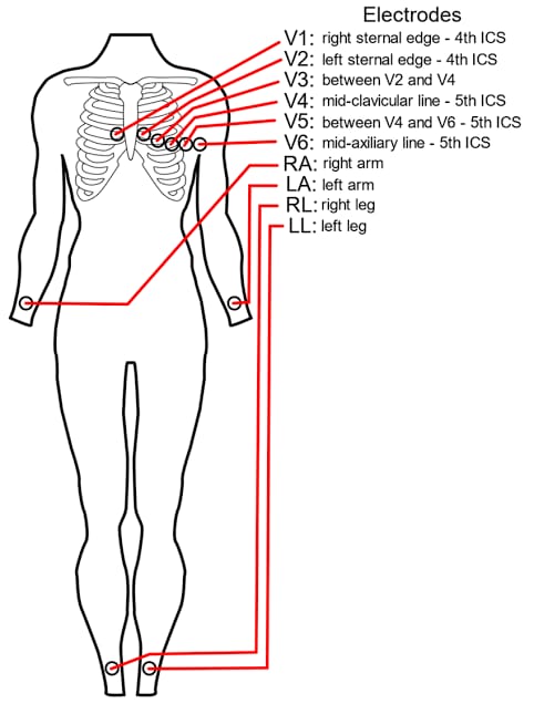
- Electrocardiogram is a non-invasive test used to record the electrical activity of the heart. In which electrodes are placed in different areas as shown in the picture below and the electrical activity of the heart is recorded on the page for a given time period and then this electrical activity is evaluated. This electrical activity depends on the conduction, rate and rhythm. Electrocardiograms are used to detect arrhythmias, heart attacks, electrolyte imbalances, and other cardiac conditions.
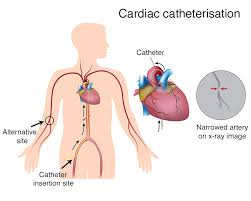
Cardiac catheterization
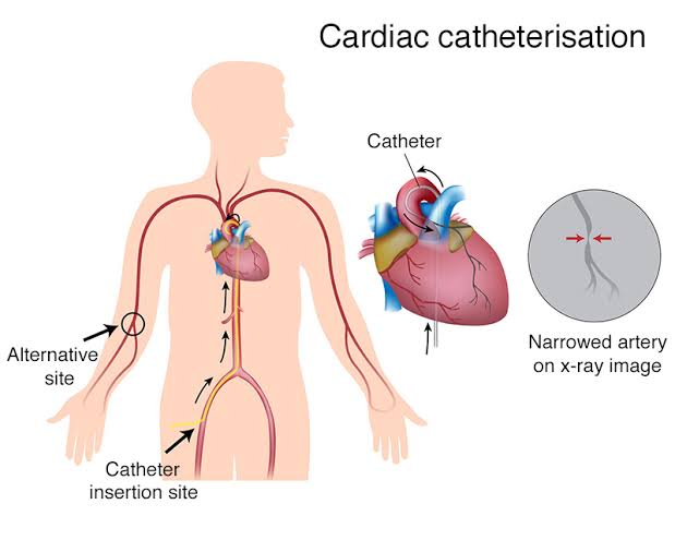
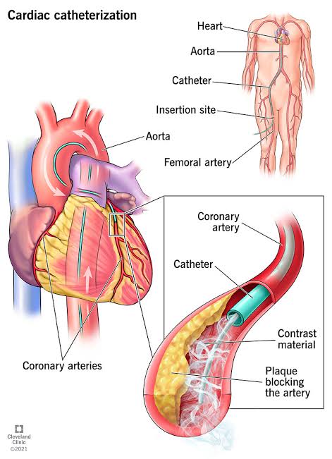
- Cardiac catheterization is an invasive diagnostic test used to diagnose and treat heart conditions. In this test, a thin flexible tube called a catheter is inserted into the heart through blood vessels in the groin or arm. Which measures the blood pressure of the heart chambers, great blood vessels, coronary arteries and provides information about the output. Cardiac catheterization is used to evaluate coronary artery disease, heart valve problems and other heart related problems. In addition, cardiac catheterization is used in procedures like angioplasty, stent replacement. Cardiac catheterization includes right side catheterization and left side catheterization.
Electrophysiology study

- Electrophysiology studies are used to evaluate the electrical activity of the heart and diagnose arrhythmias. During this test, thin flexible wires (catheters) and electrodes are inserted into the heart through blood vessels and the electrical signals are recorded and evaluated to help select the best treatment options such as medication, cardiac ablation, implantation of a pacemaker, etc.
Holter monitoring
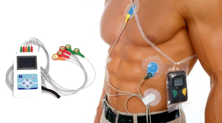
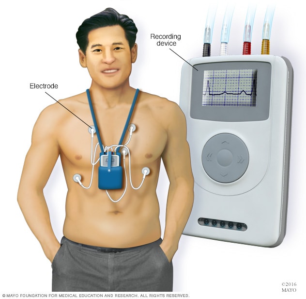
- This is a type of diagnostic test. In which the electrical activity of the heart is monitored for 24 to 48 hours with the help of a Holter monitor. A Holter monitor is a battery-operated portable electrocardiogram device. It can be easily worn and the electrodes in it are placed as shown in the picture and the electrical activity of the heart is monitored. During this test, the patient is asked to wear loose clothes and the patient is given a sponge bath, so the patient is prohibited from bathing during this test. Holter monitoring test is used to detect arrhythmia
Hemodynamic monitoring
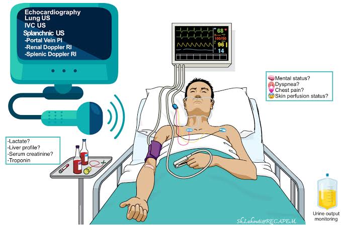
- Hemodynamic monitoring is a type of invasive test in which continuous blood flow and blood pressure are monitored. So that cardiac function and tissue perfusion can be evaluated. Hemodynamic monitoring uses invasive methods such as catheterization and non-invasive methods such as echocardiography. This method is used in conditions such as shock, heart failure, and sepsis.
Central venous pressure monitoring
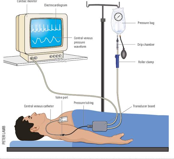
- In central venous pressure monitoring, blood is measured directly from the right atrium or vena cava. In this method, a central venous catheter is placed in a central vein such as the jugular vein or subclavian vein. Which performs pressure measurement. Which provides information about volume status, cardiac function. It provides information about hemodynamic stability in conditions such as shock, heart failure and sepsis. Normal CVP is 2-6 mm hg.
Pulse volume recording – PVR (Pulse volume recording)
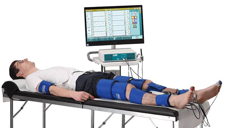
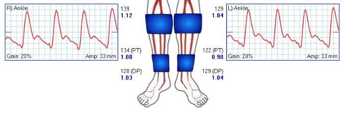
- Pulse volume recording is a non-invasive diagnostic method used to measure blood flow and pressure in an artery in the limbus. In this, a pressure cuff is placed on the patient’s leg and inflated, then rapidly deflated, and changes in venous volume are recorded. If there is a decrease in venous volume, it indicates thrombus.
- Pulse volume recording is used to diagnose deep vein thrombosis, pulmonary embolism, and peripheral vascular disease.
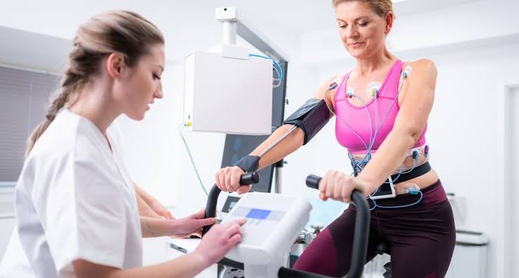
Cardiac stress test
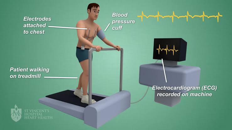
- Cardiac stress test is also known as ‘exercise stress test’ and ‘treadmill test’. In this test, the patient is asked to walk on a treadmill or ride a stationary bike and at this time the patient’s heart rate, blood pressure and electrical activity are noted and the heart’s response to stress is checked. This test is used to detect coronary artery disease and assess the condition of the heart after a heart attack or heart surgery.
Tilt table test
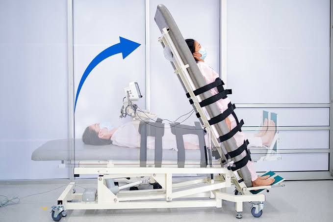
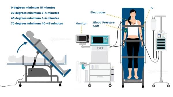
- The tilt table test is a type of diagnostic test used to evaluate the cause of fainting or syncope. In this test, the patient is made to lie on a table and the table is tilted at different angles as shown in the picture and during this, heart rate, blood pressure and other symptoms are noted.
Peripheral vascular stress test
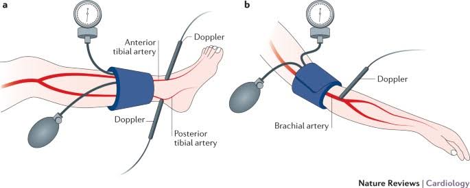
- Peripheral vascular stress test is also known as ‘Ankle-Brachial Index (ABI) test’. With the help of this test, the circulation and blood flow of the arm and leg can be assessed during physical activity. In this test, the patient is asked to walk on a treadmill or pedal on a stationary bicycle. Heart rate and blood pressure are measured before, during and after this exercise. Peripheral vascular stress test is used to identify peripheral vascular disease.
Exercise tolerance test

- An exercise tolerance test is also known as a ‘stress test’ or ‘exercise electrocardiogram’. It is a diagnostic test used to assess how well the heart works during physical activity. During this test, the patient is asked to run or walk on a treadmill and at that time the patient’s heart rate, blood pressure, and ECG are monitored. This test is used to detect reduced blood flow in the heart muscles, irregular heart rhythms, and symptoms of heart failure.