ENGLISH ANATOMY UNIT 13 JOINTS
JOINTS
- Joint and Types of joint.
Two or more bones in the body join together to form a joint. Different movements are seen at the joint. The range of motion found in a joint depends on the bone, cartilage, connective tissue and muscular structures that make up the joint.
It is divided into different types based on its mobility in this joint. Based on the movement observed at the joint, the classification of the joint is given as follows.
- Freely movable joint.
This joint is also called synovial joint. In which two or more bones are connected with each other with the help of articular cartilage to form this type of joint. In this joint, a cavity or space is formed around the joint, it is called a synovial cavity. Maximum movement is seen in this type of joint. Hence it is called a freely movable joint. Synovial joint is also known as other Diarthroses.
- Characteristics of synovial joint. (Characteristics of Synovial Joint).
A synovial joint means maximum movement occurs where two bones form a joint. That type of joint is called a synovial joint or a freely movable joint. A synovial joint has the following characteristics
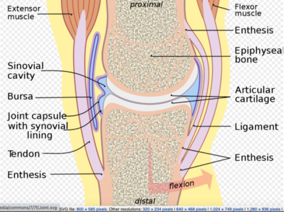
HYLIENE CARTILAGE (Hyline Cartilage)..
This cartilage is also called articular cartilage. It is located at the end of the bone. This cartilage is found between two bones where they join each other.
This cartilage prevents the ends of the bones from rubbing. It gives ability to withstand pressure. Due to this cartilage, the movement between the two bones becomes smooth and painless.
INTRA CAPSULAR STRUCTURE (capsular structure).
Where the two bones of a synovial joint meet, a structure forms around the cavity of the joint. The structure inside this capsule is known as capsular structure or intracapsular structure. The capsule is a double layer membrane. In which the outer layer is made of fibrous tissue and the inner layer is made of synovial membrane. The structure inside the capsule is called intracapsular structure. Which includes the following structure.
- SYNOVIAL MEMBRANE (synovial membrane).
This membrane covers all the membrane lining around the joint in the lining of the capsule. This membrane is not located near the hyaline cartilage. All but these capsules have a membrane covering the inner structure called the synovial membrane. It is a membrane made of loose connective tissue.
This membrane secretes a fluid called synovial fluid.
- SYNOVIAL FLUID (synovial fluid).
Synovial fluid is a viscous fluid. Which acts as a lubricant for the joint. The cavity where the synovial fluid resides is called the synovial cavity.
This synovial fluid contains hyaluronic acid. This fluid contains some phagocytic cells which protect the joint by removing the microorganisms and some cellular debris in the cavity of the joint.
This fluid acts as a nourishment by supplying nutrient material to the inner structure of the joint. This fluid also performs a wear-and-tear function on the joint. It is also important for maintaining joint stability.
- INTRA CAPSULAR LIGAMENT (intracapsular ligament).
There are bands of connective tissue on the inside of the capsule and on the ends covering the two bones of the joint. Which is known as intracapsular ligament. Ligaments within the capsule provide stability to the joint and help hold the two ends of its bones together.
- BURSE (Burse).
These tiny bumps are filled with synovial fluid. Which is called Burse. This sac is located around certain joints such as the knee joint. It is not located near the pressure bearing part but is surrounded by a shell which acts like a cushion. Which also plays an important role in preventing friction.
- FIBRO CARTILEGINIOUS DISC.
Some synovial joints have a disc made of fibrous cartilage near the articulating surface. This disc acts as a shock absorber at the joint. Which gives the joint additional pressure bearing capacity and stability.
EXTRA CAPSULAR STRUCTURE (Extra capsular structure).
The structures outside the capsule near both ends of the bone are called extracapsular structures. Which includes the following structure.
- LIGAMENT (ligaments).
Fibrous connective tissue structures are located at the ends of both bones near the joint. Which is known as ligament. Both of which pass through opposite ends of the bone and are associated with strength and movement. Due to this ligament, the stability and position of the joint is maintained.
- MUSCLES (Muscles).
Muscles pass through the structures surrounding the joint. Which are skeletal muscles. These muscles are connected to provide support and support to the joint. These muscles are also useful for performing different types of movement at the joint.
- TENDON (TENDON).
At the joint, the ends of the muscles are connected to the bones by tendons. These tendons are bands of connective tissue. Which attaches the muscles to the bone and allows movement of the joint.
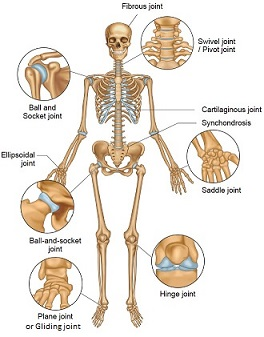
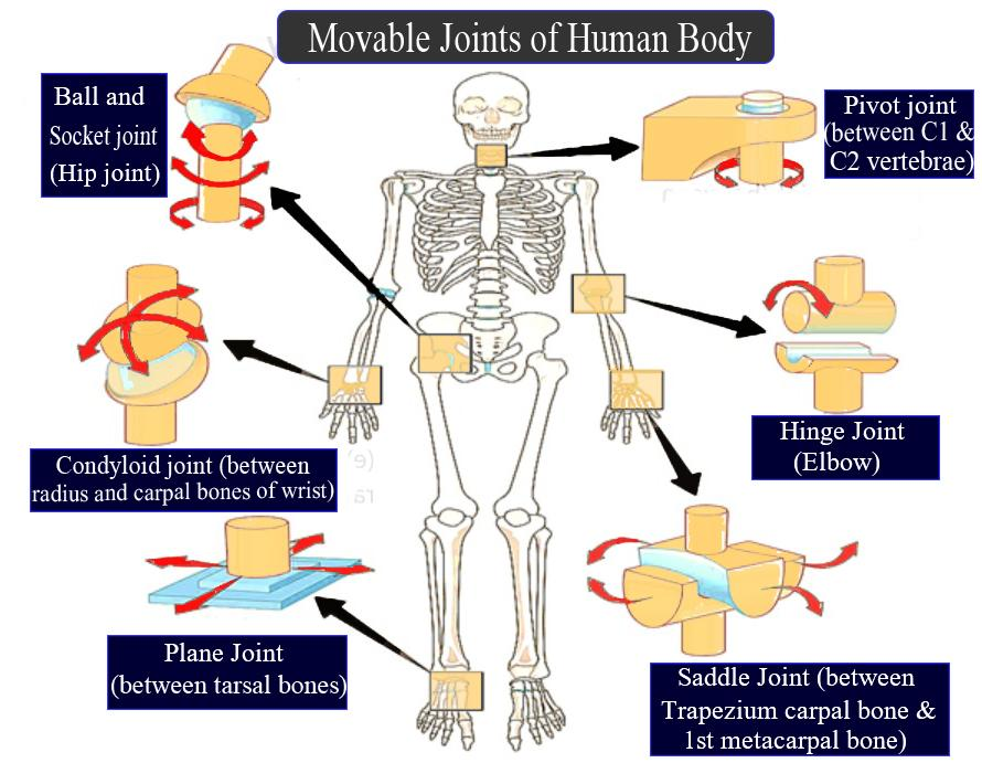
Types of synovial joint
Important synovial joints of the body.
- Slightly movable joints.
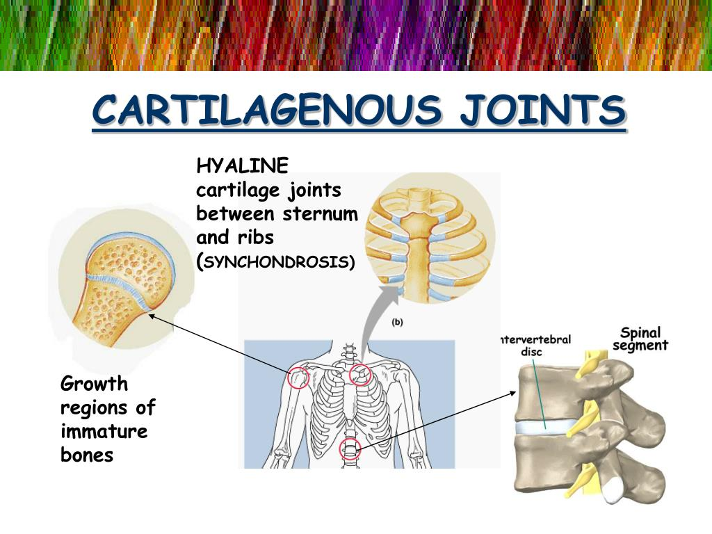
This joint is also known as second cartilaginous joint. In which the bones are connected with each other with the help of cartilage. Hence a small moment is observed at this joint. It can also be known as other Amphiarthroses.
The joint between the bodies of two vertebrae, the joint between the sternum bone and the rib are such joints. Where there is little movement of the joint.
3.Fixed joint.
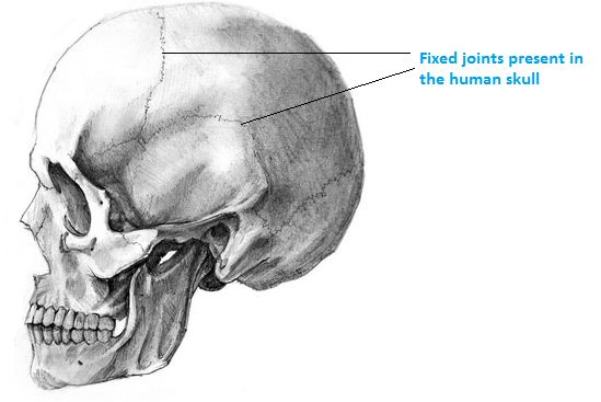
This type of joint is also known as another fibrous joint. In which the bones are closely connected with each other and no movement is seen at the joint. It is also known by another name Synarthrosis.
These are the joints formed between the cranium bones of the skull. This joint forms a suture such as the coronal suture.