ENGLISH-INTEGUMENTARY -PART-2(UPLOADED)
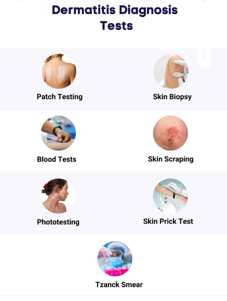
INTEGUMENTARY DIAGNOSTIC TEST :-
Culture :-
- Bacterial, viral and fungal infections through culture Identified. Culture can help determine the type of bacterial infection so that specific treatment can be provided. In a culture test, the collected sample is mixed with a special material – culture and the microorganisms present in it are detected. Special care has to be taken to take its sample
Wood lamp examination (Wood lamp examination) :
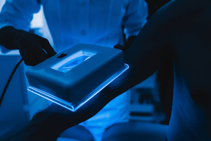
- A Wood’s lamp examination uses ultraviolet light to detect bacterial and fungal infections. In addition, skin pigmentation is also determined. This UV light is passed over the suspected area on the skin and the fungus present there is visualized. This light does not cause any harm to the skin and eyes.
Patch testing :
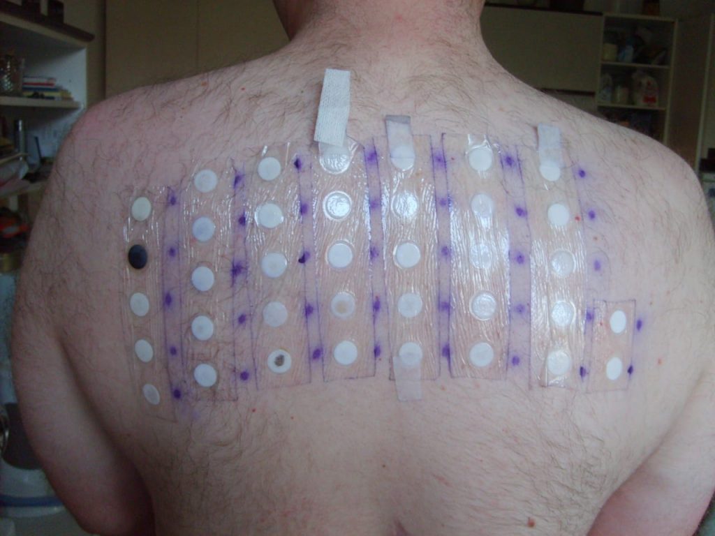
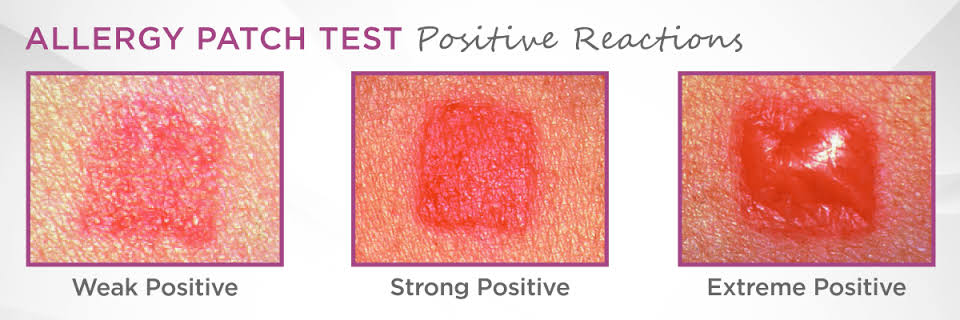
- Patch testing is a diagnostic method in which the patient is determined to be allergic to the substance causing inflammation. In patch testing, some suspected allergens are applied to the skin as a patch and the resulting reaction is noted. A positive reaction is characterized by redness and itching. A strong positive reaction involves blistering papules and severe itching. While blisters, pain and ulceration are seen in extreme positive reactions.
Skin biopsy (skin Biopsy) :
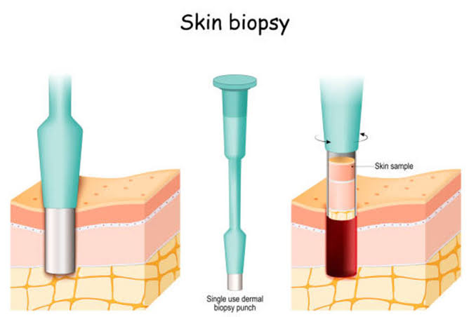
- A skin biopsy involves taking a small sample of tissue from a skin lesion or suspected area. A piece is taken and microscopic examination is done to check whether malignancy is present or not. So that an exact diagnosis can be known. This biopsy is collected from nodules, plaques, blisters and lesions.
There are three main types of skin biopsy:
i) Shave biopsy:
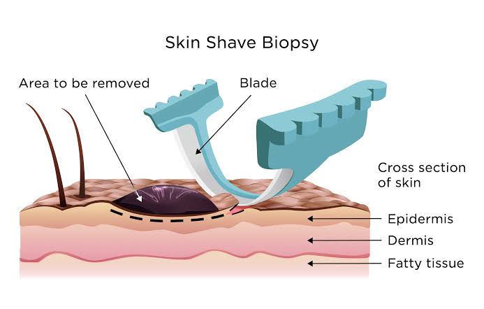
- In shave biopsy, a biopsy is collected using a tool like a razor (blade). In shave biopsy, a biopsy is collected from the upper epidermal layer.
ii) Punch biopsy (punch biopsy):
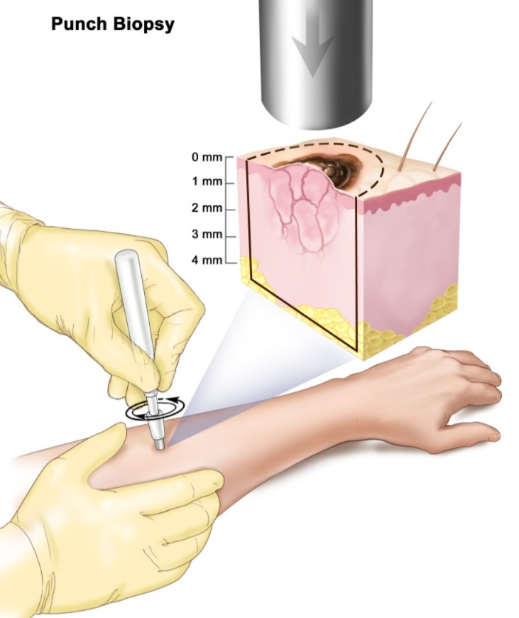
- In a punch biopsy, a small piece of skin is taken using a punch instrument, which includes the epidermis, dermis, and fat layer. In which a circular blade is attached to a pencil-like instrument and a skin core is collected by rotating it deep.
iii) Excisional biopsy:
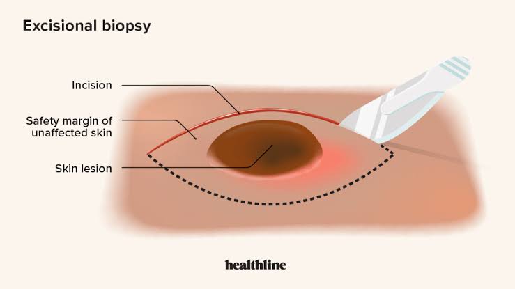
- In an excisional biopsy, the entire lump and irregular skin is removed using a scalpel, along with the surrounding healthy skin. And it is sent for microscopic examination.
Tzanck’s smear (Tzanck’s smear) :
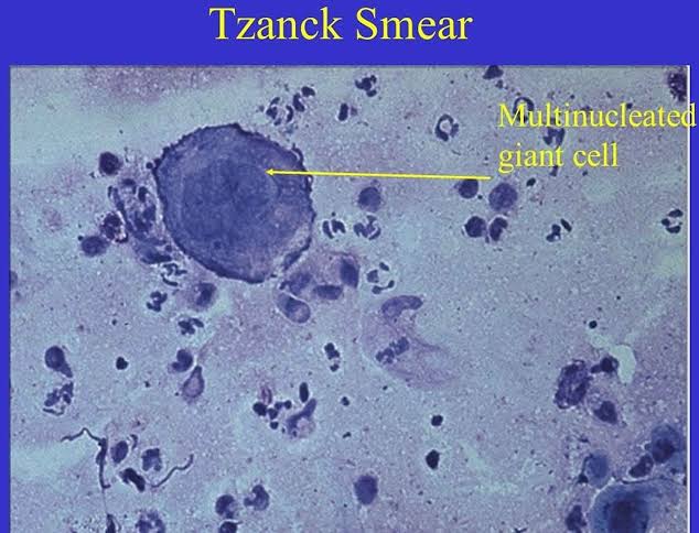
- Tzanck smear is a cytological diagnosis method. In which the blister is broken down and the cellular components are collected and examined with the help of a microscope and it is checked whether Tzanck cells are present in it or not. Tzanck cells are acantholytic cells that are present in the blisters of herpes simplex, herpes zoster, pemphigus vulgaris.
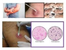
Skin scraping (skin scraping):
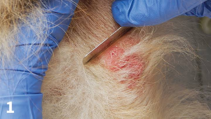
- In skin scraping, a sample is collected by scraping (scraping) the suspected area or lesion with a scalpel blade. This blade is coated with oil so that the sample sticks to the blade while scraping. The collected sample is transferred to a glass slide and a few drops of potassium hydroxide or mineral oil are added to it, covered with a cover slip and examined under a microscope. This procedure is used to diagnose fungal infections.
Immunofluorescence:
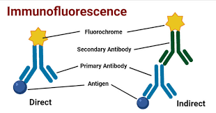
- Immunofluorescence is a technique by which specific proteins or antigens present in tissues are visualized. In this technique, a specific antibody is conjugated with a fluorescent dye such as fluorescent isothiocyanate, with the help of which we can visualize the specific antigen under a microscope under UV light. Thus, the antigen present in the skin can be detected. With the help of immunofluorescence technique, IgG antibodies found in pemphigus vulgaris can be identified. Also, varicella can be identified in skin cells in herpes zoster.

Published
Categorized as Uncategorised